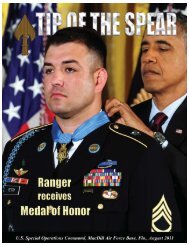
Spring 07 front cover - United States Special Operations Command
Spring 07 front cover - United States Special Operations Command
Spring 07 front cover - United States Special Operations Command
- No tags were found...

You also want an ePaper? Increase the reach of your titles
YUMPU automatically turns print PDFs into web optimized ePapers that Google loves.
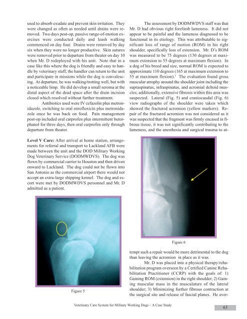
used to absorb exudate and prevent skin irritation. Theywere changed as often as needed until drains were removed.Two days post-op, passive range-of-motion exerciseswere conducted daily and leash walkingcommenced on day four. Drains were removed by daysix when they were no longer productive. Skin sutureswere removed prior to departure from theater on day 10when Mr. D redeployed with his unit. Note that in acase like this where the dog is friendly and easy to handleby veterinary staff, the handler can return to the unitand participate in missions while the dog is convalescing.At departure, he was walking/trotting well, but witha noticeable limp. He did develop a small seroma at thedistal aspect of the dead space after the drain incisionclosed which resolved without further treatment.Antibiotics used were IV cefazolin plus metronidazole,switching to oral enrofloxicin plus metronidazoleonce he was back on food. Pain managementpost-op included oral carprofen plus intermittent butorphanolfor three days, then oral carprofen only throughdeparture from theater.Level V Care: After arrival at home station, arrangementsfor referral and transport to Lackland AFB weremade between the unit and the DOD Military WorkingDog Veterinary Service (DODMWDVS). The dog wasflown by commercial carrier to Houston and then drivenonward to Lackland. The dog could not be flown intoSan Antonio as the commercial airport there would notaccept an extra-large shipping kennel. The dog and escortwere met by DODMWDVS personnel and Mr. Dadmitted as a patient.Figure 6Figure 5The assessment by DODMWDVS staff was thatMr. D had obvious right forelimb lameness. It did notappear to be painful and the lameness diagnosed to befunctional in its etiology. This was attributable to significantloss of range of motion (ROM) in his rightshoulder, specifically loss of extension. Mr. D’s ROMwas measured to be 75 degrees (130 degrees at maximumextension to 55 degrees at maximum flexion). Ina dog of his breed and size, normal ROM is expected toapproximate 110 degrees (165 at maximum extension to55 at maximum flexion). 1 The evaluation found grossmuscular atrophy around the shoulder joint including thesupraspinatus, infraspinatus, and acromial deltoid muscles;additionally, extensive fibrosis within this area wassuspected. Lateral (Fig. 5) and craniocaudal (Fig. 6)view radiographs of the shoulder were taken whichshowed the fractured acromion (yellow markers). Repairof the fractured acromion was not considered as itwas suspected that the fragment was firmly encased in fibroustissue, it was not significantly contributing to thelameness, and the anesthesia and surgical trauma to attemptsuch a repair would be more detrimental to the dogthan leaving the acromion in place as it was.Mr. D was placed into a physical therapy/rehabilitationprogram overseen by a Certified Canine RehabilitationPractitioner (CCRP) with the goals of: 1)Gaining ROM (extension) in the right shoulder; 2) Gainingmuscular mass in the musculature of the lateralshoulder; 3) Minimizing further fibrous contraction atthe surgical site and release of fascial planes. He aver-Veterinary Care System for Military Working Dogs – A Case Study63