biarticular muscles influence postural responses - Reinbolt ...
biarticular muscles influence postural responses - Reinbolt ...
biarticular muscles influence postural responses - Reinbolt ...

Create successful ePaper yourself
Turn your PDF publications into a flip-book with our unique Google optimized e-Paper software.
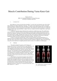
XIII International Symposium on Computer Simulation in BiomechanicsJune 30 th - July 2 nd 2011, Leuven, BelgiumBIARTICULAR MUSCLES INFLUENCE POSTURAL RESPONSES:IMPLICATIONS FOR TREATMENT OF STIFF-KNEE GAITAshley E. Clark 1 , Ajay Seth 2 , & Jeffrey A. <strong>Reinbolt</strong> 11Mechanical, Aerospace, & Biomedical Engineering, The University of Tennessee, Knoxville, TN, USA2Bioengineering, Stanford University, Stanford, CA, USAEmail: aclark54@utk.edu, Web: http://rrg.utk.eduINTRODUCTIONStiff-knee gait is a prevalent and troublesomemovement abnormality among children withcerebral palsy, where peak knee flexion isdiminished during swing phase. This diminishedflexion has been attributed to the excessiveknee extension moment produced by the rectusfemoris muscle [1,2]. This muscle is a<strong>biarticular</strong> muscle crossing the hip and knee.Rectus femoris transfer surgery, a commontreatment for stiff-knee gait, reattaches thedistal tendon of this <strong>biarticular</strong> muscle to a newsite, such as the sartorius insertion on the tibia.This treatment aims to decrease the muscle’sknee extension moment while preserving its hipflexion moment by altering the original, presurgical<strong>biarticular</strong> function.Biarticular <strong>muscles</strong> play a unique role in motorcontrol. Some suggest they function as energyconveying straps between joints [3,4]. Otherssuggest <strong>biarticular</strong> <strong>muscles</strong> may contribute tocontact control tasks by distributing the netmoments over multiple joints [4]. As a <strong>biarticular</strong>muscle, rectus femoris may offer unrecognizedbenefits that <strong>influence</strong> <strong>postural</strong> <strong>responses</strong>.In this study, we used musculoskeletalmodeling and forward dynamic simulation toinvestigate the <strong>influence</strong> of <strong>biarticular</strong> <strong>muscles</strong>in <strong>postural</strong> <strong>responses</strong>. Our goal was to compareposterior, whole-body center of mass (CoM)displacements in response to anterior supportsurfacetranslations for separate simulations ofpre- and post-surgical transfer of the rectusfemoris to the sartorius. We hypothesized thatthe pre-surgical simulation has larger peakrecovery of the original CoM location relative tothe support surface compared with the postsurgicalone.METHODSWe assessed the <strong>influence</strong> of rectus femoristransfer surgery on <strong>postural</strong> <strong>responses</strong> bycreating dynamic simulations of a patient withcerebral palsy. A three-dimensionalmusculoskeletal model with 92 muscle-tendonactuators and 23 degrees of freedom wascreated in OpenSim (Fig. 1). The model wasscaled to represent the size of the patient usingpreviously collected gait analysis data [5,6,7]. Apre-surgical simulation was altered to representsurgical transfer of the rectus femoris to thesartorius (Fig. 1a and 1b) [7]. The foot-groundinterface was modeled using elastic foundationcontact and the feet were based on cadaverfoot geometry [8].The mechanism involved in maintaining anerect posture was based on a muscle stretchreflexcontrol model [9]. The controller’s staticand dynamic thresholds and reflex gain weredetermined using MATLAB’s nonlinear leastsquaresoptimizer algorithm for a 16s-longsimulation of a quiet-standing condition for thepre-surgical model. This controller is analogous(but not identical) to monosynaptic reflexes andafferent mechanisms (e.g., muscle spindles andGolgi tendon organs) responsible for lower-levelmotor control, and should not be affectedfollowing surgical transfer. Thus, the samestretch-reflex controller was used for both thepre- and post-surgical simulations of quietstanding and support-surface translations.a) preb) postFig. 1: Musculoskeletal model of a patient with cerebralpalsy on an anteriorly translating support surface and<strong>biarticular</strong> attachments for the rectus femoris muscle (a) preand(b) post-surgical transfer to the sartorius.
The support-surface was translated anteriorly10 cm over a 0.5s time range with a peakvelocity of 28 cm/s and peak acceleration of0.23g. This translation was based onexperimental methods for adult subjects [10].The adult-sized translational distance wasscaled (83%) to be proportional to the size ofchild in this study. The simulation of supportsurfacetranslation was roughly 1.5s long,including 0.25s of quiet standing, 0.5s oftranslational motion, and 0.75s of recovery.We evaluated our hypothesis regarding thepeak recovery of CoM location relative to thesupport surface by quantifying this recovery as:where d minimum was the minimum CoMdisplacement during recovery (marked as ‘x’ inFig. 2) and d maximum was the CoM displacementat the instant the translation ended (t=0.75s).Both displacements were measured from theoriginal CoM location relative to the supportsurface. For example, a peak recovery of 50%would be indicated by a d minimum of 4 cmfollowing a d maximum of 8 cm. Moreover, a d minimumof 0 cm would result in 100% recovery.RESULTS AND DISCUSSIONThe pre-surgical simulation of <strong>postural</strong>response to support-surface translation had a2.5x larger peak recovery of the original CoMlocation relative to the support surfacecompared with the post-surgical one (Fig. 2).Both cases reached a peak recovery before theend of the 1.5s simulation (t pre =1.4s andt post =1.2s). The pre-surgical simulation had apeak recovery of 35% and the post-surgical onehad 14%. This finding supports our hypothesisthat the pre-surgical simulation has larger peakrecovery compared with the post-surgical one.Fig 2: Posterior displacements of the original CoM locationrelative to the support-surface, and anterior recoveries inresponse to anterior support-surface translations forseparate simulations of pre- and post-surgical transfer of therectus femoris to the sartorius. The support-surfacetranslation time is highlighted in gray. The minimum CoMdisplacement during recovery, dminimum, is marked by an ‘x’.Both pre- and post-surgical simulations used astretch-reflex controller with static (5.41) anddynamic (0.02) thresholds and a reflex gain(8.95) optimized for a 16s-long simulation of thepre-surgical model during quiet-standing. Thissame controller maintained an erect posture forthe post-surgical model during a quiet standingcondition for about 4s, or more than twice thetime (1.5s) for the translational simulations.Successful development of a stretch-reflexcontroller and application to a musculoskeletalmodel for support-surface translations hasallowed for evaluation of the <strong>influence</strong> of the<strong>biarticular</strong> muscle, rectus femoris, in lower-limb<strong>postural</strong> response tasks. Our findings suggestthat distal transfer of the rectus femoris resultsin diminished <strong>postural</strong> response compared tothe original, pre-transfer case, illustrating thebiomechanical advantage that <strong>biarticular</strong><strong>muscles</strong> have in contact control tasks.CONCLUSIONSPatient-specific simulation is a powerful tool toinvestigate the role of <strong>biarticular</strong> <strong>muscles</strong> incontrol tasks. Future investigations will look atthe <strong>influence</strong> of unilateral versus bilateral rectusfemoris transfers on <strong>postural</strong> <strong>responses</strong> andpotential applications to investigate <strong>biarticular</strong><strong>muscles</strong> in other treatments (e.g., hamstringslengthening) for other movement abnormalities(e.g., crouch gait).REFERENCES1. Perry J. Dev Med Child Neurol 29:153-8.1987.2. Goldberg S, et al. J Biomech 36:1111-16.2003.3. Metaxiotis D, et al. J Bone & Joint Surg (Br)86-B:102-9. 2003.4. Van Ingen Schenau GJ, et al. HumanMovement Science 13:495-517. 1994.5. <strong>Reinbolt</strong> J, et al. J Bioimech 41:2362-9.2008.6. Goldberg SR, et al. J Biomech 39:689-98.2006.7. Fox M, et al. J Biomech 42:614-619. 2009.8. Erdemir A, et al. J Biomech Eng131(9):094502. 2009.9. Feng CJ, et al. IEEE Proceedings 20-5:2317-20. 1998.10. Welch TDJ & Ting LH. J Neurophysiol99:1032-8. 2008.ACKNOWLEDGEMENTSWe are appreciative to the Center for MotionAnalysis at the Connecticut Children’s MedicalCenter in Hartford, CT. This research wassupported by The University of Tennessee.