Chem Pharm Bulletin - Synthesis of Novel Curcumin Analogues
Chem Pharm Bulletin - Synthesis of Novel Curcumin Analogues
Chem Pharm Bulletin - Synthesis of Novel Curcumin Analogues
Create successful ePaper yourself
Turn your PDF publications into a flip-book with our unique Google optimized e-Paper software.
64 <strong>Chem</strong>. <strong>Pharm</strong>. Bull. 55(1) 64—71 (2007)<br />
Vol. 55, No. 1<br />
<strong>Synthesis</strong> <strong>of</strong> <strong>Novel</strong> <strong>Curcumin</strong> <strong>Analogues</strong> and Their Evaluation as Selective<br />
Cyclooxygenase-1 (COX-1) Inhibitors<br />
Norbert HANDLER, a Walter JAEGER, b Helmut PUSCHACHER, a Klaus LEISSER, a and Thomas ERKER* ,a<br />
a Department <strong>of</strong> Medicinal <strong>Chem</strong>istry, University <strong>of</strong> Vienna; and b Department <strong>of</strong> Clinical <strong>Pharm</strong>acy and Diagnostics,<br />
University <strong>of</strong> Vienna; Althanstrasse 14, A-1090 Vienna, Austria. Received July 26, 2006; accepted October 23, 2006<br />
<strong>Curcumin</strong>, a major yellow pigment and active component <strong>of</strong> turmeric, has been shown to possess anti-inflammatory<br />
and anti-cancer activities. Recent studies have indicated that cyclooxygenase-1 (COX-1) plays an important<br />
role in inflammation and carcinogenesis. In order to find more selective COX-1 inhibitors a series <strong>of</strong><br />
novel curcumin derivatives was synthesized and evaluated for their ability to inhibit this enzyme using in vitro inhibition<br />
assays for COX-1 and COX-2 by measuring PGE 2 production. All curcumin analogues showed a higher<br />
rate <strong>of</strong> COX-1 inhibition. The most potent curcumin compounds were (1E,6E)-1,7-di-(2,3,4-trimethoxyphenyl)-<br />
1,6-heptadien-3,5-dione (4) (COX-1: IC 50 �0.06 mmM, COX-2: IC 50 �100 mmM, selectivity index�1666) and (1E,6E)methyl<br />
4-[7-(4-methoxycarbonyl)phenyl]-3,5-dioxo-1,6-heptadienyl]benzoate (6) (COX-1: IC 50 �0.05 mmM, COX-2:<br />
IC 50 �100 mmM, selectivity index�2000). <strong>Curcumin</strong> analogues therefore represent a novel class <strong>of</strong> highly selective<br />
COX-1 inhibitors and promising candidates for in vivo studies.<br />
Key words curcumin; cancer; cyclooxygenase-1; cyclooxygenase-2<br />
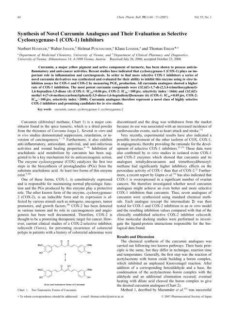
<strong>Curcumin</strong> (diferuloyl methane, Chart 1) is a major constituent<br />
found in the spice tumeric, which is a dried powder<br />
from the rhizomes <strong>of</strong> Curcuma longa L. Several in vitro and<br />
in vivo studies demonstrated suppression, retardation, or inversion<br />
<strong>of</strong> carcinogenesis. 1—3) Furthermore, it also exhibits<br />
anti-inflammatory, antioxidant, antiviral, and anti-infectious<br />
activities and wound healing properties. 4—8) Inhibition <strong>of</strong><br />
arachidonic acid metabolism by curcumin has been suggested<br />
to be a key mechanism for its anticarcinogenic action.<br />
The enzyme cyclooxygenase (COX) catalyzes the first two<br />
steps in the biosynthesis <strong>of</strong> prostaglandins (PGs) from the<br />
substrate arachidonic acid. At least two forms <strong>of</strong> this enzyme<br />
exist. 9,10)<br />
One <strong>of</strong> these forms, COX-1, is constitutively expressed<br />
and is responsible for maintaining normal physiologic function<br />
and the PGs produced by this enzyme play a protective<br />
role. The other known form <strong>of</strong> the enzyme, cyclooxygenase-<br />
2 (COX-2), is an inducible form and its expression is affected<br />
by various stimuli such as mitogens, oncogenes, tumor<br />
promoters, and growth factors. 10) COX-2 has been detected<br />
in various tumors and its role in carcinogenesis and angiogenesis<br />
has been well documented. Therefore, COX-2 is<br />
thought to be a promising therapeutic target for cancer. However,<br />
current clinical studies <strong>of</strong> a COX-2-selective inhibitor,<br />
r<strong>of</strong>ecoxib (Vioxx), for preventing recurrence <strong>of</strong> colorectal<br />
polyps in patients with a history <strong>of</strong> colorectal adenomas were<br />
Chart 1. Two Tautomeric Forms <strong>of</strong> <strong>Curcumin</strong><br />
∗ To whom correspondence should be addressed. e-mail: thomas.erker@univie.ac.at<br />
discontinued and the drug was withdrawn from the market<br />
because its use was associated with an increased incidence <strong>of</strong><br />
cardiovascular events, such as heart attack and stroke. 11)<br />
Very recently, experimental results have also indicated a<br />
possible involvement <strong>of</strong> the other is<strong>of</strong>orm <strong>of</strong> COX, COX-1,<br />
in angiogenesis, thereby providing the rationale for the development<br />
<strong>of</strong> selective COX-1 inhibitors. 12,13) These data were<br />
also confirmed by in vitro studies in isolated ovine COX-1<br />
and COX-2 enzymes which showed that curcumin and its<br />
analogues tetrahydrocurcumin and trimethoxydibenzoylmethane<br />
had significantly higher inhibitory effects on the<br />
peroxidase activity <strong>of</strong> COX-1 than that <strong>of</strong> COX-2. 2) Furthermore,<br />
a recent report by Gupta et al. 14) has also indicated that<br />
COX-1 is overexpressed in a significant number <strong>of</strong> ovarian<br />
cancers. We therefore investigated whether novel curcumin<br />
analogues might achieve an even better and more selective<br />
COX-1 inhibition than curcumin. Thus, seven analogues <strong>of</strong><br />
curcumin were synthesized using standard chemical methods.<br />
Each analogue (except the intermediate 2) was then<br />
tested for COX-1 and COX-2 inhibition in an in vitro model<br />
and the resulting inhibition values compared with that <strong>of</strong> the<br />
clinically established selective COX-2 inhibitor celecoxib.<br />
Also molecular docking studies were performed to investigate<br />
the ligand-protein interactions responsible for the biological<br />
data found.<br />
Results and Discussion<br />
The chemical synthesis <strong>of</strong> the curcumin analogues was<br />
carried out following two known pathways. Their basic principle<br />
is the same, but they differ in technique, reaction time<br />
and temperature. Generally, the first step was the reaction <strong>of</strong><br />
acetylacetone with boron oxide building a boron complex,<br />
which inhibited an unpleased Knoevenagel reaction. After<br />
addition <strong>of</strong> a corresponding benzaldehyde and a base, the<br />
condensation <strong>of</strong> the acetylacetone–boron complex with the<br />
aldehyde and an additional elimination occured; eventual<br />
heating with dilute acid cleaved the boron complex to give<br />
the desired curcumin analogues (Chart 2).<br />
Method 1, decribed by Mazumder et al. 15) was successful<br />
© 2007 <strong>Pharm</strong>aceutical Society <strong>of</strong> Japan
January 2007 65<br />
Chart 2. Basic Steps <strong>of</strong> the <strong>Synthesis</strong> <strong>of</strong> <strong>Curcumin</strong>oids<br />
Chart 3. <strong>Novel</strong> <strong>Curcumin</strong> <strong>Analogues</strong> 1—7<br />
in poor yields for compounds 2 and 7. Method 2, first described<br />
by Pabon, 16) was used for the other molecules ending<br />
up in poor yields, too. Study <strong>of</strong> the literature concerning the<br />
synthesis <strong>of</strong> curcuminoids pointed out difficulties, since only<br />
some derivatives seemed to be accessable quite well. The<br />
yields ranged from about 50% shown only for methoxy-derivatives<br />
to poor yields for p-(dimethylamino) (36%) or <strong>of</strong>uryl<br />
derivatives (8%), respectively, 15,16) and so we tried to<br />
improve our pathways. Exact analysis <strong>of</strong> our syntheses revealed<br />
that the volume <strong>of</strong> n-butylamine plays an important<br />
role in this kind <strong>of</strong> reaction and thus, in case <strong>of</strong> molecules 1<br />
and 4, its quantity was reduced to obtain the desired compounds<br />
in a somehow acceptable yield.<br />
Inhibition <strong>of</strong> COX-1 and COX-2 by curcumin analogues<br />
Table 1. Inhibitory Effect <strong>of</strong> 1—7, <strong>Curcumin</strong> and the Reference Compound<br />
Celecoxib on COX-1 and COX-2 Activity (Values Given in m M)<br />
Compound IC 50 (COX-1) IC 50 (COX-2)<br />
IC 50(COX-1)/ IC 50(COX-2)/<br />
IC 50(COX-2) IC 50(COX-1)<br />
1 0.33 12.58 0.026 38<br />
2 n.d. n.d. n.d. n.d.<br />
3 2.68 �100 �0.026 �37<br />
4 0.06 �100 �0.0006 �1666<br />
5 0.37 9.92 0.037 27<br />
6 0.05 �100 �0.0005 �2000<br />
7 1.14 5.13 0.22 4.5<br />
<strong>Curcumin</strong> 2) 50 �100 0.5 2<br />
Celecoxib 13.7 0.03 456.6 0.002<br />
Indomethacin 21) 0.018 0.026 0.692 1.444<br />
was then analyzed in a cell-free immunoassay system. Purified<br />
ovine enzyme served as the source <strong>of</strong> COX-1, while the<br />
human recombinant enzyme formed the source <strong>of</strong> COX-2.<br />
The inhibition <strong>of</strong> COX-1 by curcumin analogues (compound<br />
2, an intermediate, was not tested) is shown in Table 1. All<br />
compounds tested demonstrated a several-fold higher inhibitory<br />
activity than curcumin itself (COX-1: IC 50 �50 m M,<br />
COX-2: IC 50 �100 m M). 2)<br />
In detail, the results showed that all our curcumin derivatives<br />
had a preference towards COX-1 isoenzyme not depending<br />
on the substituent <strong>of</strong> the molecule. Even compound<br />
3 bearing a methylsulfonyl group, which can be found in<br />
some COX-2-selective compounds, exhibited an distinct<br />
affinity towards COX-1. Nurfina et al. 7) postulated that, besides<br />
olefinic double bonds, a 4-hydroxyl group at the phenyl<br />
ring <strong>of</strong> curcuminoids was essential for an antiinflammatory<br />
effect. Also position 3 <strong>of</strong> the aromatic ring played an important<br />
role for the pharmacoloigal pr<strong>of</strong>ile, since bigger 3-alkyl<br />
groups (like tert-butyl) lead to inactive molecules, whereas<br />
lower alkyl and especially 3,5-dialkyl-substituents showed<br />
high oedema inhibiting activity. Selvam et al. 17) calculated<br />
docking studies with curcumin and some derivatives, which<br />
revealed that these compounds could dock into the active site<br />
<strong>of</strong> only COX-1. In case <strong>of</strong> the COX-2 enzym complex only<br />
curcumin itself interacted with the enzyme, but no hydrogen<br />
bonding interactions were detected for the other molecules.<br />
Thus we chose derivatives without hydroxy groups but<br />
lipophilic and mainly polar groups to check possible SAR<br />
concerning COX-1 interaction.<br />
The data are presented in this paper (Table 1), where all<br />
novel curcumin derivatives showed a distinct affinity towards<br />
COX-1 isoenzyme. The corresponding IC 50 values reached<br />
from 2.68 m M (compound 3) to 0.05 m M (compound 6) including<br />
selectivity indices (IC 50 (COX-2)/IC 50 (COX-1)) from<br />
4.5 for compound 7 to �2000 for compound 6. Especially<br />
the trimethoxy derivative 4 as well as the methyl ester 6 exhibited<br />
very pronounced and selective COX-1 inhibition<br />
(IC 50 �0.06 and 0.05 m M, selectivity indices �1666 and<br />
�2000), whereas the other molecules showed just poor<br />
affinities and selectivities towards this isoenzym. However,<br />
also compound 3, bearing a methylsulfonyl group, a group<br />
found in some COX-2 inhibiting molecules, had weak effects<br />
on COX-1 but almost none towards COX-2 (IC 50(COX-<br />
1)�2.68 m M, IC 50 (COX-2)�100 m M, selectivity index �37).<br />
The other molecules exhibited only little COX-1-effects and<br />
selectivities with IC 50 values ranging from 0.33 to 2.68 m M
66 Vol. 55, No. 1<br />
and selectivity indices from 4.5 to 38, respectively.<br />
In order to better understand the anti-inflammatory activities<br />
<strong>of</strong> compounds 1, 3, 4, 6 and 7 we performed a molecular<br />
docking analysis. We tried to understand the ligand-protein<br />
interaction responsible for the perceived COX-1/COX-2 inhibitory<br />
data. Docking conformations <strong>of</strong> compounds 1, 3, 4,<br />
6 and 7 were created by a systematic conformational search<br />
with the MMFF94x as implemented in MOE. Each rotateable<br />
bond <strong>of</strong> the heptanoid part <strong>of</strong> the compounds was assigned<br />
a 60° rotation increment. Rotateable bonds <strong>of</strong> substituents<br />
<strong>of</strong> the phenyl rings were assigned a 30° rotation increment.<br />
All resulting conformers were subjected to full energy<br />
minimization using the MMFF94x force field to a gradient<br />
<strong>of</strong> 0.05 kcal/mol. Conformers having an energy <strong>of</strong><br />
15 kcal/mol above the lowest energy found were not taken<br />
into consideration. Duplicate entries in the resulting conformer<br />
database were discarded using a RMS filter <strong>of</strong> 0.1 Å.<br />
This resulted in databases <strong>of</strong> 3000 conformers on average.<br />
From these databases 1500 conformers were selected based<br />
on the dissimilarity <strong>of</strong> their internal coordinates. The resulting<br />
conformer databases were used as starting structures for<br />
a rigid docking simulation. We used the simple grid energy<br />
scoring function to score the docked conformations as implemented<br />
in program Dock 6.0. Partial charges where calculated<br />
semi-empirically using the AM1 hamiltonian. From the<br />
docked conformations only the best scored conformation was<br />
retained. The complexes were subsequently energy minimized<br />
using force field MMFF94 to a gradient <strong>of</strong> 0.05<br />
kcal/mol and were rescored using the same grid. All compounds<br />
could be docked into the active site <strong>of</strong> 1PGG successfully.<br />
The individual grid scores are shown in Table 2.<br />
All compounds showed hydrogen bond interaction to the<br />
1PGG protein. The exact numbers <strong>of</strong> hydrogen bonds can be<br />
seen in Table 3.<br />
Fig. 1. Binding <strong>of</strong> 1 into the Active Site <strong>of</strong> 1PGG<br />
The active site <strong>of</strong> 1PGG is considered to be constituted <strong>of</strong><br />
the amino acid residues ARG120, SER530, TYR385 and<br />
GLU524. 18) 1, 6 and 7 exhibited a very good DOCK 6.0 grid<br />
score showing favorable van der Waals interactions. Compound<br />
1 showed two hydrogen bonds. The two carbonyl oxygens<br />
were acceptors for hydrogen bonds forming from<br />
ARG83 (NH1). The O–N distance was found to be 3.09 and<br />
2.51 Å; the O–H distance was found to be 2.38 and 1.52 Å,<br />
respectively (Fig. 1). Although compound 3 and 4 exhibited<br />
only moderate to weak interactions calculated with DOCK,<br />
they show a tight hydrogen bond network. 3 is able to build<br />
three hydrogen bonds to 1PGG. These hydrogen bonds are<br />
formed by ARG83 and the sulfonyl oxygen <strong>of</strong> 3 with a bond<br />
length <strong>of</strong> 1.52 Å, ARG120 NH1 and the carbonyl oxygen <strong>of</strong><br />
3 with a bond length <strong>of</strong> 1.51 and ARG120 NH2 and the car-<br />
Table 2. Total Grid Score <strong>of</strong> Compounds Interacting with 1PGG<br />
Compound Grid score (Dock 6.0) 1PGG<br />
1 �24.60<br />
3 �16.09<br />
4 �11.33<br />
6 �20.10<br />
7 �24.46<br />
Table 3. Hydrogen Bond Interaction with 1PPG<br />
Compound No. <strong>of</strong> hydrogen bonds Bond lengths [Å]<br />
1 2 2.38, 1.52<br />
3 3 1.52, 1.51, 2.32<br />
4 3 1.59, 1.43, 1.80<br />
6 2 1.52, 2.38<br />
7 4 1.33, 1.47, 1.86, 1.99
January 2007 67<br />
Fig. 2. Binding <strong>of</strong> 3 into Active Site <strong>of</strong> 1PGG<br />
Fig. 3. Binding <strong>of</strong> 4 into the Active Site <strong>of</strong> 1PGG<br />
bonyl oxygen <strong>of</strong> 3 with a bond length <strong>of</strong> 2.32, respectively<br />
(Fig. 2).<br />
Compound 4 exhibited three hydrogen bonds to the 1PGG<br />
protein. These hydrogen bonds were formed by ARG120 and<br />
the methoxy group <strong>of</strong> 4, TYR355 and the carbonyl oxygen <strong>of</strong><br />
4 and SER530 and the second methoxy group <strong>of</strong> 4. The calculated<br />
bond length were 1.59 Å, 1.43 Å and 1.79 Å respec-<br />
tively (Fig. 3). We think that this accounts for the low observed<br />
IC 50 <strong>of</strong> compound 4. Compound 6 showed an excellent<br />
electrostatic and steric interaction with 1PGG and could<br />
build two hydrogen bonds with 1PGG (Fig. 4). These were<br />
formed by TYR355 and one <strong>of</strong> the carbonyl oxygens and<br />
SER530 and the other carbonyl oxygen <strong>of</strong> the heptanoid part<br />
<strong>of</strong> 6. The bond length were 1.53 Å and 2.38 Å respectively.
68 Vol. 55, No. 1<br />
Fig. 4. Binding <strong>of</strong> 6 into the Active Site <strong>of</strong> 1PGG<br />
Fig. 5. Binding <strong>of</strong> 7 into the Active Site <strong>of</strong> 1PGG<br />
Four hydrogen bonds were formed by compound 7 (Fig. 5).<br />
Three hydrogen bonds were formed with amino acids <strong>of</strong> the<br />
active site <strong>of</strong> 1PGG ARG120, TYR385 and SER530. One<br />
hydrogen bond was formed with TYR355. The hydrogen<br />
bond lengths were 1.24 Å, 1.86 Å, 1.99 Å and 1.47 Å respectively.<br />
All five compounds used for docking showed a good van<br />
der Waals interaction with 1PGG. One <strong>of</strong> the phenyl rings <strong>of</strong><br />
compound 1 was surrounded by GLU524, ARG120 and<br />
PHE470. The heptanoid part <strong>of</strong> 1 was surrounded by ILE89,<br />
ILE115 and VAL119. The second phenyl ring was surrounded<br />
by LEU93, TRP100 and LEU112. The deeper<br />
buried phenyl ring <strong>of</strong> compound 3 is surrounded by the active<br />
site amino acid residues SER530, TYR385, LEU352 and
January 2007 69<br />
Table 4. Total Grid Score <strong>of</strong> Compounds Interacting with 4COX<br />
Compound Grid score (Dock 6.0) 4COX<br />
1 �16.32<br />
3 �46.76<br />
4 n.d.<br />
6 �36.02<br />
7 �35.99<br />
Indomethacin �45.89<br />
Fig. 6. Interaction <strong>of</strong> Indomethacin with 4COX<br />
ILE523. The heptanoid part <strong>of</strong> 3 was surrounded by<br />
GLU524, ARG120 and VAL116; the second ring <strong>of</strong> 3 was<br />
surrounded by ILE89 and ARG83. Similar trends were observed<br />
for the other compounds under investigation.<br />
After docking simulations with 1PGG (COX-1) we tried to<br />
dock the compounds considered in the first simulation into<br />
the active site <strong>of</strong> 4COX (COX-2). The active site <strong>of</strong> 4COX is<br />
considered to be formed <strong>of</strong> the amino acid residues TYR385,<br />
that is supposed to abstract one hydrogen from the substrate,<br />
SER530, which is acetylated by acetylsalicylic acid and<br />
HEME. 19) All compounds except 4 could be docked into the<br />
active site <strong>of</strong> 4COX with a reasonable steric and electrostatic<br />
interaction. As a test compound, we docked the co-crystallized<br />
ligand indomethacin into the active site <strong>of</strong> 4COX. The<br />
results can bee seen in Table 4. Indomethacin showed the<br />
best electrostatic and steric interaction scores and the tightest<br />
hydrogen bond network with the active site (Fig. 6). Indomethacin<br />
formed hydrogen bonds between ARG120,<br />
TYR355 and SER530 with bond length <strong>of</strong> 1.33, 2.46 and<br />
2.26 respectively.<br />
The hydrogen bonds formed by the curcumin analogues<br />
complexes considered in this work and 4COX were much<br />
longer than those <strong>of</strong> the respective 1PGG complexes (Table 5<br />
and Fig. 7). We therefore argue, that this is the reason for the<br />
Table 5. Hydrogen Bond Interaction with 4COX<br />
Compound No. <strong>of</strong> hydrogen bonds Bond lengths [Å]<br />
1 2 1.46, 3.00<br />
3 2 2.07, 1.64<br />
4 n.d. n.d.<br />
6 2 1.49, 1.65<br />
7 3 1.54, 1.65, 2.15<br />
Indomethacin 3 1.33, 2.46, 2.26<br />
selectivity <strong>of</strong> the compounds under investigation.<br />
In conclusion, we have demonstrated that our curcumin<br />
analogues were selective COX-1 inhibitors with submicromolar<br />
to micromolar IC 50 values and promising selectivities.<br />
Especially lipophilic and polar substituents on the phenyl<br />
ring, like methoxy or methyl ester groups improved the<br />
specifity <strong>of</strong> the compounds. The biological data were also<br />
confirmed by docking studies which revealed good van der<br />
Waals interaction and hydrogen bonding towards COX-1<br />
isoenzym performed by our curcuminoid molecules. As this<br />
is<strong>of</strong>orm is overexpressed in a significant number <strong>of</strong> cancer<br />
cells and tissues our present results may yield in new candidate<br />
drugs for the treatment <strong>of</strong> these tumor entities.<br />
Experimental<br />
<strong>Chem</strong>istry Melting points were determined on a K<strong>of</strong>ler hot stage apparatus<br />
and are uncorrected. The 1 H- and 13 C-NMR spectra were recorded on a<br />
Varian UnityPlus-200 (200 MHz). <strong>Chem</strong>ical shifts are reported in d values<br />
(ppm) relative to Me 4 Si line as internal standard and J values are reported in<br />
Hertz. Mass spectra were obtained by a Shimadzu GC/MS QP 1000 EX<br />
using EI method. The elemental analysis obtained were within �0.4% <strong>of</strong> the<br />
theoretical values for the formulas given.<br />
The following compounds were synthesized via two different pathways.<br />
Method 1 15) : In a dry three-necked flask acetylacetone (5 mmol, 0.51 ml)<br />
and boron oxide (3.5 mmol, 0.244 g) were solved in absolute ethyl acetate<br />
and stirred for 30 min at 40 °C. Then the corresponding aldehyde (10 mmol)
70 Vol. 55, No. 1<br />
Fig. 7. Interaction <strong>of</strong> 1, 3, 6 and 7 with 4COX<br />
and tributyl borate (10 mmol, 2.4 ml) were added and stirred for another<br />
30 min. N-Butylamine (quantities given below) was solved in dry ethyl acetate<br />
and then added over a period <strong>of</strong> 15 min. The mixture was heated to<br />
40 °C for 24 h. Then 5 ml <strong>of</strong> HCl (10%) were added and heated to 60 °C for<br />
an additional hour. The aqueous phase was extracted with ethyl acetate several<br />
times, the organic layers were dried over Na 2 SO 4 and the solvent distilled<br />
<strong>of</strong>f. An unsoluable precipitate (part <strong>of</strong> the product) was filtered <strong>of</strong>f and<br />
recrystallized with the residue from various solvents.<br />
Method 2 16) : In a dry three-necked flask acetylacetone (5 mmol, 0.51 ml)<br />
and boron oxide (3.5 mmol, 0.244 g) were solved in 5 ml absolute ethyl<br />
acetate and heated to 75 °C for 1 h. The corresponding benzaldehyde<br />
(10 mmol) and tributyl borate (10 mmol, 2.4 ml) were mixed with ethyl acetate,<br />
stirred for 45 min and then added to the solution. The mixture was<br />
heated to 100 °C for 1 h. Then n-butylamine (quantities given below) was<br />
solved in 5 ml ethyl acetate and 3.85 ml <strong>of</strong> this solution were added drop-bydrop<br />
over a period <strong>of</strong> 90 min. The reaction was stirred for 18 h at 85 °C and<br />
then cooled to 60 °C. Then 5 ml <strong>of</strong> a HCl solution (10%) were heated to<br />
50 °C, added and the mixture was stirred at 60 °C for 1 h. The solution was<br />
extracted three times with ethyl acetate, the organic layers were dried over<br />
Na 2 SO 4 and the solvent reduced to 7—8 ml. Two and a half milliliters<br />
ethanol were added, the solution was cooled overnight, then the precipitate<br />
was filtered <strong>of</strong>f and recrystallized to obtain the purified product.<br />
(1E,6E)-1,7-Di-(3,4-difluorphenyl)-1,6-heptadien-3,5-dione (1) The<br />
compound was synthesized from 3,4-difluorobenzaldehyde (10 mmol,<br />
1.16 ml) and n-butylamin (2.5 mmol, 0.25 ml) following method 2. Crystallization<br />
from ethyl acetate/water (1�1) afforded 0.066 g (3.8%) <strong>of</strong> a yellow<br />
solid; mp 137 °C. 1 H-NMR (CDCl 3 ) d: 5.82 (1H, s), 6.52 (2H, d,<br />
J E �15.8 Hz), 7.12—7.42 (6H, m), 7.56 (2H, d, J E �15.8 Hz), 15.76 (1H,<br />
br s). 13 C-NMR (CDCl 3 ) d: 102.2, 116.1 (d, J C,F �18.0 Hz), 117.9 (d,<br />
J C,F �18.0 Hz), 124.9 (quintett), 132.1 (quartett), 138.3—138.5 (m), 148.5<br />
(d, J C,F �22.2 Hz), 153.6 (d, J C,F �39.4 Hz), 182.8; MS (EI) m/z: 348 (M � ),<br />
306, 167, 139, 119. Anal. Calcd for C 19 H 12 O 2 F 4 : C, 65.52; H, 3.47. Found:<br />
C, 65.41; H, 3.69.<br />
(1E,6E)-1,7-Di-[4-(methylmercapto)phenyl]-1,6-heptadien-3,5-dione<br />
(2) The compound was synthesized from 4-(methylmercapto)benzaldehyde<br />
(10 mmol, 1.33 ml) and n-butylamine (7.5 mmol, 0.74 ml) following<br />
method 1. Crystallization from ethanol afforded 1.14 g (62%) <strong>of</strong> a yellow<br />
solid; mp 194 °C. 1 H-NMR (CDCl 3 ) d: 2.51 (6H, s), 5.81 (1H, s), 6.58 (2H,<br />
d, J E �15.8 Hz), 7.24 (4H, A-part <strong>of</strong> AB-system, J A,B �8.7 Hz), 7.47 (4H, Bpart<br />
<strong>of</strong> AB-system, J A,B �8.7 Hz), 7.62 (2H, d, J E �15.8 Hz), enol OH signal<br />
not detected; 13 C-NMR (CDCl 3 ) d: 15.2, 101.8, 123.1, 126.0, 128.5, 140.0,<br />
183.2. MS (EI) m/z: 368 (M � ), 279, 201, 105, 77. Anal. Calcd for<br />
C 21 H 20 O 2 S 2 : C, 68.45; H, 5.47. Found: C, 68.20; H, 5.25.<br />
(1E,6E)-1,7-Di-[4-(methylsulfonyl)phenyl]-1,6-heptadien-3,5-dione (3)<br />
(1E,6E)-1,7-Di-[4-(methylmercapto)phenyl]-1,6-heptadien-3,5-dione (2)<br />
(2.5 mmol, 0.924 g) was solved in acetone; then oxone ® (10 mmol, 6.14 g) in<br />
15 ml water was added and the mixture was stirred at room temperature for<br />
24 h. After addition <strong>of</strong> 20 ml ammonia (10%) and stirring for an additional<br />
hour water was added, the precipitate formed was filtered <strong>of</strong>f and washed<br />
with water. The yellow solid was solved in DMF and ethanol was added to<br />
isolate 0.278 g (25%) <strong>of</strong> the pure compound; mp �300 °C. 1 H-NMR<br />
(DMSO-d 6 ) d: 3.24 (6H, s), 5.77 (1H, s), 7.02 (2H, d, J E �15.7 Hz), 7.48<br />
(2H, d, J E �15.7 Hz), 7.87—7.98 (8H, m), OH signal not detected; 13 C-NMR<br />
(DMSO-d 6 ) d: 43.7, 104.6, 127.7, 128.4, 134.3, 134.4, 140.6, 141.0, 181.1.<br />
MS (EI) m/z: 432 (M � ), 201, 111, 68. Anal. Calcd for C 21 H 20 O 6 S 2 : C, 58.32;<br />
H, 4.66. Found: C, 58.47; H, 4.34.<br />
(1E,6E)-1,7-Di-(2,3,4-trimethoxyphenyl)-1,6-heptadien-3,5-dione (4)<br />
The compound was synthesized from 2,3,4-trimethoxybenzaldehyde<br />
(10 mmol, 1.96 g) and n-butylamine (2.5 mmol, 0.25 ml) following method<br />
2. Purification was carried out with column chromatography (eluent<br />
toluene/ethyl acetate 8�2) and crystallization from ethanol (80%) to afford<br />
0.087 g (3.8%) <strong>of</strong> a yellow solid; mp 115 °C. 1 H-NMR (DMSO-d 6 ) d: 3.89<br />
(6H, s), 3.91 (6H, s), 3.94 (6H, s), 5.83 (1H, s), 6.63 (2H, d, J E �15.0 Hz),<br />
6.71 (2H, A-part <strong>of</strong> AB-system, J A,B �8.8 Hz), 7.30 (2H, B-part <strong>of</strong> AB-system,<br />
J A,B �8.8 Hz), 7.85 (2H, d, J E �15.8 Hz), 16.06 (1H, br s). 13 C-NMR<br />
(DMSO-d 6 ) d: 56.1, 60.9, 61.4, 101.3, 107.6, 122.2, 123.2, 123.4, 135.3,<br />
142.4, 153.4, 155.4, 183.6. MS (EI) m/z: 278 (half mass), 247, 235, 204,<br />
131, 43. Anal. Calcd for C 25 H 28 O 8 : C, 65.78; H, 6.18. Found: C, 65.49; H,<br />
6.17.<br />
(1E,6E)-1,7-Diferrocenyl-1,6-heptadien-3,5-dione (5) The compound<br />
was synthesized from ferrocenealdehyde (10 mmol, 2.18 g) and n-butylamine<br />
(7.5 mmol, 0.74 ml) following method 2. Crystallization from ethyl<br />
acetate afforded 0.205 g (8.3%) <strong>of</strong> a purple solid; mp 210 °C. 1 H-NMR<br />
(CDCl 3 ) d: 4.17 (10H, s), 4.44 (4H, s), 4.52 (4H, s), 5.62 (1H, s), 6.21 (2H,<br />
d, J E �15.5 Hz), 7.54 (2H, d, J E �15.5 Hz), enol OH signal not detected. 13 C-<br />
NMR (CDCl 3) d: 68.5, 69.7, 71.0, 79.7, 100.1, 121.3, 141.5, 182.7. MS (EI)<br />
m/z: 492 (M � ), 280, 246, 239, 121, 56. HR-MS m/z: 492.0476 (Calcd for<br />
C 27 H 24 O 2 Fe 2 : 492.0459).<br />
(1E,6E)-Methyl 4-[7-(4-methoxycarbonyl)phenyl]-3,5-dioxo-1,6-heptadienyl]benzoate<br />
(6) The compound was synthesized from (4-formyl)methylbenzoate<br />
(10 mmol, 1.64 g) and n-butylamine (7.5 mmol, 0.74 ml) following<br />
method 2. Crystallization from ethyl acetate afforded 0.334 g
January 2007 71<br />
(16.8%) <strong>of</strong> a yellow solid; mp 215 °C: 1 H-NMR (CDCl 3) d: 3.94 (6H, s),<br />
5.90 (1H, s), 6.71 (2H, d, J E �15.9 Hz), 7.62 (4H, A-part <strong>of</strong> AB-system,<br />
J AB �8.2 Hz), 7.69 (2H, d, J E �15.9 Hz), 8.07 (4H, B-part <strong>of</strong> AB-system,<br />
J AB�8.2 Hz), 15.74 (1H, br s). 13 C-NMR (CDCl 3) d: 52.3, 102.5, 126.1,<br />
127.9, 130.1, 131.2, 139.1, 139.4, 166.4, 182.9; MS (EI) m/z: 392 (M � ),<br />
145, 115, 102, 59. Anal. Calcd for C 23 H 20 O 6 : C, 70.40; H, 5.14. Found: C,<br />
70.18; H, 5.04.<br />
(1E,3E,8E,10E)-1,11-Di-(4-methoxyphenyl)-1,3,8,10-undecatetraen-<br />
5,7-dione (7) The compound was synthesized from 4-methoxycinnamaldehyde<br />
(10 mmol, 1.62 g) and n-butylamine (7.5 mmol, 0.74 ml) following<br />
method 1. Crystallization from ethanol (70%) afforded 0.219 g<br />
(11.3%) <strong>of</strong> an orange solid; mp 210 °C. 1 H-NMR (CDCl 3 ) d: 3.83 (6H, s),<br />
5.66 (1H, s), 6.12 (2H, d, J E �15.0 Hz), 6.72—6.93 (8H, m), 7.36—7.51<br />
(6H, m), 15.90 (1H, br s). 13 C-NMR (CDCl 3 ) d: 55.4, 101.5, 114.3, 125.0,<br />
126.6, 128.7, 129.1, 139.9, 141.1, 160.4, 183.1. MS (EI) m/z: 388 (M � ),<br />
187, 121, 57. Anal. Calcd for C 25 H 24 O 4 ·0.25H 2 O: C, 76.31; H, 6.29. Found:<br />
C, 76.33; H, 6.45.<br />
Cyclooxygenase Assay The effects <strong>of</strong> the test compounds on COX-1<br />
and COX-2 were determined by measuring prostaglandin E 2 (PGE 2 ) using a<br />
COX Inhibitor Screening Kit (Catalog No 560131) from Cayman <strong>Chem</strong>icals,<br />
Ann Arbor, Michigan, U.S.A. Reaction mixtures were prepared in<br />
100 mM Tris–HCl buffer, pH 8.0 containing 1 m M heme and COX-1 (ovine)<br />
or COX-2 (human recombinant) and pre-incubated for 10 min in a waterbath<br />
(37 °C). The reaction was initiated by the addition <strong>of</strong> 10 ml arachidonic acid<br />
(final concentration in reaction mixture 100 m M). After 2 min the reaction<br />
was terminated by adding 1 M HCl and finally PGE 2 was quantified by an<br />
ELISA method. The test compounds were dissolved in DMSO and diluted to<br />
the desired concentration with 100 mM potassium phosphate buffer (pH 7.4).<br />
Following transfer to a 96 well plate coated with a mouse anti-rabbit IgG,<br />
the tracer prostaglandin acetylcholine esterase and primary antibody (mouse<br />
anti PGE 2 ) were added. Plates were then incubated at room temperature<br />
overnight, reaction mixtures were removed, and the wells were washed with<br />
10 mM potassium phosphate buffer containing 0.05% Tween 20. Ellman’s<br />
reagent (200 ml) was added to each well and the plate was incubated at room<br />
temperature (exclusion <strong>of</strong> light) for 60 min, until the control wells yielded an<br />
OD�0.3—0.8 at 412 nm. A standard curve with PGE 2 was generated from<br />
the same plate, which was used to quantify the PGE 2 levels produced in the<br />
presence <strong>of</strong> test samples. Results were expressed as a percentage relative to<br />
a control (solvent-treated samples). All determinations were performed in<br />
duplicate and values generally agreed within 10%.<br />
Docking Studies All calculations were performed on a SUN Ultra 40<br />
workstation using program DOCK 6.0 (Kuntz, I.D., DOCK, UCSF Box<br />
2240, UCSF, San Francisco, CA 94143-2240) and MOE (Molecular Operating<br />
Environment, <strong>Chem</strong>ical Computing Group Inc., Montreal, Quebec,<br />
Canada). The crystal structures <strong>of</strong> COX-1 and COX-2 co-crystallized with<br />
indomethacin (1PGG.pdb and 4COX.pdb respectively) 19,20) were taken from<br />
the Brookhaven Protein Databank 18) and used for docking. Regions with a<br />
7.0 Å radius from the complexed inhibitor were marked as interesting.<br />
References<br />
1) Weber W. M., Hunsaker L. A., Roybal C. N., Bobrovnikova-Marjon E.<br />
V., Abcouwer S. F., Royer R. E., Deck L. M., Vander Jagt D. L.,<br />
Bioorg. Med. <strong>Chem</strong>., 14, 2450—2461 (2006).<br />
2) Hong J., Bose M., Ju J., Ryu J., Chen X., Sang S., Lee M., Yang C.,<br />
Carcinogenesis, 25, 1671—1679 (2004).<br />
3) Woo H. B., Shin W., Lee S., Ahn C. M., Biorg. Med. <strong>Chem</strong>. Lett., 15,<br />
3782—3786 (2005).<br />
4) Venkateswarlu S., Ramachandra M. S., Subbaraju G. V., Bioorg. Med.<br />
<strong>Chem</strong>., 13, 6374—6380 (2005).<br />
5) Goel A., Boland C. R., Chauhan D. P., Cancer Lett., 172, 111—118<br />
(2001).<br />
6) Lev-Ari S., Strier L., Kazanov D., Madar-Shapiro L., Dvory-Sobol H.,<br />
Pinchuk I., Marian B., Lichtenberg D., Arber N., Clin. Cancer Res.,<br />
11, 6738—6744 (2005).<br />
7) Nurfina A. N., Reksohadiprodjo M. S., Timmermann H., Jenie U. A.,<br />
Sugiyanto D., van der Goot H., Eur. J. Med. <strong>Chem</strong>., 32, 321—328<br />
(1997).<br />
8) Joe B., Vijaykamur N., Lokesh B. R., Crit. Rev. Food Sci. Nutr., 44,<br />
97—111 (2004).<br />
9) Xie W. L., Chipman J. G., Robertson D. L., Erikson R. L., Simmons D.<br />
L., Proc. Natl. Acad. Sci. U.S.A., 88, 2692—2696 (1991).<br />
10) Fournier D. B., Gordon G. B., J. Cell Biochem. Suppl., 34, 97—102<br />
(2000).<br />
11) Onn A., Tseng J. E., Herbst R. S., Clin. Cancer Res., 7, 3311—3313<br />
(2001).<br />
12) Kitamura T., Itoh M., Noda T., Matsuura M., Wakabayashi K., Int. J.<br />
Cancer, 109, 576—580 (2004).<br />
13) Sano H., Noguchi T., Miyajima A., Hashimoto Y., Miyachi H., Bioorg.<br />
Med. <strong>Chem</strong>. Lett., 16, 3068—3072 (2006).<br />
14) Gupta R. A., Tejada L. V., Tong B. J., Das S. K., Morrow J. D., Dey S.<br />
K., DuBois R. N., Cancer Res., 63, 906—911 (2003).<br />
15) Mazumder A., Neamati N., Sunder S., Schulz J., Pertz H., Eich E.,<br />
Pommier Y., J. Med. <strong>Chem</strong>., 40, 3057—3063 (1997).<br />
16) Pabon H. J. J., Rec. Trav. Chim. Pays-Bas, 83, 379—386 (1964).<br />
17) Selvam C., Jachak S. M., Thilagavathi R., Chakraborti A. K., Bioorg.<br />
Med. <strong>Chem</strong>. Lett., 15, 1793—1797 (2005).<br />
18) Abloa E. E., Bernstein F. C., Bryant S. H., Koetzle T. F., Weng J., “Protein<br />
Data Bank in Crystallographic Databases–Information Content,<br />
S<strong>of</strong>tware Systems, Scientific Applications,” ed. by Allen F. H., Berjerh<strong>of</strong>f<br />
G., Sievers R., Data Commision <strong>of</strong> the International Union <strong>of</strong><br />
Crystallography, Bonn, 1987, p. 171.<br />
19) Kurumbail R. G., Stevens A. M., Gierse J. K., McDonald J. J., Stegeman<br />
R. A., Pak J. Y., Gildehaus D., Miyashiro J. M., Penning T. D.,<br />
Seibert K., Isakson P. C., Stallings W. C., Nature (London), 384, 644—<br />
648 (1996).<br />
20) Loll P. J., Picot D., Ekabo O., Garavito R. M., Biochemistry, 35,<br />
7330—7340 (1996).<br />
21) Riendeau D., Percival M. D., Boyce S., Brideau C., Charleson S.,<br />
Cromlish W., Ethier D., Evans J., Falgueyret J.-P., Ford-Hutchinson A.<br />
W., Gordon R., Greig G., Gresser M., Guay J., Kargman S., Leger S.,<br />
Mancini J. A., O’Neill G., Ouellet M., Rodger I. W., Therien M., Wang<br />
Z., Webb J. K., Wong E., Chan C. C., Brit. J. <strong>Pharm</strong>acol., 121, 105—<br />
117 (1997).


