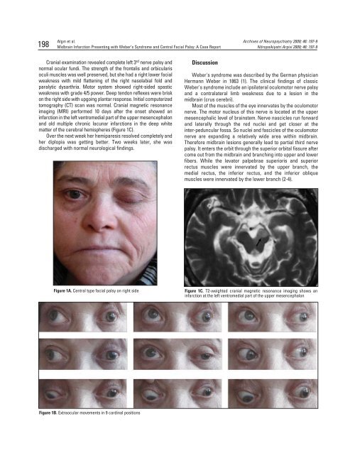
198Alg›n et al.<strong>Midbrain</strong> <strong>Infarction</strong> <strong>Presenting</strong> <strong>with</strong> Weber’s <strong>Syndrome</strong> <strong>and</strong> <strong>Central</strong> Facial Palsy: A Case ReportArchives of Neuropsychiatry 2009; 46: 197-9Nöropsikiyatri Arflivi 2009; 46: 197-9Cranial examination revealed complete left 3 rd nerve palsy <strong>and</strong>normal ocular fundi. The strength of the frontalis <strong>and</strong> orbicularisoculi muscles was well preserved, but she had a right lower facialweakness <strong>with</strong> mild flattening of the right nasolabial fold <strong>and</strong>paralytic dysarthria. Motor system showed right-sided spasticweakness <strong>with</strong> grade 4/5 power. Deep tendon reflexes were briskon the right side <strong>with</strong> upgoing plantar response. Initial computerizedtomography (CT) scan was normal. Cranial magnetic resonanceimaging (MRI) performed 10 days after the onset showed aninfarction in the left ventromedial part of the upper mesencephalon<strong>and</strong> old multiple chronic lacunar infarctions in the deep whitematter of the cerebral hemispheres (Figure 1C).Over the next week her hemiparesis resolved completely <strong>and</strong>her diplopia was getting better. Two weeks later, she wasdischarged <strong>with</strong> normal neurological findings.Discussion<strong>Weber's</strong> syndrome was described by the German physicianHermann Weber in 1863 (1). The clinical findings of classic<strong>Weber's</strong> syndrome include an ipsilateral oculomotor nerve palsy<strong>and</strong> a contralateral limb weakness due to a lesion in themidbrain (crus cerebri).Most of the muscles of the eye innervates by the oculomotornerve. The motor nucleus of this nerve is located at the uppermesencephalic level of brainstem. Nerve nascicles run forward<strong>and</strong> laterally through the red nuclei <strong>and</strong> get closer at theinter-peduncular fossa. So nuclei <strong>and</strong> fascicles of the oculomotornerve are exp<strong>and</strong>ing a relatively wide area <strong>with</strong>in midbrain.Therefore midbrain lesions generally lead to partial third nervepalsy. It enters the orbit through the superior orbital fissure aftercome out from the midbrain <strong>and</strong> branching into upper <strong>and</strong> lowerfibers. While the levator palpebrae superioris <strong>and</strong> superiorrectus muscles were innervated by the upper branch, themedial rectus, the inferior rectus, <strong>and</strong> the inferior obliquemuscles were innervated by the lower branch (2-4).Figure 1A. <strong>Central</strong> type facial palsy on right sideFigure 1C. T2-weighted cranial magnetic resonance imaging shows aninfarction at the left ventromedial part of the upper mesencephalonFigure 1B. Extraocular movements in 9 cardinal positions
Archives of Neuropsychiatry 2009; 46: 197-9Nöropsikiyatri Arflivi 2009; 46: 197-9Alg›n et al.<strong>Midbrain</strong> <strong>Infarction</strong> <strong>Presenting</strong> <strong>with</strong> Weber’s <strong>Syndrome</strong> <strong>and</strong> <strong>Central</strong> Facial Palsy: A Case Report 199The preganglionic parasympathetic fibers of the 3 rd nervewhich are transported by the nerve to the inferior obliquemuscle arrive to the ciliary ganglion <strong>and</strong> from here thepost-ganglionic parasympathetic fibers emerge. The ciliarymuscle <strong>and</strong> the muscles of the iris are innervated by thesepost-ganglionic parasympathetic fibers. The nerve fibers tolevator palpebrae muscle <strong>and</strong> the pupilloconstrictor fibers forthe muscles of the iris are located in a superficial <strong>and</strong> dorsalposition on the nerve relaying in the ciliary ganglion. Beforeexternal ophthalmoplegia develops, a fixed dilated pupil is oftenthe first sign of 3rd nerve (oculomotor) compression, <strong>and</strong> ptosisthe second, upon this anatomical characteristic (5).The clinical manifestation of the patients <strong>with</strong> isolatedmesencephalic infarct was shown up nuclear or fascicularoculomotor nerve palsy <strong>and</strong> contralateral motor deficits (6,7).In patients <strong>with</strong> isolated mesencephalic infarct, the clinicalpicture was dominated by nuclear or fascicular third-nerve palsy<strong>and</strong> contralateral motor deficits (6,8).On the other h<strong>and</strong> the corticobulbar tract (CBT) is commonlyused to describe the pathway taken by motor fibers innervatingthe cranial nerve nuclei, especially trigeminal, facial, hypoglossalmotor nuclei, nucleus ambiguus, <strong>and</strong> spinal accessorynucleus. The oculomotor, trochlear <strong>and</strong> abducensnuclei receive no input from the CBT. The cell bodies of primarymotor neurons are located in primary motor cortex (9).The motor nucleus of the facial nerve, which supplies themuscles of the facial expression, is located at the lower pontinelevel, dorsolaterally in the caudal pons (4). The CBT fibers thatconnect the motor cortex <strong>with</strong> the facial nucleus providestrongly unilateral innervation to the contralateral lower facialmuscles <strong>and</strong> bilateral innervation to the upper facial muscles.The facial CBT fibers descend at the ventromedial region of thecrus cerebri, near the corticospinal tract (10).The clinical manifestations of midbrain infarcts mostly showthe location of the lesion directly, but these features could notalways match <strong>with</strong> classical syndromes which describedpreviously in the literature, just like our case that we present inthis report (11).Posterior cerebral artery branch disease may be caused byocclusion of the arterial branch <strong>with</strong> atherothrombotic plaque(4). In these cases, hypertension is a major risk factor, as seen inour patient (10).Kumral et al. presented the topographic <strong>and</strong> clinicaldistribution of the acute posterior circulation infarcts involvingmesencephalon. They described four patients <strong>with</strong> Weber’ssyndrome <strong>and</strong> central facial palsy due to isolated midbraininfarct around ventromedial crural region <strong>with</strong>in 41 patients <strong>with</strong>mesencephalic infarction. These were the only cases in theliterature identical to our patient (6).Some minor infarctions at the midbrain which resulted inlocalized paralysis like weakness of a single extraocular muscle(12), isolated contralateral superior rectus palsy (13) have beendemonstrated previously. Some interesting cases, such aspresence of left oculomotor nerve palsy <strong>with</strong> normal pupil <strong>and</strong>right hemiparesis, were described. An ischemic lesion of thelower midbrain, which corresponds to the motor nucleus of theoculomotor nerve, was demonstrated in this case (3).Terao et al. reported two patients <strong>with</strong> contralateral centralfacial paralysis <strong>and</strong> hemiparesis of the limbs, resulted fromunilateral ventromedial medullary infarction (14). According tothese cases, they described the course of the facial corticobulbartract as consisting of looping fibers that descend at least to themedullary level <strong>and</strong> then decussate. In another study <strong>with</strong> largergroup (70 patients), they attempted to further clarify the course<strong>and</strong> the distribution of the facial corticobulbar tract (10). But theauthors could not coincide <strong>with</strong> third nerve palsy or Weber’ssyndrome <strong>with</strong>in these central facial palsy patients.Finally, this report demonstrates an extremely rare case ofcrossed hemiplegia <strong>with</strong> oculomotor <strong>and</strong> facial nerve palsy dueto an infarct in the upper part of the midbrain as documented bythe MRI scan. The other interesting feature to note in our reportis that the patient completely recovered six month later. Thisindicates that some of these patients may have a good prognosis.References1. Silverman IE, Liu GT, Volpe NJ et al. The Crossed Paralyses. TheOriginal Brain-Stem <strong>Syndrome</strong>s of Millard-Gubler, Foville, Weber <strong>and</strong>Raymond-Cestan. Arch Neurol 1995; 52:635-8. (Abstract) / (PDF)2. Liu CT, Cremer CW, Logigian EL et al. <strong>Midbrain</strong> syndrome of Benedict,Claude, Nothnagel-setting record straight. Neurology 1992 ;42:1820-2.3. Umasankar U, Huwez FU. A patient <strong>with</strong> reversible pupil-sparingWeber’s syndrome . Neurol India 2003; 51:388-9. (Abstract) / (Full Text) /(PDF)4. Hendelman WJ. Atlas of Functional Neuroanatomy. 2nd edition CRCPres Taylor&Francis Group. 2006; pp. 126.5. Sunderl<strong>and</strong> S. Neurovascular relations <strong>and</strong> anomalies at the base ofthe brain. J Neurol Neurosurg Psychiatry 1948; 11:243. (Full Text) / (PDF)6. Kumral E, Bayulkem G, Akyol A, et al. Mesencephalic <strong>and</strong> associatedposterior circulation infarcts. Stroke. 2002; 33:2224-31. (Abstract) /(Full Text) / (PDF)7. Cormier PJ, Long ER, Russell EJ. MR imaging of posterior fossainfarctions: vascular territories <strong>and</strong> clinical correlates. Radiographics1992; 12:1079-96. (Abstract) / (PDF)8. Johnson MH, Christman CW. Posterior circulation infarction: anatomy,pathophysiology, <strong>and</strong> clinical correlation. Semin Ultrasound CT MR.1995; 16:237-52. (Abstract)9. Nolte J. The Human Brain: An Introduction to its Functional Anatomy,Edition 5th, Elsevier Health Science; pp. 461.10. Terao S, Miura N, Takeda A et al. Course <strong>and</strong> distribution of facialcorticobulbar tract fibres in the lower brain stem. J Neurol NeurosurgPsychiatry 2000; 69:262-5. (Abstract) / (PDF)11. Kim JS, Kang JK, Lee SA et al. Isolated or predominant ocular motornevre palsy as a manifestation of brain stem stroke. Stroke. 1993;24:581-6. (Abstract) / (Full Text) / (PDF)12. Kwon JH, Kwon SU, Ahn HS et al. Isolated Superior Rectus Palsy Dueto Contralateral <strong>Midbrain</strong> <strong>Infarction</strong>. Arch Neurol 2003;60:1633-5.(Abstract) / (Full Text) / (PDF)13. Terao S, Takatsu S, Izumi M et al. <strong>Central</strong> facial weakness due tomedial medullary infarction: the course of facial corticobulbar fibres.J Neurol Neurosurg Psychiatry 1997; 63:391-3. (Abstract) / (Full Text) /(PDF)