Download - Optometric Extension Program Foundation
Download - Optometric Extension Program Foundation
Download - Optometric Extension Program Foundation

You also want an ePaper? Increase the reach of your titles
YUMPU automatically turns print PDFs into web optimized ePapers that Google loves.
Article 4 Press Lites – Procedures For Visual Field AwarenessLeonard J. Press, OD, Fair Lawn, New JerseyABSTRACTPatients who experience a loss of their visual field are usually unaware that their peripheral vision has been altered. The firstdistinction to be made when the periphery is altered is to differentiate traditional visual field defects from loss of peripheralawareness due to inattention or neglect. Within this population experiencing inattention/neglect, extinction is anotherproperty reflecting a de-sensitization of one side of visual space as compared to the contralateral side of space.A set of therapeutic procedures is introduced based on components of higher order cerebral function that factor into visualfield awareness. Stressing visual function in three dimensional space, a method of probing and improving sensitivity toperipheral stimuli is presented. A commercially available set of lights is used with suggested procedures that can be usedindividually or hierarchically. Emphasis is placed on developing awareness, attention, and divided attention in attemptingto broaden, if not fully restore, visual field sensitivity and scanning.Keywords: visual field, visual inattention, visual neglect, peripheral awareness, Press LitesIntroductionOne of the more difficult conditions to treat clinicallyoccurs when patients lose awareness of a portion of theirvisual field. Even individuals with homonymous hemianopiamay be unaware that half of their visual field is missing. Thiscan be considered as an elaborated version of perceptualfilling-in, a normal process which helps to explain why weare not aware of the physiological blind spot created wherethe optic nerve is situated within the retina. 1 The challengefor the clinician is to arrange conditions so that the patientdevelops more sensitivity regarding areas of field loss, andultimately to cultivate prescriptive measures including therapyprocedures that expand visual awareness into the areas offield loss or inattention. The term visual neglect is often usedinterchangeably with visual inattention, and represents a lackof awareness of visual space on one side of the visual fieldwithout overt visual pathway disease that would account forthe deficit. 2 It is indicative of a cognitive change that may bemore amenable to restoration with therapy as compared toa visual field defect. Homonymous visual field defects mayoccur with or without unilateral spatial inattention or neglect. 3Homonymous hemianopia is a common type of visualfield loss occurring in patients with acquired brain injury. 4The field loss may be complete or incomplete, and results in avariety of functional vision deficits. 5 The three primary areasof deficit are:1) Orientation and mobility2) Navigation while driving3) Reading comprehension and fluencyThe procedures reviewed here center on skills used forspatial orientation and navigation when walking or driving,more so than reading. These skills have been classified asrestorative and compensatory therapy, the former involvingvisual field expansion procedures and the latter involvingscanning strategies to improve visual search. 6 For a fulldiscussion of field loss or inattention related to reading, seethe detailed overviews provided by Schuett et al. 7 and Roweet al. 8 Patients with field loss or inattention/neglect oftenexhibit a midline shift favoring the side 9 of space on theirseeing or more attentive side. For example, a patient withleft homonymous hemianopia may veer toward the right sidewhen walking a hallway, or position material when reading orwriting on her right side. She may also exhibit postural skewswith head and/or body shifted to maximize the useful field ofview.In some instances it is difficult to differentiate betweenvisual neglect or inattention and visual field loss. Morethorough investigation of this topic can be found elsewhere, 10but for the purposes of this paper we will concentrate on thebasic challenge of increasing the patient’s sensitivity to areasof the visual field in which awareness is deficient or lacking.To set the stage for this, it is useful to re-visit the conceptof visual field measurement distinct from but related toperipheral visual awareness, followed by considerations ofvisual inattention.Visual Field vs. Peripheral AwarenessIn traditional visual field measurements, the patient isasked to maintain steady central fixation while simultaneouslyjudging when a single target has appeared in the periphery. Thepatient does not have to process any information about thecentral light, only to use it as a point of fixation. Automatedvisual field units have become the standard of care in whichthe patient has to respond with a hand-held device to indicateawareness of light presented sequentially in the peripherywithin a certain time interval.Compare this to the concept of a traditional peripheralawareness chart (Figure 1), in which the patient is asked to62 Optometry & Visual Performance Volume 1 | Issue 2
maintain central fixation on a spot while simultaneouslyidentifying objects in the periphery. 11 While the response isverbal rather than motor, and the targets are simultaneouslyin view rather than presented sequentially, the cognitivefunctions are very similar. It is fair then to say that clinicalprofiles of visual fields are based in part on peripheralawareness. To the extent that this is true, any interventionthat enhances peripheral awareness should result not only inbetter functional performance in activities of daily living, butin improved visual field measurements.Visual Field Loss Vs. Visual InattentionThere are certain visual field losses, rooted in disruptionof the visual pathways, which will not recover significantlydue to loss of neural function. However, the concept of visualneuroplasticity challenges the notion that visual field loss isabsolute and inalterable. Visual inattention or neglect mayoccur independent of absolute visual field loss and may bemore amenable to therapeutic or rehabilitative procedures.There are several grades of visual inattention or neglect.The most severe form is when the patient is simply unawareof objects in one region of space. Classical examples are thepatient who shaves one side of his face and leaves the otherunshaven, thinking that the task has been completed, or thepatient who draws a clock and squeezes all the numbers in sothat 1 – 12 occurs in the region of 1 – 6. A less pronouncedform of neglect is extinction, wherein the patient respondsto all regions of space, but not when events are occurring inmultiple regions simultaneously. An example is the patientwho eats and leaves food on one side of the plate unattended.However, if food were presented on only one side of the plate,the patient would locate it. It is as if the presence of itemsin one field dampens or inhibits awareness of items in theinattentive or neglected field.As a general rule, making the target in the unresponsiveor insensitive field more striking will help the patient becomeaware of its presence. Examples include increasing thebrightness, using color, or flickering the stimulus. To combatextinction, one can load the stimuli so that at the outset oftherapy there are minimally competing stimuli in the goodor responsive field, and strong stimuli in the inattentive field.At the outset of therapy, the patient should be asked tomaintain fixation on the midline while a target is introducedinto the inattentive field very close to the midline. If theseprocedures are being done to try to enhance awareness in thepresence of field loss, and there is incongruous or incompletefield loss on one side, begin with the patient’s seeing field andslowly progress toward the non-seeing field.There are several computer-driven approaches toincreasing visual field awareness or expansion includingVision Restoration Therapy (VRT) and the Sanet VisionIntegrator (SVI). They work on principles similar to what weare presenting here. In particular, the SVI is very versatile interms of quadrant loading, cognitive loading, and buildingperceptual speed in the eye-hand coordination responsesinvolved.It is highly probable that the patient with field loss orinattention has developed a midline shift in which the egocenterof the body is now linked with visual guidance opposite to theside of loss or inattention. Patients experiencing midline shiftwill be observed to drift or veer to one side when walking.They may bump into objects, people, or walls, and they maybe surprised that the object was present when it appears intheir unseeing field. In other instances, they have consciouslyor subconsciously learned to scan toward their side of loss orinattention.Yoked prisms are sometimes used to shift the useful fieldof view so that objects normally falling in the non-seeing fieldare re-positioned into the seeing field. 12 The bases of the prismsare generally positioned in the same direction as the field loss.For example, with a right homonymous hemianopia, or rightfield neglect, the prisms are placed with both bases right,moving objects in the patient’s non-seeing field to where theycan be seen. The procedures suggested here are not intended todisplace the application of yoked prisms, as reviewed in detailby Harris. 13 Instead, yoked prisms can be used in conjunctionwith many of these procedures.Divided AttentionStrictly speaking, any test of visual field awarenessrequires divided attention. The patient’s gaze must be directedtoward a specific region of space, while we are probing ormeasuring awareness in another region of space. Even a basicautomated visual field test is only valid when the patient canmaintain fixation on a central target while simultaneouslybeing aware of stimuli in the periphery.Extinction can be considered a more complex failure ofdivided attention when the patient maintains central gazewhile processing what is occurring on both sides of the fieldsimultaneously. This is done when, for example, a clinicianconducts confrontation visual fields using fingers during aVolume 1 | Issue 2 Optometry & Visual Performance 63
chairside examination. If two fingers are extended on onehand, and one finger is extended on the other hand, thepatient is expected to report awareness of three fingers beingextended. Motion may be required by wiggling the fingers inorder to aid divided attention and offset extinction of one ofthe fields.Patients with field loss, neglect, or compromisedperipheral awareness may have their difficulties compoundedby a fundamental deficit in divided attention. This may bedue to alterations in executive function, but irrespective of theanatomical locus, the challenge remains the same. Successfultreatment reflects improvement in divided attention at abasic level.Procedural Stimuli: The Press LitesI am using the name “Press Lites” to designate procedures,although the lights are not proprietary. They are Life GearGlow Sticks available commercially through www.lifegear.com(Figure 2). The sticks are produced as roadside safety hazarddevices. A button activates multiple settings, from a flashlightbeam to a solid colored light within the stick, which can alsoflash on and off.The lights are available ina number of different colors,but for our purposes twocolors suffice. Since the redand green colors cancel wellwith anaglyphic filters, wetypically use those two colors.There may be applicationsfor and implications ofusing specific colors of light,but that is in the realm ofsyntonics and colored fieldswhich is beyond the scope ofour discussion. Good insightson the application of color inrehabilitation can be obtainedin the overview by Stern. 14 Theprocedures introduced hereprimarily address awarenessof the two lights and whethereach is on continuously orflashing.If we want the patient to have a cue that the light ischanging, pressing the button is audible. There are othermodels of glow sticks available with no auditory cue, forwhich the light can be changed by rotating the bottom of thestick. If the patient has difficulty maintaining steady fixationon the midline, holding the target can aid fixation throughkinesthetic feedback. As a general rule, we begin rehabilitativeprocedures in the patient’s intact visual field, and move towardthe field of inattention/neglect or loss. Unless otherwisespecified, both eyes are open for all of the procedures.The progression we follow is generally to work fromthe midline slowly into the non-seeing or inattentive field.Once there is evidence of some field expansion or awareness,the light is brought inward from non-seeing field towardthe midline until it is reliably perceived. Although we areultimately working toward increasing the useful field of visualawareness, these procedures can be beneficial in primingthe patient to scan or search the neglected field even if themeasured visual field is not expanding. Lastly, we are listingthese activities as procedures, but they may also be viewed asphases in a sequence of progressions.Press Lite ProceduresProcedure One: Basic Fixation and AwarenessThe red light is positioned on the midline to establishthat the patient can maintain steady fixation. Press the buttonso that the light is flashing and ask the patient to confirmawareness of the change by saying “now.”Procedure Two: Simultaneous AwarenessPosition the red light on the midline and hold the greenlight slightly off the midline in the patient’s seeing field, or theside of attention, with the light off (Figure 3). As the patientmaintains central fixation, ask the patient to say “now” whenthe light is turned on (Figure 4). Repeat the procedure withthe light on and ask the patient to say “now” when the lightbegins flashing.Procedure Three: Simultaneous Awareness with MovementWhen the patient is able to complete procedure two,repeat the process with one light as the fixation target on themidline and the other slightly off the midline, now into thenon-seeing or inattentive field. When working on this side,64 Optometry & Visual Performance Volume 1 | Issue 2
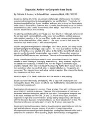
Figure 4: Press Lites separated laterally on midline both lights on.you may need to begin with the light flashing in order for itto be perceived. Probe different sectors of the field.Procedure Four: Physiological DiplopiaWe are going to take advantage of the principle thatphysiological diplopia is an example of divided visualattention. 15 Begin working in the patient’s preferred field. Forexample, if the patient has a right homonymous defect, beginby having her look slightly to the left of midline. Positionthe green light about 20 centimeters beyond the red light,directly along the line of sight. When the patient fixates thered light, there should appear to be two green lights in thebackground, and when the patient fixates the green light,there should appear to be two red lights in the foreground.If the patient has difficulty registering physiological diplopia,leave the light being fixated in the “on” position, and have theother light flashing. It is more difficult to suppress the lightsin physiological diplopia when they are flashing.As the patient is able to perceive physiological diplopiain near and/or far space, gradually move the lights laterally intandem until they are close to or at the anatomical midline,midway between the two eyes. When the lights are on themidline, have the patient fixate the front red light and beaware of two green lights beyond it. If the patient is able to dothis, she is now aware of simultaneous stimuli, on one in thepreferred field and one in the formerly neglected, inattentivefield. By definition, they split the midline in equal degrees.If the patient is experiencing a midline shift, and reportsthe lights being perceived as straight ahead when they areFigure 5: Press Lites separated sufficiently on the z-axis togenerate physiological diplopia of the light not being fixated.to the right or left of midline, the lights should initially beplaced along the perceived midline. As physiological diplopiais perceived, the lights can slowly be moved toward theanatomical midline while the patient is asked to maintainphysiological diplopia (Figure 5).Some patients may only be able to perceive the proximalor distal lights in physiological diplopia when the light isflashing. It is possible that flicker detection channels tapinto visual field regionalization. 16 First try having the patientfixate the proximal light when the light source is continuous,while having the distal light flashing. When the patient canmaintain awareness of physiological diplopia this way, tryhaving the proximal fixation light flashing and the distal lightseen in physiological diplopia continuous.Procedure Five: Reducing ExtinctionOnce the patient is able to appreciate targets on both sidesof midline simultaneously through physiological diplopia, itshould be easier to combat extinction. The patient shouldnow be ready to have one of the lights placed slightly off themidline to the left while one is slightly off the midline to theright. Begin with both lights off and illuminate one, askingthe patient to identify which of the colors is on. When bothlights are on, ask the patient to identify when the light beginsflashing. Once this is done accurately, ask the patient to reportwhen the light stops flashing. When the patient needs anauditory cue to aid sensory integration, use the Glow StickVolume 1 | Issue 2 Optometry & Visual Performance 65
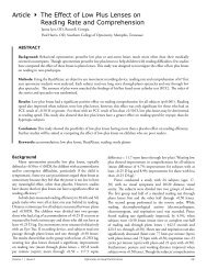
with a button so that the patient is cued that something ischanging. Once this is achieved, Glow Sticks with a twistbottom rather than a button can be used so that there is noauditory cue.As previously noted, the distinctions between visualspatialneglect, visual hemi-inattention, and visual field losscan be subtle. Margolis 17 presented an excellent overviewof the subject in the context of therapeutic strategies forvision rehabilitation. It is important to note that extinctioncan occur with two simultaneous stimuli from differentsensory modalities, one visual and the other one tactile. 18 Wetherefore engage the patient in simultaneous touch and visionprocedures. For example, have the patient reach for an objectin the seeing field while reporting awareness of the color of thelight that you will introduce into the non-seeing field.Procedure Six: Processing SynchronouslyThe lights have a flash rate that occurs at slightly differingpeak cycles. This results in the lights flashing so that atone point they are perfectly in sync blinking together, andapproximately seven flashes later they have moved 180degrees out of phase so that they flash alternately. Any of thepreceding five procedures can be done so that the patient hasto say “now” when the lights are flashing synchronously. Thisrepresents a higher level cognitive task, and is an importantcognitive load in dividing attention equally between the twofields.DiscussionThe procedures presented here fall within the category ofperceptual learning as applied to vision rehabilitation. 19 By thenature of the stimuli in Press Lites as a threshold detection task,we are applying the principle that perceptual learning is basedon sufficient numbers of repeated trials designed to improveone’s ability to detect a target in a complex environment. 20As a general principle, one begins working with patientsin a quiet environment, where both visual and auditorydistractions are kept to a minimum. 21 The procedures havebeen presented as phased sequences, from lower order thinkingto higher order cognition. Although perceptual learning is notpredicated on feedback, and theoretically does not transfer totasks that are not practiced, the utility of these procedures isenhanced when the therapist is able to guide the patient onmaking changes from what is initially perceived.Patients with rehabilitative needs vary widely regardingtheir initial capabilities and cognitive capacities. At the outset,the patient may need to work at a basic level of visual awareness,detecting when a single fixation light is on or off in the seeingfield. We then progress to whether the light is flashing onand off or is on continuously. Intersensory integration canaid peripheral localization by adding a tone or a click to thetarget, or by touch. However, neglect or inattention can existin all sensorimotor domains, and some patients may thereforeFigure 6: The Neuropsychology of Visual Spacebe confused or conflicted by the addition of non-visual cuesrather than be aided by them.We have emphasized the role of divided attentionand that inattention or neglect reflects a limitation in thepatient’s ability to divide attention. Although the Press LitesProcedures have greatest utility in aiding restoration of visualfield awareness, they may be used to accentuate peripheralawareness even when there are no visual field abnormalitiesper se. This dovetails with the concept of useful field of view.When a patient has intact binocular vision withoutsuppression, we utilize awareness of physiological diplopia as astrong stimulus that bisects visual space symmetrically. Thereis less likelihood of extinction because the simultaneity of thetwo lights splitting the midline is generated by the patient’sinternal projection of a single stimulus on the midline,not two physical objects in space competing for attentionpresented by the therapist. The awareness of physiologicaldiplopia in the background or foreground of the fixation lightcan be accentuated by leaving the non-fixated light on theflashing mode.All of these procedures may have differential effects basedon the location of the targets within the sphere of the patient’sneuropsychological visual space (Figure 6). This is where theresults of a visual field conducted in a two dimensional planewithin an instrument, with visual fixation at one fixed distance,may not be indicative of how the patient functions out of theinstrument. It also explains why chairside confrontation visualfields may be more indicative of visual function regardingperipheral awareness, as there are more stimuli competingfor visual attention. For some patients, neglect or inattentionmay be accentuated in near or personal space, for others inintermediate or peripersonal space, and for still others in faror extrapersonal space. By varying location of the targets, thetherapist will find the area of space within which the patientcan best perform initially, and work toward extending thatregion within three-dimensional space.66 Optometry & Visual Performance Volume 1 | Issue 2
ConclusionPress Lites are commercially available road hazard lightsadapted for use as versatile stimuli for vision rehabilitation andtherapy procedures. They provide the advantage of being usedunder natural conditions in three-dimensional visual space,and the fixation distance in various visual planes can be easilyadjusted. Awareness of a stimulus peripherally when a centrallight is fixated can expand the useful field of view. Inattentionor neglect can be combatted when the non-fixated light is set tothe flash mode. Physiological diplopia can be used to overcomeextinction, a feature unavailable in current computerizedtechnologies which operate within two-dimensional planesrather than on the z-axis of open visual space.These procedures are used to complement rather thandisplace existing technologies. They have the added advantageof being readily available, inexpensive, and easily dispensedfor reinforcement outside of the office. They may be used inconjunction with lens and prism procedures synergistically,and the cognitive load can be varied by using the lights invarious visual environments progressing from quiet tocluttered. Given the complexity of the visual and cognitiveprocesses involved, their use should be closely monitored bythe optometrist prescribing the procedures.References1. De Weerd P. Perceptual filling in: More than the eye can see. Prog Brain Res2006;154:227-45.2. Suchoff IB, Ciuffreda KJ. A primer for the optometric management ofunilateral spatial inattention. Optometry 2004;75:305-18.3. Margolis NW, Suter PS. Visual field defects and unilateral spatial inattention:Diagnosis and treatment. J Behav Optom 2006;17:31-7.4. Bruce BB, Zhang X, Kedar S, Newman NJ, et al. Traumatic homonymoushemianopsia. J Neurol Neurosurg Psychiatry 2006;77:986-8.5. Gianutsos R. Visual field deficits after brain injury: Computerized screening. JBehav Optom 1991;2:143-50.6. Kerkhoff G. Restorative and compensatory therapy approaches in cerebralblindness – a review. Restor Neurol Neurosci 1999;15:255-71.7. Schuett S, Heywood CA, Kentridge RW, Zihl J. The significance of visualinformation processing in reading: Insights from hemianopic dyslexia.Neuropscyhologia 2008;46:2445-62.8. Rowe F, Wright D, Brand D, Jackson C, et al. Reading difficulty after stroke:Ocular and non-ocular causes. Int J Stroke 2011;6:404-11.9. Padula W, Argyris S. Post trauma vision syndrome and visual midline shiftsyndrome. Neuro Rehab 1996;6:165-71.10. Super S. Intention, Attention, Inattention and Neglect. Santa Ana, CA:<strong>Optometric</strong> <strong>Extension</strong> <strong>Program</strong> 2004.11. Marrone MA. Peripheral Awareness. J Behav Optom 1991;2:7-11.12. Padula WV, Nelson CA, Padula WV, Benabib R, et al. Modifying posturaladaptation following a CVA through prismatic shift of visuo-spatial egocenter.Brain Inj 2009;23:566-76.13. Harris PA. The Use of Lenses to Improve Quality of Life Following BrainInjury. In PB Suter, LH Harvey, eds. Vision Rehabilitation: MultidisciplinaryCare of the Patient Following Brain Injury. Boca Raton, FL: CRC Press/Taylor& Francis 2011;259-77.14. Stern CD. Photophobia, Light, and Color in Acquired Brain Injury. In PBSuter, LH Harvey, eds. Vision Rehabilitation: Multidisciplinary Care of thePatient Following Brain Injury. Boca Raton, FL: CRC Press/Taylor & Francis2011;283-300.15. Sanet RB, Press LJ. Spatial Vision. In PB Suter, LH Harvey, eds. VisionRehabilitation: Multidisciplinary Care of the Patient Following Brain Injury.Boca Raton, FL: CRC Press/Taylor & Francis 2011;130.16. Hussey ES. Use of visual flicker in remediation of intermittent centralsuppression suggests regionalization of vision. J Behav Optom 1999;10:3-11.17. Margolis NM. Evaluation and Treatment of Visual Field Loss and Visual-Spatial Neglect. In PB Suter, LH Harvey, eds. Vision Rehabilitation:Multidisciplinary Care of the Patient Following Brain Injury. Boca Raton, FL:CRC Press/Taylor & Francis 2011;165-87.18. Feloiu FD, Marotta JJ, Vesia M, Black SE. Left-to-right reversal of hemispatialneglect symptoms following adaptation to reversing prisms. In MRM Jenkin,LR Harris eds. Cortical Mechanisms of Vision. Cambridge, UK: CambridgeUniversity Press 2009;47.19. Zihl J. Rehabilitation of Visual Disorders After Brain Injury. 2nd ed. NewYork: Psychology Press/Taylor & Francis 2011;11.20. Gilbert CD. Intermediate-Level Visual Processing and Visual Primitives. In ERKandel, JH Schwartz, TM Jessell, SA Siegelbaum, AJ Hudspeth eds. Principlesof Neural Science 5th ed. New York: McGraw Hill Medical 2013;616.21. Press LJ. Parallels Between Auditory and Visual Processing. Santa Ana, CA:<strong>Optometric</strong> <strong>Extension</strong> <strong>Program</strong> <strong>Foundation</strong> 2012;57.Correspondence regarding this article should be emailed to pressvision@aol.comor sent to Leonard J. Press, OD, 17-10 Fair Lawn Avenue, Fair Lawn, NJ07410, Tel: 201-794-7977. All statements are the authors’ personal opinionsand may not reflect the opinions of the the representative organizations, ACBO,COVD, or OEPF, Optometry & Visual Performance or any institution ororganization with which the author may be affiliated. Permission to use reprintsof this article must be obtained from the editor. Copyright 2013 <strong>Optometric</strong><strong>Extension</strong> <strong>Program</strong> <strong>Foundation</strong>. Online access is available at www.acbo.org.au,www.covd.org, and www.oepf.org.Press, L. Press lites – procedures for visual field awareness. Optom Vis Perf2013;1(2):62-7.VP Today is the quarterly news supplement to Optometry &Visual Performance. It contains product reviews, event reviews,book reviews, continuing education notices, and more!Volume 1, Issue 1, March 2013• InfantSEE ® : Patient Education• Event Review: Colorado Vision Training Conference• Book Review: “Concussions and Our Kids”• In the SpotlightGo to http://bit.ly/VPT1-1 for VP Today online only!Volume 1 | Issue 2 Optometry & Visual Performance 67