Pulmonary function in long-term survivors of hyaline membrane ...
Pulmonary function in long-term survivors of hyaline membrane ...
Pulmonary function in long-term survivors of hyaline membrane ...

Create successful ePaper yourself
Turn your PDF publications into a flip-book with our unique Google optimized e-Paper software.
<strong>Pulmonary</strong> Function <strong>in</strong> Long-<strong>term</strong>Survivors <strong>of</strong> Hyal<strong>in</strong>e Membrane Disease*James R. Shelter, M.D., EC.CP. A William D. Lucht, M.D.;tA. K. Goel, B.S.; James D. Snell, M.D., F.C.C.P,§and MildredT. Stahlman, M.D.\\In order to de<strong>term</strong><strong>in</strong>e the <strong>long</strong>-<strong>term</strong> pulmonary consequences<strong>of</strong> hyal<strong>in</strong>e <strong>membrane</strong> disease (HMD), we measuredpulmonary <strong>function</strong> and airway responsiveness to methachol<strong>in</strong>e<strong>in</strong> 22 <strong>survivors</strong> <strong>of</strong> HMD aged 18 to 22 years.N<strong>in</strong>eteen age-matched control subjects without a history <strong>of</strong>lung disease or asthma were also studied. There was nostatistically significant difference between the mean pulmonary<strong>function</strong> test results <strong>of</strong> the control vs the HMDgroup. The mean N28 <strong>of</strong> the five HMD patients whorequired assisted ventilation was significantly different fromthat <strong>of</strong> the control group. Airway responsiveness to methachol<strong>in</strong>e,as estimated by the dose <strong>of</strong> methachol<strong>in</strong>e aerosolnecessary to provoke a 35 percent fall <strong>in</strong> SCaw did notdiffer between the control and HMD groups. In the absence<strong>of</strong> prematurity, low birth weight (
Measurements <strong>of</strong> forced expiratory flow were made us<strong>in</strong>g acomputerized mass flow meter. Functional residual capacity (FRC)was measured by nitrogen washout. The s<strong>in</strong>gle breath diffus<strong>in</strong>gcapacity for carbon monoxide was de<strong>term</strong><strong>in</strong>ed. The slope <strong>of</strong> phase3 <strong>of</strong> the s<strong>in</strong>gle breath oxygen test (the nitrogen delta) was obta<strong>in</strong>edus<strong>in</strong>g standard methods. 12 The values are reported as the percentpredicted, <strong>in</strong> an effort to control for the effect <strong>of</strong> age, sex, andheight on pulmonary <strong>function</strong>. K!,fiMethachol<strong>in</strong>e <strong>in</strong>halation challenge was done by hav<strong>in</strong>g subjects<strong>in</strong>hale doubl<strong>in</strong>g concentrations <strong>of</strong> methachol<strong>in</strong>e aerosol from abreath-activated dose-meter<strong>in</strong>g device connected to a nebulizerwhich aerosolized approximately 0.010 ml <strong>of</strong> solution per breath.Measurements <strong>of</strong> specific airway conductance (SGaw) were made<strong>in</strong> triplicate <strong>in</strong> a constant volume body plethysmograph beforemeasurements <strong>of</strong> forced expiratory flow were obta<strong>in</strong>ed. 1718 Fivebreaths <strong>of</strong> methachol<strong>in</strong>e aerosol were <strong>in</strong>haled from <strong>function</strong>alresidual capacity dur<strong>in</strong>g tidal breath<strong>in</strong>g. Increas<strong>in</strong>g concentrations<strong>of</strong> methachol<strong>in</strong>e were <strong>in</strong>haled at five-m<strong>in</strong>ute <strong>in</strong>tervals until theSGaw had fallen by 35 percent from basel<strong>in</strong>e measurements, or thehighest concentration <strong>of</strong> methachol<strong>in</strong>e had been achieved. If subjectsdid not have a significant fall at a concentration <strong>of</strong> 10 mg/ml,then ten breaths <strong>of</strong> this concentration were given, and this wasconsidered to be equivalent to a concentration <strong>of</strong> 20 mg/ml forpurposes <strong>of</strong> analysis. Airway responsiveness was expressed as thedose <strong>of</strong> methachol<strong>in</strong>e (mg/ml) required to cause a 35 percent fall <strong>in</strong>SGaw (EDajSGaw) and was calculated by <strong>in</strong>terpolation from the<strong>in</strong>dividual log dose response curves. One patient had an FEV[ thatwas 53 percent <strong>of</strong> predicted, and the response to bronchodilatorswas measured <strong>in</strong>stead <strong>of</strong> the response to methachol<strong>in</strong>e. No subjectwas studied with<strong>in</strong> six weeks <strong>of</strong> a viral upper respiratory tract<strong>in</strong>fection.Standard chest roentgenograms were made <strong>in</strong> the posterioranteriorand lateral views.Review <strong>of</strong> the nursery records was undertaken to provide birthweight, gestational age, duration <strong>of</strong> assisted ventilation, pulmonarycomplications, and the oxygen score. The oxygen score was calculatedby assign<strong>in</strong>g one po<strong>in</strong>t for every hour spent at an FIo2 <strong>of</strong> .21to .39, two po<strong>in</strong>ts .40 to .59, three po<strong>in</strong>ts for .60 to .79, four po<strong>in</strong>tsfor .8 to 1.0. 3 Eighteen <strong>of</strong> the 22 HMD subjects had pulmonary<strong>function</strong> tests done at age 7 and/or ll. 5The study was approved by the Vanderbilt University Committeefor the Protection <strong>of</strong> Human Subjects.StatisticsDifferences between the control and HMD groups were testedfor by impaired one tailed Student's t-test or by Wilcoxon nonpairedrank sum test when the data were found to be nonnormal <strong>in</strong>distribution. A p
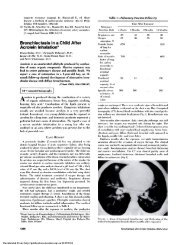
Table 2—<strong>Pulmonary</strong> Function Test Results <strong>in</strong> Selected Individual HMD Patients*BirthWeight,0,FEV]/FEFSubjectGScore FVC FEV,FVC25-75 Vmax 50% Vmax 25% TLC N2oDeoIt 1,687 1,824 82t 58+ Tit 28t 31t 18+ 75t 2,9t 74+2+ 2,665 519 92 94 105 96 99 98 76t 1.2t 80+3? 1,205 463 86t 85t 99 91 118 68 74t .8 934t 2,155 466 112 108 93 78 71 57 114 5,8f 996+ 4,196 85 94 83t 85f 55t 51+ 47 90 .8 877 1,106 320 98 102 106 117 123 97 84 1,9+ 1048 2,438 277 102 107 108 99 99 85 84 1,2+ 1109 2,381 241 108 107 108 96 97 80 94 4.2+ 9710 1,984 200 100 107 107 116 112 107 82t .6 9911 2,601 217 125 107 88t 71 70 66 105 .7 9612 2,438 108 119 106 94 76 75 48 103 .6 127Control 91 87 89 70 67 48 83 .9 80limit•Values are expressed as percent predicted except for N28, which is expressed as %N/500ml.Underl<strong>in</strong>ed values are those outside the 95 percent confidence limits <strong>of</strong> Knudson et al.""+Values outside the control values. The control limit was calculated: Mean ± 1.729 SD <strong>of</strong> the values <strong>of</strong> the 19 control subjects.iPatients who underwent assisted ventilation.terenol with no improvement <strong>in</strong> flow The third HMDpatient had a recent viral upper respiratory tract<strong>in</strong>fection and was unavailable for further test<strong>in</strong>g. Therewas no correlation between the oxygen score or birthweight and the degree <strong>of</strong> airway responsiveness tomethachol<strong>in</strong>e. The HMD and control subjects werestudied over a period <strong>of</strong> three years, but there was nocorrelation between the time <strong>of</strong> year <strong>in</strong> which theywere studied and the ED^ SGawWheez<strong>in</strong>g was reported by six <strong>of</strong> the 19 HMDLog E D 3 5SGaw(mg/ml)>1.31.20,80.40-0.4-0,8OOOOOOOO° o°°°ooo0 0oCONTROL•••••HMDFIGURE 1. Log estimated dose <strong>of</strong> methachol<strong>in</strong>e (mg/ml) required tocause a fall <strong>in</strong> SGaw <strong>of</strong> 35 percent from basel<strong>in</strong>e values <strong>in</strong> control(open circles) and HMD patients (closed circles). Each po<strong>in</strong>trepresents the value obta<strong>in</strong>ed <strong>in</strong> a s<strong>in</strong>gle subject. There was nodifference between the log EDjsSGaw <strong>of</strong> the control and HMDgroups.patients undergo<strong>in</strong>g methachol<strong>in</strong>e challenge and three<strong>of</strong> 19 control subjects, but this <strong>in</strong>crease <strong>in</strong> the report<strong>in</strong>g<strong>of</strong> wheeze was not statistically significant (p = 0,47 byFisher's exact test). Only one HMD patient had beengiven a diagnosis <strong>of</strong> asthma and had taken bronchodilators<strong>in</strong> the past. Four HMD <strong>survivors</strong> reported ahistory <strong>of</strong> asthma <strong>in</strong> parents or sibl<strong>in</strong>gs. Two <strong>of</strong> thesefour responded to methachol<strong>in</strong>e. Four <strong>of</strong> the controlsubjects had a parent or sibl<strong>in</strong>g with asthma, and three<strong>of</strong> these four responded to methachol<strong>in</strong>e.Of the 16 chest roentgenograms, one showed asevere pectus deformity that has s<strong>in</strong>ce been surgicallycorrected; patient 1 had evidence <strong>of</strong> hyper<strong>in</strong>flation,and two others had a suggestion <strong>of</strong> very mild <strong>in</strong>terstitialdisease.DISCUSSIONOur study shows that <strong>long</strong>-<strong>term</strong> <strong>survivors</strong> <strong>of</strong> HMDwho were not premature and who were treated onlywith oxygen and/or negative pressure ventilation havefew detectable abnormalities <strong>in</strong> pulmonary <strong>function</strong>.There was no difference <strong>in</strong> the mean test resultsbetween the HMD and the control group. However,the five patients with the most severe disease whorequired assisted ventilation had a mean N28 that wassignificantly different from the control group.Patient 1 had the highest oxygen score <strong>in</strong> the HMDgroup (1824) and had been identified <strong>in</strong> previousfollow-up studies as hav<strong>in</strong>g symptomatic respiratoryimpairment. His current tests suggest both restrictiveand obstructive lung disease. This patient, how1,5ever, had normal spirometry at age 7, and had normalvolumes and diffusion at age 11, with only an FEVjthat was low He has a history <strong>of</strong> recurrent acutebronchitis and is a cigarette smoker. He requirednegative pressure ventilation, and for a time, was736 Long-<strong>term</strong> Survivors <strong>of</strong> Hyal<strong>in</strong>e Membrane Disease (Shelter et al)Downloaded From: http://publications.chestnet.org/ on 03/29/2014
<strong>in</strong>tubated and ventilated with positive pressure. Likewise,patient 2 had a high 0 2 score (519), with normalspirometry at age 11. He had surgical repair <strong>of</strong> apectus deformity at age 19. His current pulmonary<strong>function</strong> test results <strong>in</strong>dicate mild restrictive lungdisease. He received negative pressure ventilation.Patient 4, who also received negative pressure ventilation,had f<strong>in</strong>d<strong>in</strong>gs consistent with mild restrictivelung disease; his prior test<strong>in</strong>g had shown only adecreased FVC at age 7, with normal spirometry atage 11. Patient 3 had tests consistent with airflowobstruction. She was the daughter <strong>of</strong> a diabetic motherand had mild to moderate HMD. She had a familyhistory <strong>of</strong> asthma and began to smoke cigarettes at age14. Her tests <strong>of</strong> lung volumes and spirometry at age11 were normal. The failure to discover more abnormalities<strong>in</strong> these patients at ages 7 and 11 may haveresulted from a wider range <strong>of</strong> normal values <strong>in</strong> thoseage groups. An alternative explanation is that thepulmonary disease <strong>of</strong> these patients may have progressed.Fourteen <strong>of</strong> the rema<strong>in</strong><strong>in</strong>g HMD group hadnormal spirometry measured at their 11 year followup.Six HMD <strong>survivors</strong> had a s<strong>in</strong>gle breath oxygen testthat was outside the 95 percent confidence range <strong>of</strong>our normal control subjects, suggest<strong>in</strong>g that HMD<strong>survivors</strong> may have subtle abnormalities <strong>in</strong> the distribution<strong>of</strong> ventilation, perhaps related to small airwaysdisease. However, the degree <strong>of</strong> impairment <strong>of</strong> thedistribution <strong>of</strong> ventilation alone did not appear to affectsymptoms.It appears that some HMD patients manifest abnormalities<strong>of</strong> pulmonary <strong>function</strong> <strong>in</strong> adult life. There isthe possibility that these abnormalities may haveprogressed s<strong>in</strong>ce childhood, perhaps <strong>in</strong> response tothe deleterious effects <strong>of</strong> environmental lung <strong>in</strong>juries,such as cigarette smok<strong>in</strong>g, viral upper respiratory tract<strong>in</strong>fections, or air pollution. It will be important tocont<strong>in</strong>ue to follow the pulmonary <strong>function</strong> <strong>of</strong> at leastthose <strong>in</strong>dividuals with identifiable abnormalities.Unlike the studies by Bertrand et al and Mac-9Luskey et al our study did not reveal an <strong>in</strong>creased10<strong>in</strong>cidence <strong>of</strong> airway responsiveness <strong>in</strong> our group <strong>of</strong>HMD <strong>survivors</strong>. However, there are differences <strong>in</strong>methodology and <strong>in</strong> patient population between ourstudy and that <strong>of</strong> Bertrand and <strong>of</strong> MacLuskey Weused the ED 3 5 SGaw to quantify airway responsiveness<strong>in</strong>stead <strong>of</strong> the FEV: used by others. The SGaw is amore sensitive <strong>in</strong>dex <strong>of</strong> airway change dur<strong>in</strong>g <strong>in</strong>halationchallenge than is the FEVX, but may not discrim<strong>in</strong>atebetween subjects with atopy and those withasthma. In the study <strong>of</strong> Bertrand et al, eight <strong>of</strong> 112123 9HMD subjects responded to <strong>in</strong>haled histam<strong>in</strong>e with afall <strong>in</strong> FEV, <strong>of</strong> 20 percent or more; 30 <strong>of</strong> 46 patientsstudied by MacLuskey et al responded to methachol<strong>in</strong>e.If our HMD patients were similar, we 10 shouldhave found an even greater proportion <strong>of</strong> respondersby us<strong>in</strong>g measurements <strong>of</strong> SGaw However, only n<strong>in</strong>e<strong>of</strong> 19 HMD patients tested responded to methachol<strong>in</strong>e.It might be argued that our small control groupconta<strong>in</strong>ed an <strong>in</strong>ord<strong>in</strong>ate number <strong>of</strong> hyperreactivesubjects. We do not believe this to be the case, because<strong>in</strong> 22 mild asthmatic subjects receiv<strong>in</strong>g no <strong>long</strong>-<strong>term</strong>medications who responded to an identical challengeprotocol <strong>in</strong> our laboratory with a 20 percent or greaterfall <strong>in</strong> FEV,, the mean ED 3 5 SGaw was 3.3±5.1,which is significantly less than that <strong>of</strong> the 11 responders<strong>in</strong>ourcontrolgroup(10.5±6.0, p0.8).Rather, we believe that the difference <strong>in</strong> our f<strong>in</strong>d<strong>in</strong>gsand those <strong>of</strong> Bertrand et al and Macluskey et al lies9 10<strong>in</strong> the populations studied. Our subjects were older atthe time <strong>of</strong> the study, and therefore, reflect a differentage and management approach at the time <strong>of</strong> their<strong>in</strong>itial illness. They were treated with oxygen and/orassisted ventilation. Infants received negative pressureventilation only when oxygen therapy had failed andwere <strong>in</strong>tubated and ventilated by hand us<strong>in</strong>g ananesthesia circle respirator when negative pressureventilation had failed and the patient was <strong>in</strong> extremis.The mean birth weights <strong>of</strong> our patients was242,244 ± 734 g (mean±SD), which is larger than thatreported by Bertrand et al (1,900 ±615) or Mac9Luskey et al (1,166 ±193). Likewise, the oxygen10scores <strong>of</strong> the patients <strong>in</strong> our study were slightly lowerthan those <strong>of</strong> Bertrand et al (365 ±352 vs 554), and9only four <strong>of</strong> our 22 HMD subjects had a gestationalage <strong>of</strong> 32 weeks or less. Positive pressure ventilationwas not rout<strong>in</strong>ely used <strong>in</strong> our neonatal <strong>in</strong>tensive careunit <strong>in</strong> the treatment <strong>of</strong> HMD at the time this cohort<strong>of</strong> patients was born. Thus, it may still be true thatHMD patients receiv<strong>in</strong>g positive pressure ventilationor other newer modes <strong>of</strong> <strong>in</strong>tensive supportive therapyand <strong>in</strong>fants <strong>of</strong> very low birth weight (
sive support may exhibit abnormal lung <strong>function</strong> <strong>in</strong>childhood.ACKNOWLEDGMENTS: The authors thank Dr. Ryszard Dworskifor use <strong>of</strong> unpublished observations, Dr. William Vaughn for helpwith statistical analysis, Dr. Dan L<strong>in</strong>dstroni for data analysis,Marguerite Douglas and Sherry Taylor for expert technical assistance,and Carol Cauthron and Reg<strong>in</strong>a Stanley for secretarialassistance.REFERENCES1 Stahiman M, Hedvall G, Dolanski E, Faxelius G, Burko H,Kirk V A six year follow-up <strong>of</strong> cl<strong>in</strong>ical hyal<strong>in</strong>e <strong>membrane</strong> disease.Ped Cl<strong>in</strong> N A 1973; 20:422-462 Lamarre A, L<strong>in</strong>sao L, Reilly BJ, Sawyer PR, Levison H. Residualpulmonary abnormalities <strong>in</strong> <strong>survivors</strong> <strong>of</strong> idiopathic respiratorydistress syndrome. Am Rev Respir Dis 1973;108:56-613 Coates AL, Desmond K, Willis D, Nogrady B. Oxygen therapyand <strong>long</strong>-<strong>term</strong> pulmonary outcome <strong>of</strong> respiratory distress syndrome<strong>in</strong> newborns. Am J Dis Child 1982; 136:892-954 Heldt CP, Mcllroy MB, Hansen TN, Tooley WH. Exerciseperformance <strong>of</strong> the <strong>survivors</strong> <strong>of</strong> hyal<strong>in</strong>e <strong>membrane</strong> disease.J Pediatr 1980; 96:995-995 Stahiman M, Hedvall G, L<strong>in</strong>dstrom D, Snell J. Role <strong>of</strong> hyal<strong>in</strong>e<strong>membrane</strong> disease <strong>in</strong> production <strong>of</strong> later childhood lung abnormalities.Pediatrics 1982; 69:572-766 Smyth JA, Tabchnik E, Duncan WJ, Reilly BJ, Levison H.<strong>Pulmonary</strong> <strong>function</strong> and bronchial hyperreactivity <strong>in</strong> <strong>long</strong>-<strong>term</strong><strong>survivors</strong> <strong>of</strong> bronchopulmonary dysplasia. Pediatrics 1982;68:336-407 O'Brodovich HM, Mell<strong>in</strong>s RB. State <strong>of</strong> the art: bronchopulmonarydysplasia: unresolved neonatal acute lung <strong>in</strong>jury. Am RevRespir Dis 1985; 132:694-7098 Northway WH Jr, Rosan RC, Porter DY. <strong>Pulmonary</strong> diseasefollow<strong>in</strong>g respiratory therapy <strong>of</strong> hyal<strong>in</strong>e-<strong>membrane</strong> disease,N Engl J Med 1967;"276:357-689 Bertrand J-M, Riley SP, Popk<strong>in</strong> J, Coates AL. The <strong>long</strong>-<strong>term</strong>pulmonary sequelae <strong>of</strong> prematurity: the role <strong>of</strong> familial airwayhyperreactivity and the respiratory distress syndrome. N EnglJ Med 1985; 312:742-4510 MacLusky IB, Str<strong>in</strong>ger D, Zarfen J, Smallhorn J, Levison H.Cardiorespiratory status <strong>in</strong> <strong>long</strong>-<strong>term</strong> <strong>survivors</strong> <strong>of</strong> prematurity,with and without hyal<strong>in</strong>e <strong>membrane</strong> disease. Pediatr Pulmonol1986; 2:94-10211 Nickerson BG, Taussig LM. Family history <strong>of</strong> asthma <strong>in</strong> <strong>in</strong>fantswith bronchopulmonary dysplasia. Pediatrics 1980; 65:1140-4412 Buist AS, VanFleet DI, Ross BB. Quantitative analysis <strong>of</strong> thealveolar plateau <strong>in</strong> the diagnosis <strong>of</strong> early airflow obstruction. AmRev Respir Dis 1973; 107:735-4313 Goldman HI, Becklake MR. Respiratory <strong>function</strong> tests: normalvalues at median altitudes and the prediction <strong>of</strong> normal results.Am Rev Tuberc 1959; 79:457-6714 Burrows B, Kasik JE, Niden AH, Barclay WR. Cl<strong>in</strong>ical usefulness<strong>of</strong> the s<strong>in</strong>gle breath pulmonary diffus<strong>in</strong>g capacity test. AmRev Respir Dis 1961; 84:789-80615 Knudson RJ, Slat<strong>in</strong> RC, Lebowitz MD, Burrows B. The maximalexpiratory flow volume-curve: normal standards, variability, andeffects <strong>of</strong> age. Am Rev Respir Dis 1976; 113:587-60016 Knudson RJ, Lebowitz MD. Maxima! mid-expiratory flow(FEFavsw): normal limits and assessment <strong>of</strong> severity. Am RevRespir Dis 1978; 117:609-1017 DuBois AB, Botelho SY, Bedell GN, Marshall R, Comroe J. Arapid plethysmographic method for measur<strong>in</strong>g thoracic gasvolume. J Cl<strong>in</strong> Invest 1956; 35:322-2618 DuBois AB, Botehlo SY, Comroe JH. A new method formeasur<strong>in</strong>g airway resistance <strong>in</strong> man us<strong>in</strong>g a body plethysmograph.J Cl<strong>in</strong> Invest 1956; 35:327-3519 Steel RGD, Torrie JH. Pr<strong>in</strong>ciples and procedures <strong>of</strong> statistics.New York: McGraw-Hill Book Co, 1980:113-1820 Zar JH. Biostatistical analysis. Englewood Cliffs, NJ: Prentice-Hall, Inc, 1974:413.21 Fish JE, Rosenthal RR, Batra G, Menkes H, Summer W,Permutt S, et al. Airway responses to methachol<strong>in</strong>e <strong>in</strong> allergicand nonallergic subjects. Am Rev Respir Dis 1977; 113:579-8622 OrehekJ, Nicoli MM, Delpierre S, Beaupre A. Influence <strong>of</strong> theprevious deep <strong>in</strong>spiration on the spirometrie measurement <strong>of</strong>provoked bronchoconstriction <strong>in</strong> asthma. Am Rev Respir Dis1981; 123:269-7223 Michoud MC, Ghezzo H, Amyot R. A comparison <strong>of</strong> pulmonary<strong>function</strong> tests used for bronchial challenges. Bull Eur PhysiopatholRespir 1982; 18:609-2124 Stahiman MT, Malan AF, Shephard FM, Blankenship WJ, YoungWC, Gray J. Negative pressure assisted ventilation <strong>in</strong> <strong>in</strong>fantswith hyal<strong>in</strong>e <strong>membrane</strong> disease. J Pediatr 1970; 76:174-8225 Galdes-Sebaldt M, Sheller JR, Grogaard J, Stahiman MT.Extreme prematurity, not hyal<strong>in</strong>e <strong>membrane</strong> disease (HMD), isl<strong>in</strong>ked with <strong>in</strong>creased airway responsiveness and abnormalpulmonary <strong>function</strong> at 10 to 13 years <strong>of</strong> age. Pediatr Res 1987;21:501.738 Long-<strong>term</strong> Survivors <strong>of</strong> Hyal<strong>in</strong>e Membrane Disease (Sheller et al)Downloaded From: http://publications.chestnet.org/ on 03/29/2014