Chapter 1 - Dellon Institutes for Peripheral Nerve Surgery
Chapter 1 - Dellon Institutes for Peripheral Nerve Surgery
Chapter 1 - Dellon Institutes for Peripheral Nerve Surgery
- No tags were found...

You also want an ePaper? Increase the reach of your titles
YUMPU automatically turns print PDFs into web optimized ePapers that Google loves.
<strong>Chapter</strong> OneWhy <strong>Nerve</strong>s Cause Pain
“It is okay tolose your nerve.”
Pain SolutionsPain SolutionsPain Solutions is a book to help you understand there is hope that your paincan be greatly relieved, and, sometimes, completely eliminated by the appropriateperipheral nerve surgery. <strong>Surgery</strong>, of course, is the last resort.If you are reading this book, then most likely you have already had all theusual non-surgical treatments <strong>for</strong> your pain. By describing how individuals,like you, have been helped, I hope to make this book personally importantto you or someone you know.In this chapter, I outline the basic mechanisms of pain related to conditions<strong>for</strong> which I have developed pain solutions. In the chapters that followspecific solutions are discussed <strong>for</strong> your pain.If you have pain, then impulses are traveling along a nerve into yourspine. From your spine those impulses continue to your brain. Pain is amessage that there is a problem somewhere in your body to which you needto pay attention. You may not like the pain message, but it calls your attentionto a problem. Pain Solutions will help you find the answers that perhapsyour own caring physician(s) have not been able to find <strong>for</strong> you.The central nervous system consists of the brain and spinal cord. Problemsin the brain are usually related to tumors, bleeding, or lack of bloodsupply (stroke). These usually cause headache and loss of some function,but they do not usually cause pain in your arms or legs, or your body. Problemsin the spinal cord can cause pain in these areas of your body by havingsome part of the boney or ligamentous spine cause pressure on the spinalcord or nerve roots. It is usually pretty clear that this pain is coming fromyour neck or back. Traditional x-rays, the newer mris (special imagingstudies), or traditional electrodiagnostic studies usually can identify thisproblem. An example of nerve root compression and imaging <strong>for</strong> the spinein the neck, the cervical spine is given in Figure 3-1. These symptoms can betreated often without surgery, but sometimes portions of the vertebralcolumn must be removed, like a disc, or the bone alongside the nerve (alaminectomy). If the bone is not stable, the spine may have to be fused atsome level. These operations are done by Neurosurgeons or Orthopedic<strong>Chapter</strong> 1 6
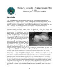
Pain SolutionsThe <strong>Peripheral</strong> Nervous System consists of all the nerves that are outside of thebrain and the spinal cord: the nerves in your arms and legs, and in your face andin your chest and abdomen. In general, there are three main problems or eventsthat happen to peripheral nerves that cause them to send a pain message to yourbrain. These three categories are neuroma, nerve compression, and neuropathy.Let us first define what these are and give you some common examples so thatyou may see that there is hope to stop the pain message by correcting directly theproblem with the nerve itself at the point at which the pain message starts. Whenthe pain is coming from the peripheral nerves, there is usually a spot along thepath of a nerve that you can touch which causes that pain (see Figure 1-3).The <strong>Peripheral</strong> <strong>Nerve</strong> surgeons of the <strong>Dellon</strong> <strong>Institutes</strong> <strong>for</strong> <strong>Peripheral</strong><strong>Nerve</strong> <strong>Surgery</strong>® are especially trained to identify these sources of pain.(Visit us at <strong>Dellon</strong>.com). Let us understand each pain source better.Figure 1-3. The brownish region is where this upper arm and elbow were injured at work. The* on the skin is where pain occurs when that spot is touched. The dotted area is where thepain travels when the painful spot is touched. There is also less feeling in this region. Thisindicates injury to a peripheral nerve. The painful spot has a neuroma. This patient can behelped by removing the neuroma (see Figure 1-4.).NeuromaFor the rest of Pain Solutions, a peripheral nerve will be called simply a“nerve.” A nerve begins in the spinal cord and extends to somewhere in thebody, <strong>for</strong> example the index finger tip. Whenever a nerve is injured, it triesto grow back to where it originally was. This is called nerve regeneration.<strong>Peripheral</strong> nerves do regenerate. They are wrapped by small cells described<strong>Chapter</strong> 1 8
A Lee <strong>Dellon</strong>, MD, PhD<strong>Nerve</strong> CompressionCompression of a nerve is very common. The name of the commonest siteof nerve compression is now almost a household word: Carpal TunnelSyndrome. Almost everyday, you will see someone wearing a splint on theirwrist to keep the wrist straight, preventing it from bending over andcompressing the median nerve. Almost everyone knows someone who hashad carpal tunnel surgery. Decompression of the median nerve at the wristmay be the most common operation done in the United States. About500,000 of these operations are done almost every year. About 125 out ofevery 100,000 people in the United States will get carpal tunnel syndromeduring their lifetime. The surgery is successful in about 85% of people inrelieving their symptoms.The commonest symptom of carpal tunnel syndrome is that you wakeup at night with your thumb, index and middle fingers asleep, but sometimesit seems as if the whole hand is asleep. With time, these three fingersbecome numb most of the day. In the advanced condition, some of thethumb muscles become weak, and may atrophy.The initial treatment of chronic nerve compression is not surgery.First, daily activity that prolongs wrist flexion is altered. For example, theposition in which you hold your wrist while typing on the computershould be altered so it is not so bent. Next, you will take an anti-inflammatorymedication to reduce swelling of the tissues that surround thetendons within the carpal tunnel (there are nine such tendons that movethe fingers). This tissue can become swollen and stuck to the mediannerve with injury or arthritis or over-use. You will wear a splint to keepthe wrist from bending, especially at night. You may receive an injection ofsteroid into the carpal tunnel to shrink the swollen tissues (but do nothave the nerve itself injected!).Why <strong>Nerve</strong>s Cause Pain 13
A Lee <strong>Dellon</strong>, MD, PhDIf you are being choked, your brain does not get oxygen, and you willcollapse and become unconscious. When a nerve does not get oxygen, itstops conducting normal electrical impulses. When this occurs , it is like theelectricity going off in your house; the lights flicker, and then go out.When you awaken at night with your “hand asleep,” or buzzing, it isbecause the nerves at the wrist or elbow are compressed, and the lack ofoxygen to the nerve sends the message that awakens you.When you cross your legs, and your top leg “goes to sleep,” it is becausethe nerve on the outside of the knee is getting compressed and sends themessage to warn you of this problem. If this problem persists, you haveweakness in your foot, and it feels as if you can hardly take a step.When the pressure on the nerve is sudden and heavy, you experiencepain as well as buzzing. But if the pressure comes on slowly, and lasts a longtime, and continues <strong>for</strong> many months, you do not have pain. Just numbnessthat comes and goes, and then the skin supplied by the nerve stays numband loses its feeling. This is chronic nerve compression.The <strong>Dellon</strong> <strong>Institutes</strong> <strong>for</strong> <strong>Peripheral</strong> <strong>Nerve</strong> <strong>Surgery</strong>® specialize in decompressionof nerves in both the upper and lower extremities. Descriptions ofsome of these operations are available to download from our website at<strong>Dellon</strong>.com. Brochures are available on these subjects:Carpal Tunnel SyndromeCubital Tunnel SyndromeRadial <strong>Nerve</strong> EntrapmentsBrachial Plexus Compression (Thoracic Outlet Syndrome)Tarsal Tunnels SyndromeFoot DropHeel Pain SyndromesWhy <strong>Nerve</strong>s Cause Pain 15
Pain SolutionsThe research models I helped to develop in the early 1980’s demonstratedthat within 2 months of nerve compression, fluid begins to leak fromblood vessels into the nerve, that by 6 months of compression, the myelinprotein covering the nerve fibers begins to get damaged, and that by oneyear, nerve fibers have begun to die. Scar tissue <strong>for</strong>ms between the bundleswithin the large nerve.* The nerve itself, may become stuck to thesurrounding ligaments. Once this degree of scar tissue <strong>for</strong>ms, only surgerycan relieve pressure on the nerve sufficiently to relieve symptoms.<strong>Surgery</strong> must relieve pressure on the compressed nerve. Either the rockor the hard place must be removed, or the nerve itself must be moved toplace without a rock or a hard place to compress it.*References to research on nerve compression from the 1980’s:*<strong>Dellon</strong> AL, Kallman CH: Evaluation of functional sensation in the hand. J Hand Surg 8:865-870, 1983.*Mackinnon SE, <strong>Dellon</strong> AL, Hudson AR, Hunter D: Chronic nerve compression – an experimentalmodel in the rat. Ann Plast Surg 13:112-120, 1984.*Mackinnon SE, <strong>Dellon</strong> AL, Daneshvar A: Histopathology of the tarsal tunnel syndrome: Examinationof a human tibial nerve. Contemp Orthop 9:43-48, 1984.*Mackinnon SE, <strong>Dellon</strong> AL, Hudson AR, Hunter DA: A primate model <strong>for</strong> chronic nerve compression.J Reconstr Microsurg 1:185-194, 1985.*Mackinnon SE, <strong>Dellon</strong> AL, Hudson AR, Hunter DA: Histopathology of compression of the superficialradial nerve in the <strong>for</strong>earm. J Hand Surg 11A:206-209, 1986.*<strong>Dellon</strong> AL: Musculotendinous variations about the elbow. J Hand Surg 11B:175-181, 1986.<strong>Chapter</strong> 1 16
A Lee <strong>Dellon</strong>, MD, PhDAn example of removing one of the hard places is given <strong>for</strong> the carpal tunnelin Figure 1-9. An example of moving the nerve to a new place is given <strong>for</strong> ulnarnerve compression at the elbow, Cubtial Tunnel Syndrome, in Figure 1-10.Figure 1-10. Cubital Tunnel Syndrome is the name given to ulnar nerve compression at theelbow. Symptoms of numbness in the little and ring finger, weakness of pinch and grasp, and ofclumsiness or dropping objects are corrected by moving the ulnar nerve from between the twobones and ligament that cause the compression. The operation I developed <strong>for</strong> this purpose isillustrated here. The ulnar nerve is seen moving from its location behind the elbow in (5) to anew place created <strong>for</strong> it in front of the elbow (6). The muscles are lengthened (4) to provide alarge place <strong>for</strong> the ulnar nerve. Immediate elbow movement is permitted to prevent scar tissuefrom making the nerve stuck in the new location. The most recent report of success with thisoperation notes more than 600 patients treated without recurrence of symptoms (<strong>Dellon</strong> AL,Coert JH: Technique of musculofascial lengthening <strong>for</strong> treatment of ulnar nerve compression atthe elbow. J Bone Joint <strong>Surgery</strong>, 86A: 169-179, 2004., with permission from http://ww.<strong>Dellon</strong>.com)Why <strong>Nerve</strong>s Cause Pain 17
Pain SolutionsFigures 1-11 and 1-12 show examples of different nerve decompressions.Figure 1-11. Example of ulnar nerve compression at the right elbow. Left: This nerve isentrapped in scar tissue overlying the site where the elbow was bruised in a fall. Right: Thesite of indentation of the ulnar nerve is noted by the white arrow, with swelling of the nervein either side of the entrapment. After completion of this neurolysis stage of the surgery, theulnar nerve will be transposed to lie beneath the lengthened flexor-pronator muscle massusing Dr. <strong>Dellon</strong>’s operation (Figure 1-10).Figure 1-12. Example of nerve compression of the common peroneal nerve at the knee.Left: overall view to orient the surgical view. The incision is located at the boney prominenceof the fibular, where the site of compression is. This patient injured the outside of this knee.Center: The metal retractor is underneath the large nerve, which is white in color in contrastthe appearance (yellow) noted in patients with diabetes or some <strong>for</strong>ms of neuropathy. Theband that is compressing the nerve is noted by the white arrow. Right: the compressive bandhas been removed. The white arrow points to the indentation or notch in the commonperoneal nerve at the site of compression by the band. With pressure gone from the nerve,sensation and strength will return to the leg and foot.<strong>Chapter</strong> 1 18
A Lee <strong>Dellon</strong>, MD, PhDDocumentation of <strong>Nerve</strong> Compression“Doctor <strong>Dellon</strong>,” my medical doctor sent me to a neurologist to see if mysymptoms of numbness were due to a nerve compression. The neurologistsaid I had to have nerve conduction testing and electromyography. The ncvand emg really hurt! The test cost about $1,600. And after all that theNeurologist said I was ‘normal! But I really have a problem Doctor <strong>Dellon</strong>.Isn’t there some test that can identify my nerve problem?This patient’s experience is all too common. The traditional electrodiagnostictesting was developed in the 1950’s. It gives electrical stimulus, orshocks to the nerves through the skin, and sometimes actual needles areinserted into the skin and the response to the shock is recorded through theneedle. Of course this hurts, and is very expensive. Un<strong>for</strong>tunately, becausethis test measures the speed of electrical activity in the fastest nerve fibers, alot of nerve fibers can be injured or not working and the test still shows anormal measurement. This electrical test is simply not sensitive enough toidentify many nerve compressions. The electromyography is still necessaryif your doctor is evaluating a nerve root compression in your neck or aprimary muscle disease. An mri, a special <strong>for</strong>m of x-ray will be necessary toimage your spinal cord.This is a subject I have written extensively about <strong>for</strong> many years.My most recent writing on this subject compares traditional electrodiagnostictesting to a test that I developed in 1989 and have been proving thevalue of ever since.* This neurosensory test is non-painful, has no needles,and is not expensive. The testing instrument is called the Pressure-SpecifiedSensory Device (pssd). Here is how it works.You are seated com<strong>for</strong>tably in a chair, and the pssd is touched to yourfinger tip, your toe, or your lip (see Figure 11-4). The two rounded metalprongs are pressed gently into the skin. You press a button when you can feelthe pressure <strong>for</strong> the first time and when you can tell whether one or two tipsare pressing the skin. This does not hurt. By comparing how hard you had*<strong>Dellon</strong> AL: Measuring <strong>Peripheral</strong> <strong>Nerve</strong> Function: Neurosensory Testing versus ElectrodiagnosticTesting, in Atlas of the Hand Clinics: <strong>Nerve</strong> Repair and Reconstruction, D. Slutsky,editor, Elsevier, Philadelphia, <strong>Chapter</strong> 1, pp 1-31, 2005.Why <strong>Nerve</strong>s Cause Pain 19
Pain Solutionsto be touched to feel the prongs, and how close together you could tell youwere being touched, the pssd results give us the in<strong>for</strong>mation to knowwhether you have nerve compression and whether the nerve is dying (seeFigure 1-16.) If the nerve has begun to die, which means you cannot tellwhen two points are touching the skin close together, then it is time <strong>for</strong>surgery to decompress the nerves.Neuropathy“Neuropathy” is best understood to be a problem with the nerves in your body,in contrast to a neuroma or a nerve compression, which is a problem with asingle nerve in your body. If your median nerve is injured, you can have aneuroma of the median nerve, somewhere along its path, on either your left oryour right side (see Figure 1-7 right). If you have a compression of your mediannerve, you have an area at which this nerve is compressed, <strong>for</strong> example at yourwrist (carpal tunnel syndrome, see Figure 1-9). About 50% of people have carpaltunnel syndrome bilaterally, which is on both sides of your body, your right andyour left hand. The carpal tunnel syndrome on the right side may be worse thanyour left side. If all the fingers of both your right and left hands are equally numband/or painful, then you have a neuropathy. Something systemic in your body isaffecting your peripheral nerves. You have a peripheral neuropathy.The most common cause of a peripheral neuropathy is diabetes. Othercommon causes of neuropathy are thyroid disorders: if the thyroid function islow, then water accumulates in your nerves, making them swell and causingthem to become compressed in regions with tight anatomic tunnels like thewrist and ankle. Another cause of neuropathy are diseases in which the bodyattacks itself with antibodies, like lupus and rheumatoid arthritis; the inflammationalong the blood vessels (vasculitis) in the nerves makes them susceptibleto compression at known sites of anatomic narrowing like the wrist andankle. Another cause of neuropathy is poisoning by heavy metals, like arsenic,lead, and mercury: these cause fluid to leak from the blood vessels into theinside of the nerve, making it susceptible to compression at known sites ofanatomic narrowing like the wrist and ankle.<strong>Chapter</strong> 1 20
A Lee <strong>Dellon</strong>, MD, PhDChemotherapy drugs used to fight cancer, like, vincristine, taxol,cisplatin, and thalidomide, can slow down the transport of critical moleculeswithin the nerve. Again, this makes the nerve susceptible to compression.It is clear that neuropathy, what ever its cause, may create symptomsthrough mechanisms similar to those that give symptoms with a singlenerve compression, and this gives us a cause <strong>for</strong> optimism, a cause <strong>for</strong> hope.In many people with neuropathy, there are compressed nerves that areresponsible <strong>for</strong> most of the symptoms. If this is true, then these symptoms,attributed to neuropathy, can be relieved by decompression of nerves.Stocking and GloveFigure 1-13. A peripheral neuropathy causes loss of sensation and or pain in a specific pattern.In the legs the pattern is that of a stocking. In the arms it is the pattern of a glove. A stockingpattern can be created by compression of several nerves in the leg and ankle, and a glovepattern can be created by compression of several nerves in the arm and wrist. Compressednerves can be decompressed, creating the possibility of hope <strong>for</strong> neuropathy.Usually, with a peripheral neuropathy, your feet become involved, symptomatic,first. The feet are involved in both the top and bottom of your feet,and the symptoms extend up the ankle, in what is the pattern of a stocking.When the neuropathy is in the upper extremities, it occurs in the patternyou would have if you were wearing gloves.In the upper extremity, if you combine compression of the ulnar nerve atthe elbow (cubital tunnel syndrome), compression of the radial nerve in the<strong>for</strong>earm (radial sensory nerve compression), and compression of themedian nerve at the wrist (cubital tunnel syndrome) you will have a patternWhy <strong>Nerve</strong>s Cause Pain 21
Pain Solutionsof sensory loss that fits a glove. As you now know, from reading above, acompressed nerve can be decompressed by surgery, relieving pain andnumbness in most patients. The same thinking applies to the leg and foot.In the lower extremity, if you combine compression of the commonperoneal nerve at the knee (fibular tunnel syndrome), compression of thedeep peroneal nerve over the top of the foot (described by me in 1990), andcompression of the tibial nerves and its branches in the four medial (insideof) ankle tunnels (tarsal tunnels syndrome), you will have a pattern ofsensory loss that fits like a stocking. As you now know, compressed nervescan be decompressed with surgery, relieving pain and numbness in mostpatients. This is discussed in detail in <strong>Chapter</strong> 2.New “Ropathy”“I have neuropathy?,” the patient with diabetes asked her doctor. “I havenumbness and burning in my hands and feet. Is it going to get better? Canyou help me?”“ I am sorry Mrs. Brown,” her doctor answered, “Certainly I can help youkeep your blood sugar under control, and I can give you medicine <strong>for</strong> thepain, but neuropathy is progressive and irreversible” he in<strong>for</strong>med her.“progressive and irreversible.” For decades, this was the correct answerin medical school, the correct answer on a medical exam, and the answer givento patients. “Neuropathy is progressive and irreversible” means there is no hope.If your medical problem is hopeless, depression and disability is likely.Figure 1-14. Examples of progressive neuropathy. Ulcer beneath the bottom of the second toe(left) and at tip of fifth toe (right) develop because of lack of sensation. Muscle wasting in thehand is noted in the center. The fingers begin to <strong>for</strong>m a “claw” and this can happen in the footas well. On the right, the toes are beginning to curl up.<strong>Chapter</strong> 1 22
A Lee <strong>Dellon</strong>, MD, PhD“Progressive and irreversible” is the old view of neuropathy.“Doctor <strong>Dellon</strong>, I have neuropathy. Can you help me?” This is thee-mail question that comes through my website, <strong>Dellon</strong>.com so often eachday, and through my phone line 1 877-dellon-1. “Yes, I can help you. Inmost people who have a nerve compression associated with theirneuropathy, the nerve can be decompressed. Symptoms can be relieved in80% of people.”“Doctor <strong>Dellon</strong>, will I still be at risk <strong>for</strong> getting an ulcer or having anamputation,” the questions go on.“If sensation is restored to your feet, you will not have an ulcer, youwill not have an amputation, and even your balance can be improved,” Ianswer. That is the new neuropathy, or “new-ropathy” as I like to call it.New-Ropathy is the first good news about neuropathy. It is optimistic.There is hope. “Restore sensation. Relieve Pain.”Figure 1-15. Adam, Bob, and Clark, shown above, have had each arm and each leg operatedupon to decompress nerves. Every three months <strong>for</strong> one year, they each had an operation tillall four extremities was decompressed. Each has neuropathy still. But each no longer haspain, and each has recovered sensation. They each now have…“New-Ropathy,” a systemicdisease without the symptoms of nerve compression.Why <strong>Nerve</strong>s Cause Pain 23
Pain SolutionsNeurosensory Testing and the PSSD“Doctor <strong>Dellon</strong>,” the man sitting in my office said, “I know I have neuropathy.I have been to so many doctors. They have done electrical testing that showI have neuropathy, but they say nothing can be done. How did you decidethat I have a nerve compression and that I can be helped? How did you decidethat I would most likely be better in about three months?”My new approach to neuropathy, or new-ropathy, is based upon:The concept that the symptoms of neuropathy can be due to thepresence of nerve compression;The ability to measure peripheral nerve function with thePressure-Specified Sensory Device;index finger pulprightleft100806040pressure: gm/sqmmradial hand dorsumrightleft100806040pressure: gm/sqmmlittle finger pulprightleft100806040pressure: gm/sqmmthenar eminencerightleft100806040pressure: gm/sqmm202020201pt2pt1pt2pt1pt2pt1pt2ptstaticstaticstaticstaticFigure 1-16. pssd report demonstrating mild carpal tunnel syndrome. The blue bars are <strong>for</strong>the left side and the red bars are <strong>for</strong> the right side. Normal bar height is below the blacklines, representing low pressure. The far left graph is the index finger, the left-center is theback of the hand, the right-center is the little finger, and the far right is the palm. The rightindex finger bar is elevated indicating abnormal pressure on the right median nerve. Sincethere is no * (asterisk) next to the bar, no nerve fibers are dying. This test is consistent withmild right carpal tunnel syndrome. Splinting is advised, not surgery.<strong>Chapter</strong> 1 24
A Lee <strong>Dellon</strong>, MD, PhDIdentification of the site of nerve compression along the length of thenerve by knowing where the anatomy can create the tight area, and theability to determine if the nerve can still regenerate by tapping on the nerve(the presence of a positive Tinel sign);If there is a tingling into the skin when the nerve is tapped at the siteof compression, and the pssd shows a moderate degree of degeneration,there is an 80% chance of recovery and this recovery usually occurs bythree months after surgery. If the pssd shows more advanced degeneration,then recovery can take up to one year <strong>for</strong> the nerves to regenerateinto the toes.index finger pulprightleft*100806040pressure: gm/sqmmradial hand dorsumrightleft100806040pressure: gm/sqmmlittle finger pulprightleft100806040pressure: gm/sqmmthenar eminencerightleft100806040pressure: gm/sqmm202020201pt2pt1pt2pt1pt2pt1pt2ptstaticstaticstaticstaticFigure 1-17. pssd report demonstrating severe carpal tunnel syndrome. The blue bars are <strong>for</strong>the left side and the red bars are <strong>for</strong> the right side. Normal bar height is below the blacklines, representing low pressure. The far left is the index finger, the left-center is the back ofthe hand, the right-center is the little finger, and the far right is the palm. The right indexfinger bar is elevated indicating abnormal pressure on the right median nerve, and there isan * (asterisk) next to the bar; nerve fibers are dying. This test is consistent with severe rightcarpal tunnel syndrome. <strong>Nerve</strong> decompression is advised.Why <strong>Nerve</strong>s Cause Pain 25
Pain SolutionsFigure 1-16 and 1-17 should be compared to Figure 1-18 below to see thedifference in appearance of the results of testing with the pssd. It is clear thatchronic nerve compression can easily be differentiated from a neuropathy,and the degree of these nerve problems can be determined as well. This painlesstesting with the pssd documents your peripheral nerve problem andhelps your doctor plan your treatment.great toe pulprightleft**100806040pressure: gm/sqmmheel, medialrightleft**10080100* 801008060 * 60 * 6040pressure: gm/sqmmdorsal web spacerightleft40pressure: gm/sqmmsuperficial peronealrightleft*40pressure: gm/sqmm202020201pt2ptstatic1pt2ptstatic1pt2ptstatic1pt2ptstaticFigure 1-18. pssd report demonstrating neuropathy. The blue bars are <strong>for</strong> the left side and the redbars are <strong>for</strong> the right side. Normal bar height is below the black lines, representing low pressure.The four skin territories tested represent the big toe (far left) and the heel (left-center), innervatedby the tibial nerve, in the tarsal tunnel, and the two areas on the top of the foot (rightcenterand far right), innervated by the peroneal nerve. Note that the bars are elevated <strong>for</strong> theleft and the right side of the top and the bottom of the foot, indicating a problem withmultiple nerves, to the same degree on each side of the body. This is the pattern of aneuropathy, such as that seen in diabetes. The elevated bars are <strong>for</strong> two-point static touch, thefirst test to become abnormal with nerve compression or neuropathy. Other traditional testingwould still indicate the nerves are normal. The small plastic filaments, called Semmes-Weinsteinnylon monofilaments, attempt to give a measure of one point static touch, which is similarto the low bars on the left of each graph. Note that this measurement is still normal, so that thenylon filament test would still say this patient had normal sensation even though the pssddemonstrates neuropathy. The asterisks indicate that nerve fibers are dying at each sitetested. This result demonstrates a sensory neuropathy with axonal loss, but is also consistentwith nerve entrapments at the knee, the top of the foot, and the ankle region. Decompressionof these nerves offers hope <strong>for</strong> relief of the symptoms attributed to neuropathy.<strong>Chapter</strong> 1 26
A Lee <strong>Dellon</strong>, MD, PhDPain Solutions SummaryThere are three categories of problems that can occur with a peripheralnerve, and each can be helped by approaches I have developed. The threecategories of nerve problems are:1. Neuroma which is an actual injury to the nerve.2. <strong>Nerve</strong> Compression which is localized area of pressure.3. Neuropathy which is systemic disease that affects the nerves in thebody, usually the legs and feet worse, then the hands, but which also canmake the nerves more likely to become compressed at predictable locations.My research into peripheral nerve problems over the past 25 years hasdemonstrated that:Painful neuromas can be removed. Scarring can be removed fromcompressed nerves.Even in the presence of neuropathy, areas of tightness, causing chronicnerve compression, can be opened about the nerves, restoring sensation,relieving pain, preventing ulceration and amputation, and permittingbalance to recover. This is the new news about neuropathy.Go to <strong>Dellon</strong>.com or call +1 877-dellon-1 (+1 877-335-5661) <strong>for</strong>more in<strong>for</strong>mation.Why <strong>Nerve</strong>s Cause Pain 27