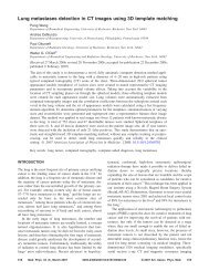
440 O’DELL MCCULLOCHAnnu. Rev. Biomed. Eng. 2000.2:431-456. Downloaded from arjournals.annualreviews.orgby UNIVERSITY OF ROCHESTER LIBRARY on 07/27/09. For personal use only.?Volumes from Three-Dimensional Surface Reconstructionsmodels cannot as readily approximate RV volumes, because their geometry doesnot possess such symmetry (44).The Simpson’s-rule method computes the cavity volume by summing the areasof 2-D planar slices, scaled by the slice separation. The implied geometric assumptionfor this method is that the area of the measured cross-section remains constantover the distance between slices (or at least is representative of the average area),which becomes increasingly error-prone as the slice separation increases. Thereare also the previously mentioned errors associated with the identification of themitral-valve plane, through-plane motion, and registration artifacts. A commonmodification to the volume equation involves reducing the volume contributionof the most apical and/or most basal slices by 50%. Many other variations arecommonly used.Regardless of the modality used to acquire the anatomical data, once these areattained, 3-D surface reconstructions allow more exact descriptions of the heart geometrythan simple elliptical models. Many 3-D surface reconstruction techniquesare also able to incorporate anatomical data from multiple views/projections. Manyrecent advances in 3-D surface reconstruction have been driven by computer visionresearch (45, 46). Piecewise mathematical constructs have also been proposed, including3-D splines and finite-element basis <strong>function</strong>s (47, 48). Deformable sheetor balloon models (3-D extensions of “snakes”) are able to interact directly withfeatures of the images to satisfy both the need to match image data and to smoothout noisy image data. The smoothing is accomplished by incorporating energies associatedwith stretching and bending into the deformable sheet or balloon (49–51).Global polynomial representations are also applicable (52, 53). For example, wehave used a surface model expressed as a polynomial series in prolate spheroidalcoordinates. Here the base geometry is an ellipsoid, to which are superposed spatialmodulations as a <strong>function</strong> of angular position, analogous to spherical harmonics.Mathematically, the radial coordinate λ is represented as a <strong>function</strong> of the circumferentialand longitudinal angles (θ,φ):Here P mlλ =L∑l∑l=0 m=−la j P ml (cos(θ))e imφ (1)are the associated Legendre polynomials. The coefficients a i in the polynomialare fit, in a least-squares sense, simultaneously to contour data pointsfrom multiple views. The goodness/smoothness of the reconstruction can be adjustedto match the expected uncertainty in the contour data, by admitting oromitting higher-order terms in the series, which is achieved by adjusting the serieslimit “L.”Figure 1A (see color insert) shows the fitted endocardial surfaces for a zerothorderfit, that is, the best-fit prolate spheroid. From left to right in the figure,
Annu. Rev. Biomed. Eng. 2000.2:431-456. Downloaded from arjournals.annualreviews.orgby UNIVERSITY OF ROCHESTER LIBRARY on 07/27/09. For personal use only.?Left ventricular endocardial surface-fitted data sets for canine heart. The light lines areFigure 1the raw contour data, and the dark lines are generated from the fitting equation (Equation 1). Top:Top view; bottom: side view. The order of fitting goes from zero-th to third, sixth, and tenth fromleft to right.