Cervical Spine Trauma: Pearls and Pitfalls
Cervical Spine Trauma: Pearls and Pitfalls
Cervical Spine Trauma: Pearls and Pitfalls

Create successful ePaper yourself
Turn your PDF publications into a flip-book with our unique Google optimized e-Paper software.
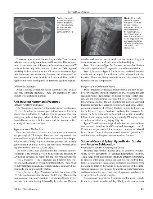
<strong>Cervical</strong> <strong>Spine</strong> <strong>Trauma</strong>Fig. 3—24-year-oldman with hangman’sfracture. Bilateral C2pars interarticularisfractures are seen.(Reprinted withpermission from [1])Fig. 4—28-year-oldman with atypicalhangman’s fracture.C2 body fracturewith anteroposteriordisplacement offracture fragments(fat C2 body sign) isseen. Fracture disruptsHarris ring posteriorly<strong>and</strong> causes posterioroffset of spinolaminarline from C1 to C3(dotted line).Transverse separation of fracture fragments by 7 mm or moreindicates transverse ligament injury <strong>and</strong> instability. This measurement,known as the rule of Spence, can be made on transverse CTbut is applicable only in the presence of a fracture. Other signs ofinstability include avulsion of the C1 tubercle (transverse ligamentinsertion), two anterior ring fractures, <strong>and</strong> atlantodental intervalgreater than 3 mm in adults or 5 mm in children. MRI ishighly sensitive for the diagnosis of transverse ligament rupture.Differential Diagnosis<strong>Pitfalls</strong> include congenital fusion anomalies <strong>and</strong> aplasiasthat may simulate fractures. These are identified by theirsmooth well-corticated margins.Axis Injuries: Hangman’s FracturesRelevant Anatomy <strong>and</strong> CauseThe “hangman’s fracture,” or traumatic spondylolisthesis ofC2 (Fig. 3), refers to bilateral pars interarticularis fractures,which bear similarities to findings seen in persons who haveundergone judicial hanging. Most of these fractures resultfrom falls <strong>and</strong> motor vehicle crashes, <strong>and</strong> the fractures reflecta variety of injury mechanisms.Appearance <strong>and</strong> MechanismPars interarticularis fractures are best seen on transverse<strong>and</strong> parasagittal CT images. They are often asymmetric <strong>and</strong>are considered atypical when the fracture extends into the posteriorvertebral body. Atypical fracture patterns are actuallyquite common <strong>and</strong> may involve the transverse foramen, placingthe vertebral artery at risk for injury.The most widely used classification for traumatic spondylolisthesisis the system devised by Effendi <strong>and</strong> modified byLevine <strong>and</strong> Edwards, as outlined in the following subsections.Type 1 fractures—Type 1 fractures are bilateral pars fractureswithout angulation or significant translation. These resultfrom hyperextension <strong>and</strong> axial loading <strong>and</strong> are considered mechanically<strong>and</strong> neurologically stable.Type 2 fractures—Type 2 fractures include disruption of theC2–3 disk with anterior translation of the C2 body. These are themost common hangman’s fracture type <strong>and</strong> result from hyperextensionwith axial loading followed by hyperflexion. They areunstable <strong>and</strong> may produce a small posterior fracture fragmentthat can narrow the canal <strong>and</strong> cause spinal cord injury.Type 2A fractures—Type 2A fractures are unstable flexiondistractioninjuries with C2 angulation but without translation.Type 3 fractures—Type 3 fractures are combined anteriortranslation <strong>and</strong> angulation with facet subluxation or frank dislocation.These are highly unstable injuries that result fromhyperflexion <strong>and</strong> compression.Diagnostic <strong>and</strong> Differential PointsType 1 fractures are radiographically subtle <strong>and</strong> may be easilyoverlooked but should be identified on CT with multiplanarreconstructions. Prevertebral soft-tissue swelling is often present,<strong>and</strong> the spinolaminar line from C1 to C3 may show posteriordisplacement of the C2 spinolaminar junction. Atypicalfractures disrupt the Harris ring posteriorly <strong>and</strong> show anteroposteriorseparation of C2 body fracture fragments, known asthe fat C2 sign (Fig. 4). Fractures involving the transverse foramen,or those associated with neurologic deficit, should befollowed with angiographic imaging, usually CT angiography,to exclude vertebral artery injury (Fig. 5).Types 2A <strong>and</strong> 3 require surgical reduction <strong>and</strong> internal fixation<strong>and</strong> must therefore be differentiated from types 1 <strong>and</strong> 2.Concurrent upper cervical fractures are common <strong>and</strong> shouldbe excluded. These include odontoid fractures, posterior C1arch fractures, <strong>and</strong> hyperextension teardrop fractures.Subaxial <strong>Cervical</strong> <strong>Spine</strong>: Hyperflexion Sprain<strong>and</strong> Anterior SubluxationRelevant Mechanism, Anatomy, <strong>and</strong> CauseSubaxial hyperflexion injuries (Fig. 6) comprise progressivelysevere disruptions of cervical ligamentous integrity.These range from hyperflexion sprain, to anterior subluxation,to bilateral interfacetal dislocation <strong>and</strong> flexion teardrop fracture.Corresponding ligamentous disruption progresses fromposterior to anterior, beginning with disruption of the supraspinousligament, interspinous ligaments, capsular ligaments,<strong>and</strong> ligamentum flavum. This group of ligaments is referred toas the posterior ligament complex.Disruption of the posterior ligament complex alone is insufficientto generate spinal instability. The hyperflexion sprain is<strong>Pitfalls</strong> in Clinical Imaging 23