1 General Biology II Lab Lab #10: Circulatory & Respiratory Systems
1 General Biology II Lab Lab #10: Circulatory & Respiratory Systems
1 General Biology II Lab Lab #10: Circulatory & Respiratory Systems
Create successful ePaper yourself
Turn your PDF publications into a flip-book with our unique Google optimized e-Paper software.
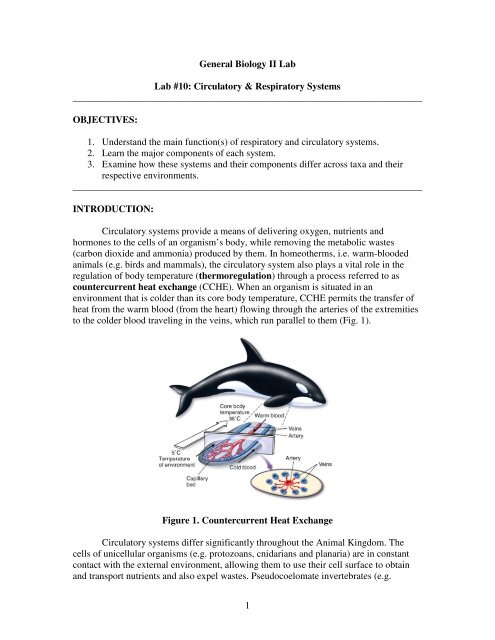
<strong>General</strong> <strong>Biology</strong> <strong>II</strong> <strong>Lab</strong><strong>Lab</strong> <strong>#10</strong>: <strong>Circulatory</strong> & <strong>Respiratory</strong> <strong>Systems</strong>________________________________________________________________________OBJECTIVES:1. Understand the main function(s) of respiratory and circulatory systems.2. Learn the major components of each system.3. Examine how these systems and their components differ across taxa and theirrespective environments.________________________________________________________________________INTRODUCTION:<strong>Circulatory</strong> systems provide a means of delivering oxygen, nutrients andhormones to the cells of an organism’s body, while removing the metabolic wastes(carbon dioxide and ammonia) produced by them. In homeotherms, i.e. warm-bloodedanimals (e.g. birds and mammals), the circulatory system also plays a vital role in theregulation of body temperature (thermoregulation) through a process referred to ascountercurrent heat exchange (CCHE). When an organism is situated in anenvironment that is colder than its core body temperature, CCHE permits the transfer ofheat from the warm blood (from the heart) flowing through the arteries of the extremitiesto the colder blood traveling in the veins, which run parallel to them (Fig. 1).Figure 1. Countercurrent Heat Exchange<strong>Circulatory</strong> systems differ significantly throughout the Animal Kingdom. Thecells of unicellular organisms (e.g. protozoans, cnidarians and planaria) are in constantcontact with the external environment, allowing them to use their cell surface to obtainand transport nutrients and also expel wastes. Pseudocoelomate invertebrates (e.g.1
Ascaris), in contrast, utilize the fluids present inside their body cavities not only as ahydrostatic skeleton (for movement) but also as a means of circulation. However,transport of nutrients, gases, and waste products presents a major challenge for large,multicellular organisms (Why?). As a result, these animals developed specialized organsand organ systems, i.e. circulatory and respiratory systems, for this purpose.In an open circulatory system (Fig. 2A), as found in mollusks and arthropods, amuscular heart pumps hemolymph (blood and fluid of the body tissues) through anetwork of channels and cavities in the body which eventually drain back into a centralcavity. On the other hand, in a closed circulatory system (Fig. 2B), as found in annelidsand vertebrates, the heart pumps blood through a series of blood vessels (arteries, veinsand capillaries) that transport oxygen and nutrients to the rest of the body whilesimultaneously removing wastes from the tissues.A) B)Figure 2. A) Open and B) Closed circulatory systemsOrganisms respire (breathe) to obtain the oxygen necessary for cellular respirationand to eliminate carbon dioxide. Depending on their body size, oxygen demands andenvironment, organisms have developed a variety of respiratory structures (Fig. 3).Arthropods, for example, do not possess a single respiratory organ. Instead, air enters thebody through spiracles (specialized openings in the exoskeleton) and into small,branched, cuticle-lined trachea (air-ducts) which transmit gases to the entire body.Aquatic vertebrates, in contrast, use gills to extract oxygen from their surroundingenvironment. Within the gills, lamellae (membranous plates within the gill arches) arearranged to create countercurrent flow, where blood flows in the opposite direction ofwater movement, thus maximizing blood oxygenation. Meanwhile, amphibians respirevia the lungs and the skin. Their highly vascularized epidermis allows exchange ofoxygen and carbon dioxide in a process referred to as cutaneous respiration. Finally, interrestrial organisms, lungs minimize water evaporation by moving air through tubuleswhere gas exchange takes place. <strong>Respiratory</strong> systems of most terrestrial organisms aremost efficient because they need to maintain the high metabolic demands of endothermyand increased cellular respiration.2
Figure 3. <strong>Respiratory</strong> structures of different organisms________________________________________________________________________Task 1: Measuring heart rate, blood pressure and tidal volumeThe human circulatory (cardiovascular) system is composed of (1) a heart, (2)blood and (3) blood vessels. The heart pumps blood through the blood vessels to variousorgans, supplying the tissues with oxygen and nutrients while concomitantly removingcarbon dioxide and other wastes produced by them. In this system, oxygenated bloodflows away from the heart to the rest of the organs (with the exception of the lungs) andtissues through arteries. Deoxygenated blood, on the other hand, leaves the tissues andorgans through veins and returns to the heart.The cardiovascular system is divided into two parts, the systemic circuit and thepulmonary circuit. The pulmonary circuit is composed of the lungs and all the vesselsthat connect the lungs with the heart while the systemic circuit consists of the entire body(excluding the lungs) and the remaining vessels. The heart is also separated into right andleft halves, where the right half (right atrium and ventricle) supplies blood to thepulmonary circuit and the left half (left atrium and ventricle) supplies blood to thesystemic circuit. In mammals, birds and crocodiles, the septa between the atria(interatrial septum) and the ventricles (interventricular septum) prevent oxygenatedand deoxygenated blood from mixing. Furthermore, multiple valves within the heartensure that blood flows in the right direction (Fig. 4) and also prevent backflow. Theatrioventricular valves (AV valves), namely the bicuspid /mitral (left) and tricuspid(right) valves, allow blood in the atria to empty into the ventricles. The semilunar valves(aortic and pulmonary), on the other hand, are located between ventricles and arteries.When the left ventricle contracts, the aortic valve opens pushing blood from the ventricleinto the aorta, the main artery supplying oxygen-rich blood to the entire body (systemiccirculation). The pulmonary valve, in contrast, forces blood from the right ventricle intothe pulmonary artery, which transports deoxygenated blood from the heart back to thelungs (pulmonary circulation).3
Figure 4. Heart and circulation of mammals and birdsIn summary, blood flow alternates between the pulmonary and systemic circuits.The left ventricle pumps oxygenated blood into the aorta and to the rest of the body. Asblood flows through tissues and organs, it becomes deoxygenated and is eventuallyreturned to the right atrium of the heart via the venae cavae. As the right atriumcontracts, deoxygenated blood flows into the right ventricle and then into the pulmonaryarteries. The pulmonary arteries carry blood to the lungs, where it supplied with oxygen.This oxygenated blood is then returned to the left atrium of the heart by the pulmonaryveins. Thus, with every heartbeat, a highly coordinated series of contractions push bloodfrom the atria into the ventricles and then either to the lungs or to the rest of the body.To accommodate the increased metabolic demands of endothermy, mammalsevolved lungs that are composed of millions of alveoli that function in gas exchange.During inhalation, the diaphragm contracts, pushing down against the abdomen, whichresults in an increased volume of the thorax. Opening of the thorax allows air that hasbeen inhaled through the mouth and nose to pass through the pharynx into the larynx(voice box) and then into the trachea. The trachea then bifurcates to form left and rightbronchi in the left and right lung, respectively (Fig. 5).4
Figure 5. Human lungsIn this task you will examine the structure and function of your circulatory andrespiratory systems. You will also measure the effects of exercise on these systems. Forthis task, you will need to choose two people from your group whose pulse rate, bloodpressure and tidal volume will be measured both before and after exercise. The other twogroup members will administer the tests and record the data.===============================================================Procedure 1: Measuring pulse rate1. Find your pulse by placing your second and third fingers on the side of your innerwrist that is closest to the thumb (the radial artery passes into the hand there).2. Press down slightly and count your pulse (the number of beats you feel) for 15seconds. Record your results in Table 1.3. Multiply this value by 4 to get your pulse rate in beats/minute. Record yourresults in the “Pulse Rate” column of Table 1.4. Repeat steps 1-3 three times.5. Average your results for the three trails and record this value in Table 1.Table 1:Sampling time Beats in 15 seconds Pulse Rate (beats/min)123AVERAGE5
6. Measure your pulse at the common carotid artery (on either side of your neck):Pulse Rate = _______________beats/min7. Run or jump in place for 5 min. Then sit down and immediately measure yourpulse rate.Pulse Rate = ______________beats/min===============================================================Procedure 2: Measuring the effect of exercise on blood pressureFor this procedure, you will work in pairs, serving as subject and experimenter.1. Have your partner sit down and relax for 2min. Attach the inflatable cuff aroundhis/her arm above the elbow (Fig. 6). Tuck the flap of the bag under the fold.Figure 6. Measuring blood pressure2. Inflate the cuff to about 200 mm Hg. This pressure will collapse the brachialartery, causing the blood flow to stop. At this point, you should not feel a pulse inyour partner’s wrist.3. Place the stethoscope over the brachial artery (underneath the cuff as shown inFigure 6). You should not hear anything with the stethoscope.4. If the pressure has gone below 200mm Hg, inflate the cuff again.5. Slowly begin releasing the pressure in the cuff. As the pressure falls, the bloodwill begin to spurt through the artery, producing vibrations and turbulence that areaudible through the stethoscope. You should hear loud, tapping sounds as theheart contracts. The pressure at which you begin hearing these sounds is termedsystolic pressure.Systolic pressure =______mm Hg6
2. Inhale and exhale three times through your mouth only.• You will need to do this before and after exercise3. Read the reading off of the dial and record the tidal volume (volume is measuredas cubic centimeter, cc) in the appropriate Table.Table 4: Student 11NameInitial pulserate(beats/min)Pulse rateafterexercise(beats/min)Initialbloodpressure(mm Hg)Bloodpressure afterexercise(mmHg)Initialtidalvolume(cc)Tidal volumeafter exercise23AverageWhile reclinedWhile StandingBlood pressure (mm Hg)_________________________________________________Pulse rate (beats/sec)____________________________________________________Table 5: Student 2 (that’s you)1NameInitial pulserate(beats/min)Pulse rateafterexercise(beats/min)Initialbloodpressure(mm Hg)Blood pressureafter exercise(mmHg)Initialtidalvolume(cc)Tidal volumeafter exercise23AverageWhile reclinedWhile StandingBlood pressure (mm Hg)__________________________________________________Pulse rate (beats/sec)_____________________________________________________9
Questions:1. How does exercise affect pulse rate, blood pressure and tidal volume?2. Explain what happens to the circulatory system during exercise. Include themajor organs involved in your explanation.i. Why does increased physical activity increase heart rate?ii. Why is heart rate lower in an individual who does aerobic exerciseregularly?iii. From your study of the circulatory system, how would youdescribe a "fit" individual?3. Explain what happens to the respiratory system during exercise. Include themajor organs involved.10
Figure 8. Comparison of respiratory and circulatory systems of vertebrates------------------------------------------------------------------------------------------------------------NOTES:• You will examine the same organisms for Tasks 2 and 3. Many of theseorganisms you have observed previously to understand the digestive andnervous systems.• Pay special attention to the dogfish shark, fetal pig, frog and pigeon. Theseorganisms are double injected with red and blue dyes marking the arteriesand veins, respectively.------------------------------------------------------------------------------------------------------------<strong>General</strong> Procedures:1. Examine the positions of the organs listed in Tables 6 and 7 within the animal. Makesure that you understand how these organs connect to the rest of the body.2. Once you feel comfortable, carefully cut out the hearts, gills, lungs and a piece ofleopard frog skin.a. Examine all of these structures under a dissecting microscope.b. Examine the external morphology of each organ paying attention to thestructures noted in Tables 6 and 7.Cut open the heart and lungs to identify the structures noted in the Tables.------------------------------------------------------------------------------------------------------------Task 2: <strong>Respiratory</strong> systemsIn this task, you will examine the different respiratory structures (Table 6) in organismsbelonging to various animal phyla. Sketch your observations in the space provided. Referto the figures indicated below each organism in your Dissection Atlas.12
Table 6:Organism Main respiratory organ DrawingPerchFig. 17.19, 17.21GillsLocate the operculum, gillarches and gill rakersShark (by TA)Fig. 17.5, 17.12GillsLocate the gill arches andgill rakersLeopard frogSkin and lungsPig (by TA)Fig. 17.59, 17.61LungsRatFig. 17.45, 17.44LungsCut a piece of the lung andobserve it under adissecting microscope.PigeonFig. 17.42Lungs, Air sacksand Trachea13
Questions:1. Examine the lungs of the pigeon. How do they compare to the lungs of othermammals? (Use the Figures listed in Table 6 to help you)a. The avian respiratory system is considered to be the mostefficient. Based on this organism’s lung anatomy and what youknow about gas exchange in birds (from lecture) can you explainwhy it is so efficient?2. Compare and contrast the respiratory systems of the rat and pig with that ofthe shark and perch?3. What is the benefit of respiring cutaneously?a. Are there possible disadvantages for this type of respiration?Consider the role of temperature regulation in your answer.b. Why are terrestrial animals unable to respire cutaneously?14
________________________________________________________________________Task 3: <strong>Circulatory</strong> systemsIn this task, you will examine the heart anatomy in animals from different phyla. Sketchyour observations in the space provided. Use Figures 9a and 9b to guide you.As you examine the various hearts, locate the following structures:1) Ventricles (note quantity)2) Atria (note quantity)3) Aorta (if present)4) Pulmonary arteries and veins (if present)Fig. 9a Comparative anatomy of the vertebrate hearts15
A) INTERNAL EXTERNALB) INTERNAL EXTERNALC) INTERNAL EXTERNALFig. 9b Comparative anatomy of vertebrate hearts: A) Amphibian, B) Teleost, andC) Mammalian & Avian16
Table 7:OrganismSheep heartFig. 17.92, 17.93,17.94# of chambers inheart (if any)DrawingRatPig (by TA)Fig. 17.61Leopard frogFig. 17.22PerchFig. 17.21Shark (by TA)Fig. 17.617
Questions:1. Consider the size of the hearts you examined. How does heart size relate tothe size of the animal?a. What does this relationship tell you about the animals’ ability tosustain normal function? (Hint: consider the main function of theheart)2) <strong>Lab</strong>el the structures of the heart on the figure below.3) Place a number on eachline which represents thepath of blood as it movesfrom the body, through theheart to the lungs, back tothe heart and then to therest of the body.4) Fill in the blanks with the electrical events of the cardiac cycle in the order theyoccur when blood arrives from the body.Blood arrives from the body or the lungs entering the heart at the__________________. The ____________ node fires, sending a wave of18
depolarization across the atria, causing them to _________________ forcingblood into the ______________________. The _____________ node firescausing a wave a depolarization across the ______________________causing them to contract sending blood to the lungs or to the body.5) Consider Task 1 performed at the beginning of the lab period. How did yourcirculation and blood pressure change during exercise?a. What happened to your heart as you exercised?________________________________________________________________________REFERENCES:Pictures of hearts were taken fromhttp://www.peteducation.com/article.cfm?c=16+2160&aid=2951Germann WJ and Stanfield CL. 2001. Principles of Human Physiology. PearsonEducation.19


