1 GENERAL BIOLOGY LAB 1 (BSC1010L) Lab #10: Biotechnology ...
1 GENERAL BIOLOGY LAB 1 (BSC1010L) Lab #10: Biotechnology ...
1 GENERAL BIOLOGY LAB 1 (BSC1010L) Lab #10: Biotechnology ...
- No tags were found...

You also want an ePaper? Increase the reach of your titles
YUMPU automatically turns print PDFs into web optimized ePapers that Google loves.
Table 1:LB -Condition Expected result (growth or no growth) Reason for expectationLB/Amp -LB +LB/Amp +The circles below represent each of the four petri dishes listed in Table 1. Based on yourpredictions, draw what you expect to occur in each.3
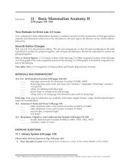
Develop a Hypothesis:Based on your expectations (Table 1), state null and alternative hypotheses regarding what youexpect to see if the bacteria plated on an ampicillin rich medium were successfully transformed.Procedure:1. Obtain two sterile Eppendorf tubes.a. Mark one tube “+ DNA” – this tube will receive the plasmid.b. Mark the other “- DNA”.2. On the side of both tubes, write your group number.3. Add 250µL of ice-cold calcium chloride (CaCl 2 ) to each tube using a sterile transferpipette. Addition of CaCl 2 to the host cells, followed by heat shock (see step 14),increases the likelihood of plasmid uptake. Cells that are able to take up the plasmidDNA are referred to as competent cells.4. Place both tubes on ice.5. Using one side of a plastic sterile loop, transfer a cell mass of E. coli cells from the starterplate into the + DNA tube by sweeping the cells superficially.a. Note: You only need a small quantity of bacteria. Do NOT completely fill theloop.Notes:• Be careful not to transfer any agar from the plate along with the cell mass• Immerse the cells on the loop in the calcium chloride solution in the + DNA tubeand vigorously tap the loop against the wall of the tube to dislodge the cell mass.Hold the tube up to the light to observe that the cell mass has fallen off the loop.6. Immediately suspend the cells by repeatedly pipetting in and out with a sterile transferpipette. Be careful not to spill the contents.• Examine the tube against the light to confirm that no visible clumps of cellsremain in the tube or are lost in the bulb of the transfer pipette. The + DNAtube should be appear cloudy while the - DNA tube should be clear (Fig. 1).4
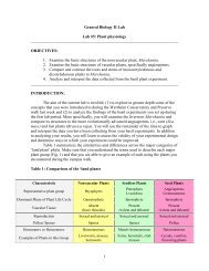
Figure 17. Return the + DNA tube to ice.8. Using the opposite end of the plastic sterile loop, transfer a mass of cells to the - DNAtube and suspend as described in steps 5 and 6 above.9. Return the - DNA tube to ice. Both tubes should now be on ice.10. Your instructor will add 10µL of plasmid DNA (concentration = 0.005 µg/µl) to the+ DNA tube. Gently tap the tube with your fingers to mix the DNA with the cells. Tokeep consistency between the two treatments, gently mix the – DNA tube as well.11. Centrifuge both tubes in a microcentrifuge for approximately 5 seconds before returningboth tubes to ice. BE SURE TO NOTE THE SLOT NUMBERS WHERE YOUPLACED YOUR GROUP’S TUBES SO YOU DON'T GET THEM CONFUSEDWITH THOSE OF ANOTHER GROUP. ALSO MAKE SURE THAT THE TUBESARE BALANCED INSIDE THE CENTRIFUGE BEFORE SPINNING (Fig. 2). Ifyou are unsure about this step, speak to your instructor before continuing.12. Incubate the tubes on ice for 30 min.Figure 25
13. While the tubes are incubating, label your media plates with your group name, section #and date, as follows (See Fig. 3):a. <strong>Lab</strong>el one LB/Amp "+". This is an experimental plate. This plate containsnutrients and ampicillin. If cells were transformed you should see cell growth onthis plate since the plasmid was added to the + DNA cell suspension being addedto this plate.b. <strong>Lab</strong>el another plate LB/Amp "-". This is a negative control. This plate alsocontains nutrients and ampicillin. Cell growth should not occur on this plate sinceno plasmid was added to the – DNA cell suspension being added to this plate.c. <strong>Lab</strong>el one of the remaining two plates as LB "+" and the other as LB "–". Theseplates, which only contain nutrients, serve as positive controls to test the viabilityof the cells after they have gone through the transformation process.Figure 3Note: If the following step is not performed exactly as stated the host cells will not beable to uptake the plasmid and transformation will not occur.14. Following the 30 min incubation period, “heat shock” the cells by removing both tubesfrom ice and immediately immersing them in the 42°C water bath for 90 seconds. Gentlyagitate the tubes while they are in the water bath. Immediately return both tubes to icefor 2 min. The purpose of this step is to expose the host cells to one temperature extremequickly followed by another. This process is required for a successful transformationbecause heat shock (along with addition of the CaCl 2 – step 3) increases the cell’scompetency.15. Centrifuge both tubes in a microcentrifuge for 4 min at 6000 rpm.16. Decant the supernatant carefully without disturbing the pellet of cells at the bottom of thetube. Do NOT attempt to do this before you observe the proper procedure demonstratedby your TA.6
17. Using a sterile transfer pipette, add 250µL of Luria broth (LB) to each tube. Luria brothis a nutrient-rich media containing vitamins and peptides essential for E. coli growth.18. Gently re-suspend the pellet in LB by repeatedly pipetting in and out. Make sure to use aseparate sterile pipette tip for each tube. Incubate the tubes at room temperature for 30min.19. At the end of the incubation period, remove 100µL from each transformation tube (Fig.4) and spread them on the LB and LB/Amp plates (Fig. 5). Do one plate at a time, fromstart to finish. Cells from the - DNA tube should be spread on the "-" plates, and cellsfrom the + DNA tube should be spread on the "+" plates.a. Use a sterile transfer pipette to add 100µL of cell suspension from the + DNAtube onto the LB/Amp+ plate. This is your experimental plate. Then add another100µL onto the LB + plate for the + DNA positive control.b. Use another sterile transfer pipette to add 100µL of cell suspension from the -DNA tube onto the LB/Amp- plate, as described in Step 16a. Then add another100µL onto the LB - plate for the - DNA negative control.Figure 4***Use the procedure below to spread the cells over the surface of the plate***c. Dip the cell spreader in ethanol, shake off the excess ethanol and briefly pass itthrough a Bunsen flame to ignite the alcohol. While the spreader is burning keepit away from the Bunsen flame and the alcohol container. NEVER dip a spreaderback into alcohol immediately after burning because invisible flames may ignitethe alcohol in the container.7
d. Let the spreader cool for about a minute. Note: If the cell spreader is too hot, youwill kill the E. coli host cells. Once cool, use the spreader to distribute the cellsuspension evenly over the surface of the plate (See Fig. 5) and then replace thelid of the Petri dish.Figure 5.e. Return the cell spreader to the ethanol container without flaming. Repeat steps 16c and d for EACH PLATE.20. Allow the plates to dry for several minutes. Tape the 4 plates together and place themupside down in the incubator for 24-48 hours. Your TA will transfer them to therefrigerator until next week’s lab._____________________________________________________________________________TASK 2 - Recording Results (To be completed during the next lab session)1. Retrieve your group’s plates from the refrigerator.2. Open each plate, one by one, to determine if growth of E. coli occurred. If growth didoccur please note the type of growth - lawn vs. colonial (See Figs. 6A and B).8
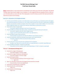
A. B.Figure 6. Plates illustrating A. Lawn Growth and B. Colonial growth of E.coli3. The circles below represent each of the four petri dishes tested in this lab. Draw yourgroup’s results for each petri dish. Make sure to label the plates appropriately.9
4. Record your results Table 2.Table 2:ConditionLB -Results(growth/no growth)Type of GrowthObserved(lawn/colonial)Did your results agree with yourpredictions in Table 1?(if not, provide a possibleexplanation)LB/Amp -LB +LB/Amp +5. Calculate transformation efficiency.Transformation efficiency is a measure of how successful host cells are in uptaking theplasmid, where uptake is expressed as the number of transformed colonies per µg ofplasmid DNA.Note: For this exercise, you will need to count the number of colonies present on yourLB/Amp+ plate. Follow steps a through e to calculate the transformation efficiency ofyour E.coli strain.a. The total amount (µg) of plasmid DNA used can be calculated with the followingformula.µg DNA = concentration (µg/µL) of DNA x volume of DNA (µL)b. Calculate the total volume of cell suspension prepared in the + DNA tube.Total volume (µL) = amount (µL) of CaCl 2 + amount (µL) of plasmid + amount (µL) of LB10
c. Since only a portion of the + DNA solution was added to the LB/Amp + plate, youwill need to calculate the fraction of DNA spread onto this plate using the formulabelow:Fraction of DNA spread = Volume (µL) spread on LB/Amp+ plateTotal sample volume (µL) in + DNA tubed. Using the values obtained for parts a and c, determine the actual amount of DNA (µg)present on your plate.Total amount (µg) of DNA = µg of DNA x Fraction of DNA spreade. Calculate transformation efficiency.Transformation Efficiency = Total number of cells on the LB/Amp+ plate__Total amount of DNA spread on LB/Amp+ plateQuestions:1. What role did the following compounds or steps play in bacterial transformation?a. Calcium Chlorideb. Heat shockc. Agard. LB Brothe. Ampicillin11
f. Centrifugationg. Plasmidh. Aseptic Techniques2. What factors could have affected transformation efficiency?3. What transformation efficiency would you expect for all the other treatments?4. How would you improve or extend the experiment above?5. In nature, DNA uptake by different organisms can impart advantages as well asdisadvantages to the host organism.a. When would genetic transformation be advantageous to a host organism?b. When would genetic transformation be maladaptive to a host organism? Whatconsequences would there be for the host cells?12
5. Can you think of a case where the uptake of foreign DNA would be advantageous forthe organism, but disruptive for the surrounding ecosystem?______________________________________________________________________________LOOK AHEAD:Before coming to lab next week, make sure to read the Population Genetics task sheet as well asChapter 18 (pgs 191-197) in your lab manual.13