Target Volume delineation - AROI
Target Volume delineation - AROI
Target Volume delineation - AROI
- No tags were found...

You also want an ePaper? Increase the reach of your titles
YUMPU automatically turns print PDFs into web optimized ePapers that Google loves.
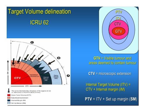
<strong>Target</strong> <strong>Volume</strong> <strong>delineation</strong>ICRU 62PTVITVCTVGTVGTV = Visible tumour andareas deemed to contain tumourCTV = microscopic extensionInternal <strong>Target</strong> <strong>Volume</strong> (ITV) =CTV + Internal margin (IM)PTV = ITV + Set up margin (SM(SM)
Imp points in Imaging for brain tumours CT scan alone should not be used, try to use MRI as well MRI – T1 contrast; T2/FLAIR sequences, thin slices, if preop MRIavailable- great for fusion; sag and coronal also, if possible 3D FSPGR sequence 1-313 mm slices with excellent resolution Equal matrix size (256x256), not oblique and equal spacing for MRIM important to check accuracy in CT/MRI fusion/co-registration (basilarartery, lens, IIIrd ventricle as common anatomical structures) Biological imagingMagnetic Resonance spectroscopy (MRS)Positron Emission Tomography (PET)
Image Fusion
Important points before contouring Ensure high resolution planning scans, preferably withadequate contrast administration Review all pre-operative and post-operative operative scans in detail Good knowledge and experience in the tumourcharacteristics seen on radiology and anatomy When in doubt, discuss with the neurosurgeon and theradiologist high grade tumours (Gliomas, medullo, ependymoma),MUST do a post op scan within 24-48 48 hours For benign tumours, TREAT WHAT YOU SEE, so do ascan and plan RT 6-868 weeks post op.
Results after SurgeryAfter surgery alone, the regrowth rates are between 10 to 75%RT reserved for large/invasive/recurrent cases
RadiotherapyLarge residual diseaseCavernous sinus invasionSuprasellar invasionAggressive histologyRecurrent tumours
Modern surgery – best cases132 patients, 98 were advisedpostoperative observation no radiologic or surgical evidence ofparapituitary invasion complete surgical removal absence of suprasellar extension2 months postoperatively no histologic feature of aggressive tumourbehaviour (mitoses or poor cellulardifferentiation)21 of 65 (32%)patientsprogressed atfollow up of 20yearsBradley Clin Endocrinol 1994
Need for RT?Role of radiotherapy has been questioned Fear of RT complications Fear of increased morbidity for re-operations Lack of randomised data Will re-excise excise at recurrence (?morbidity/mortality) Better results with modern radiological and surgicaltechniques
Best non randomised dataComparative study of 2 surgicalinstitutions; same RT set upRTNo RTPFS n=63 n=635 yr 93% 68%10 yr 93% 47%15 yr 93% 33%Administration of RT was the onlysignificant factorGittoes Clin Endocrinol 1998
RT in functioning tumoursacromegaly Largest series of 884 patients with acromegaly treated with RT Mean GH levels declined from 13.5 ng/ml to5.3 ng/ml at 2 years2 ng/ml at 10 years1.1 ng/ml at 20 years 63% of patients achieved IGF-1 1 levels by 10 yearsJenkins JCEM 2006 (in press)
Pituitary adenomaCavernous sinus involvement
Erich Steiner et al. Radiology 1992Teng et al – AJNR 1988Immediate post op scan
Packing material / post operative changes2 Mar 044 Mar 04
EvolutionPre-opDay 26 mths 24 mths
Sphenoid/Inferior extent uncommon but canhappen
Late effects Hypopituitarism Optic Neuropathy Neurocognitive dysfunction Cerebrovascular Accidents Second Tumours
SRSNecrosisMitsumori 1998Misumori IJROBP 1998;42:431-6
Stereotactic Conformal RadiotherapyAccurate ImmobilisationFractionated RTTight ConformationIndividualised planningHigh Quality AssurancePrecise treatment delivery
French SRT data110 consecutive patients treated with SRT to 50.4 Gy75 with persistent macroscopic tumor, 47 with persistent hormonal l secretionsTreated At minimum FU of 48 months, only 1 patient progressed (109/110(controlled/stable)Of functioning adenomas, 42% had complete response and 100% had objective responseProbability of requiring hormonal replacement at 4 and 8 years28.5% and 35%No other late sequelaeColin IJROBP 2005
TMH audit of 3D CRT in pituitaryadenomasStarted in 200062 patients (48 non functioning and 14 secretory)Carefully implemented protocolGTV: residual disease on planning scanCTV: 5mm margin, edited appropriatelyPTV: 5 mm margin three dimensionally as per departmentalstudy of assessment of random and systematic errors
Seller tumours: 3DCRT with 3-434 field conformal non-coplanarbeamsGantryTable100 10260 35020-5090
Results of 3DCRT34 females and 28 malesMedian follow up = 28 months (12 months to 62 months)All 62 patients had radiologically verified tumour control (3year actuarial PFS 100%)Vision stable/improved in all patients1 1 patient developed second tumour
MeningiomaMRI (post Gadolinium) - MUSTDural tail sign
Gross Total Resection: High Late RelapseAuthorMirimanoff (MGH)Condra (U Fl)Stafford (Mayo)5-yr7%7%12%Local Recurrence10-yr20%20%25%15-yr32%24%-May sometimes cause significant morbidity in certain sitesMirimanoff, Neurosurg 62:18, 1985Condra, , IJROBP 39:427, 1997Stafford, Mayo Clin Proc 73:936, 1998
Subtotal ResectionAuthorLocal RecurrenceWara (UCSF)Condra (U Fl)Stafford (Mayo)5-yr47%47%39%10-yr63%60%61%15-yr-70%-20-yr75%--Wara, , Am J Reontgenol Ther Nucl Med 123:453, 1975Stafford, Mayo Clin Proc 73:936, 1998Condra, , IJROBP 39:427, 1997
Adjuvant Radiotherapy
5 Yr Actuarial PFSAuthor (year)nGTRSTRSTR+ RTMirimanoff (1985)22593% (n=145)63% (n=80)Taylor (1988)13296% (n=90)43% (n=42)85% (n=13)Glaholm (1990)11784%Miralbell (1992)11548% (n=79)88% (n=17, 8yPFS)Mahmood (1994)25498% (n=183)54% (n=65)4/6 stable diseaseGoldsmith (1994)11789% (98% p1980, n=77)Condra (1997)246 a95% (n=174)83% (n=55)86% (n=17, 5 atypical)Stafford (1998)58188% (n=465) b61% (n=116) cNutting (1999)8292%Vendrely (1999)15689% (12 >WHO grade 1)Dufour (2001)3193%223688-98%43-83%84-98%
Anaplastic/Malignant MeningiomaPFS 2yrsPFS 5yrsSubtotal resection 44% 0%STR + XRT 87% 0%Total resection 70% 28%TR + XRT 100% 57%Dziuk J. NeuroOnc 37: 177, 1998
Meningiomas – Conformal RTSCRTIMRT Sharply delineated, Not infiltrating the brain, Complex shapes Often Adjacent to eloquent structures, 54 to 60 Gy required
Impact of modern RT planning1.00.80.698%: RT with CT/MR (n=77)77%: RT without CT/MR (n=40)0.40.2Progression-Free SurvivalSTR + postop RT; p=0.0020.0Goldsmith J Neurosurg 80:195-201, 19940 60 120 180 240Months
Fractionated Stereotactic/conformal radiotherapyIndicated for residual tumoursAlso for tumours close to critical structures such asoptic apparatus/brain stem (optic sheath meningioma)80-100%local control and PFSLess doses to the normal brain than conventionalradiotherapy and so less toxicityEmerging impressive “prospective” data ofmaintained cognitive and neuropsychologicalfunction (SteinworthRadioth & Oncol 2003;69:177-8282)Debus IJROBP 2001Jalali Clin Oncol 2002;14:103-9Selch IJROBP 2004;59:101-1111
Brain stem + spinal cord
NTCP organ modelsSerial model (e.g spinal cord, cranial nerves)Any break causes complication,volume effect less importantParallel Model (e.g(pituitary hypothalmicaxis)Damaging a few - intact organ function,volume effect present
Optic chiasm
Hypothalamus (PHA)
CTV generationTumour histology; benign Vs low-grade Vs malignantPatterns of failureResolution of neuro-imagingConfidence to delineate GTV accuratelyAny known anatomical barrier such as falx, tentorium, , boneand base of skullWell studied and carefully implemented uniform margingeneration protocol