

Foam and liquid sclerotherapy for varicose veins - CO2mmander
Foam and liquid sclerotherapy for varicose veins - CO2mmander
Foam and liquid sclerotherapy for varicose veins - CO2mmander

Create successful ePaper yourself
Turn your PDF publications into a flip-book with our unique Google optimized e-Paper software.
<strong>Foam</strong> <strong>and</strong> <strong>liquid</strong> <strong>sclerotherapy</strong> <strong>for</strong><br />
<strong>varicose</strong> <strong>veins</strong><br />
P Coleridge Smith<br />
Consultant Vascular Surgeon, London Vein Institute, 4 Upper Wimpole Street,<br />
London WIG 6 LF, UK<br />
Correspondence: Philip Coleridge Smith DM FRCS, The Bridge Clinic, Bridge Road,<br />
Maidenhead SL6 8DG, Berks, UK. Email: p.coleridgesmith@ucl.ac.uk<br />
Abstract<br />
Objective: The objective of this study is to review the methods <strong>and</strong><br />
outcome of ultrasound-guided foam <strong>sclerotherapy</strong> (UGFS) <strong>for</strong> the<br />
treatment of superficial venous incompetence.<br />
Method: Medical literature databases including Medline were searched <strong>for</strong><br />
recent literature concerning UGFS. Papers describing methods <strong>and</strong><br />
outcome have been assessed <strong>and</strong> their main findings included in this<br />
summary. A detailed description of the methods used by the author has<br />
been included as an example of how successful the treatment may be<br />
achieved.<br />
Results: A diverse range of practice is described in published literature in<br />
this field. Each group of authors used their own variation of the methods,<br />
described in the published literature, with good results. It is clear that<br />
foam <strong>sclerotherapy</strong> is far more effective than <strong>liquid</strong> <strong>sclerotherapy</strong> <strong>and</strong> that<br />
ultrasound imaging allows the treatment to be delivered accurately to<br />
affected <strong>veins</strong>. There is evidence that 3% policocanol foam is no more<br />
effective than 1% polidocanol foam. The optimum ratio of gas to <strong>liquid</strong> is<br />
4:1, although a range of ratios is reported in published work. There is a<br />
wide variation in the volume used as well as the method by which it is<br />
injected. The use of carbon dioxide foam reduces the systemic<br />
complications, particularly visual disturbance, when compared with air<br />
foams. Very few serious adverse events have been reported in the<br />
literature despite the widespread use of this method. Rates of<br />
recanalization of saphenous trunks following UGFS are similar to those<br />
observed after endovenous laser <strong>and</strong> endovenous radiofrequency ablation<br />
of <strong>veins</strong>, as well as the residual incompetence after surgical treatment.<br />
Conclusions: UGFS is a safe <strong>and</strong> effective method of treating <strong>varicose</strong><br />
<strong>veins</strong>. The relative advantages or disadvantages of this treatment in the<br />
longer term are yet to be published.
Key Words: <strong>varicose</strong> <strong>veins</strong> • telangiectases • reticular varices •<br />
ultrasound-guided foam <strong>sclerotherapy</strong> • duplex ultrasound imaging<br />
Introduction<br />
Sclerotherapy gained a reputation <strong>for</strong> lack of efficacy in the latter part of<br />
the 20th century, at least in part, thanks to the work of Hobbs. 1 His 10year<br />
r<strong>and</strong>omized controlled study showed that the clinical recurrence of<br />
varices was common in patients with truncal saphenous reflux managed<br />
by <strong>sclerotherapy</strong>, as described by Fegan. 2 Hobbs found that after 10<br />
years, 71% of patients treated surgically <strong>for</strong> truncal saphenous<br />
incompetence had a good outcome compared with only 6% of patients<br />
treated by <strong>sclerotherapy</strong>. Recent scientific evidence has shown that <strong>liquid</strong><br />
<strong>sclerotherapy</strong> is not very effective at eliminating truncal saphenous<br />
incompetence <strong>and</strong> failure to achieve abolition of reflux leads to early<br />
recurrence of varices.<br />
Advances in technology led to improvements in the practice of<br />
<strong>sclerotherapy</strong>. In the 1980s ultrasound was introduced <strong>for</strong> the diagnosis<br />
of venous disease of the lower limb. In France this led Schadeck <strong>and</strong> Vin<br />
to improve the efficacy of their treatment using ultrasound imaging to<br />
guide the placement of injections into incompetent saphenous trunks. 3,4<br />
However, this treatment was not as successful as had been hoped. Bishop<br />
examined a series of 55 patients in whom 89 legs had been managed by<br />
ultrasound-guided <strong>liquid</strong> <strong>sclerotherapy</strong>. He found that 57% showed<br />
residual saphenofemoral reflux <strong>and</strong> 75% of patients had great saphenous<br />
vein (GSV) reflux. 5 The problem of recanalization of <strong>veins</strong> was<br />
encountered in up to one-quarter of patients at one year according to<br />
Kanter <strong>and</strong> Thibault. 6<br />
The next significant advancement came in 1995 when Cabrera et al. 7<br />
suggested that foam could be created using carbon dioxide mixed with a<br />
polidocanol (POL), a detergent sclerosant. This invention built on the work<br />
of several previous authors who had experimented with various types of<br />
foam. Foote 8 described a method of foam <strong>sclerotherapy</strong> in 1944, which<br />
was improved by Orbach in 1950 9 who published a paper describing the<br />
use of a foam which he created by vigorously shaking a syringe containing<br />
air <strong>and</strong> sclerosant to produce a froth. Cabrera used <strong>sclerotherapy</strong> with<br />
foam, guiding his injections by ultrasound imaging. He called his invention<br />
‘microfoam’, comprising very small bubbles in contrast to the large bubble<br />
froths that had been used previously. Cabrera et al. 10 published a further<br />
article in 1997 describing his experience in 261 legs with great saphenous<br />
varices <strong>and</strong> eight patients with vascular mal<strong>for</strong>mations. Some of the<br />
<strong>varicose</strong> <strong>veins</strong> reached 20 mm in diameter. He considered that foam<br />
greatly extended the range of vein sizes which could be managed by<br />
<strong>sclerotherapy</strong>. He felt that the increased efficacy of foam was attributable
to it displacing blood from the treated vein <strong>and</strong> increasing the contact<br />
time between the sclerosant <strong>and</strong> the vein.<br />
In the intervening years, several clinical series <strong>and</strong> one r<strong>and</strong>omized<br />
clinical trial have confirmed that foam <strong>sclerotherapy</strong> is effective in<br />
managing truncal saphenous incompetence. No detailed follow-up beyond<br />
three years has been published.<br />
Commencing a practice of <strong>sclerotherapy</strong><br />
Sclerotherapy has not been widely taught during vascular surgical training<br />
in the UK in recent years <strong>and</strong> differs significantly in its conduct from<br />
surgery. Diagnostic ultrasonography of the venous system is largely<br />
carried out by vascular technologists in the UK (in contrast to many other<br />
European countries) <strong>and</strong> surgeons may have only passing familiarity with<br />
this technology. As a result it is common to find that surgeons are illequipped<br />
to undertake ultrasound-guided foam <strong>sclerotherapy</strong>.<br />
Appropriate training is essential be<strong>for</strong>e commencing clinical practice. This<br />
can be broken down into the components of <strong>sclerotherapy</strong>, ultrasound<br />
imaging <strong>and</strong> ultrasound-guided cannulation of vessels.<br />
Sclerotherapy. Injecting the right volume of the right sclerosant into the<br />
right vein is essential to getting a good outcome. Complications may arise<br />
if excessive concentrations or volumes of sclerosant are used. Lack of<br />
efficacy may be encountered when not enough is given. The right ‘recipe’<br />
<strong>for</strong> success seems to vary greatly between practitioners, but consensus<br />
guidelines have been published. 11,12 Theoretical knowledge is useful, but<br />
there is no substitute <strong>for</strong> practice! A skilled sclerotherapist will be able to<br />
put a needle into any vein visible from the surface of a limb, from the<br />
finest thread vein to large varix. This is best learnt under the supervision<br />
of an expert.<br />
Vascular imaging. Diagnosis of abnormality in the venous system takes a<br />
great deal of practice <strong>and</strong> training. However, the anatomy of the<br />
superficial <strong>veins</strong> is readily assimilated <strong>and</strong> most patterns of <strong>varicose</strong> <strong>veins</strong><br />
can be assessed by surgeons familiar with the surgical anatomy of the<br />
superficial <strong>and</strong> per<strong>for</strong>ating <strong>veins</strong>. Learning the skills required with the<br />
assistance of an experienced vascular technologist or radiologist is<br />
probably the most effective strategy.<br />
Ultrasound guided cannulation. This is an essential component of foam<br />
<strong>sclerotherapy</strong> <strong>and</strong> is the single most difficult aspect of the treatment <strong>for</strong> a<br />
beginner. Ultrasound phantoms are available with simulated <strong>veins</strong> to<br />
facilitate the learning process. These are not very life-like <strong>and</strong> are easy to<br />
master. The next stage involves patients <strong>and</strong> can be undertaken on
suitably consented patients undergoing surgical treatment <strong>for</strong> <strong>varicose</strong><br />
<strong>veins</strong>. The procedure of cannulating the saphenous trunk can be<br />
practised, but without injecting any foam (there is a high risk of deep vein<br />
thrombosis [DVT] in patients who have foam <strong>sclerotherapy</strong> under general<br />
anaesthesia).<br />
Combining these skills into a complete treatment <strong>for</strong> a patient requires<br />
some further training in the best strategy to use in order to obtain good<br />
results. Again this is best done under the supervision of a skilled<br />
practitioner.<br />
Facilities <strong>and</strong> equipment required <strong>for</strong> <strong>sclerotherapy</strong><br />
In comparison with surgery <strong>and</strong> other endovenous methods of treating<br />
<strong>varicose</strong> <strong>veins</strong>, very little is required in the way of equipment <strong>and</strong> facilities<br />
to treat <strong>varicose</strong> <strong>veins</strong> by foam <strong>sclerotherapy</strong>. The room used <strong>for</strong> this<br />
purpose may be a consulting room, a clinic room or treatment room. The<br />
facilities offered by an operating theatre are not required <strong>and</strong> may be<br />
intimidating to patients.<br />
An adjustable <strong>and</strong> tiltable examination couch is essential <strong>for</strong> com<strong>for</strong>table<br />
working. Most <strong>sclerotherapy</strong> is done with the patient lying recumbent, but<br />
it is useful to be able to tilt the couch during foam <strong>sclerotherapy</strong> to empty<br />
distal <strong>veins</strong> <strong>and</strong> to deal with occasional instances of syncope.<br />
A modern ultrasound machine with imaging frequencies in the range of 5–<br />
15 MHz is required. A linear array transducer of about 40 mm length is<br />
the most appropriate <strong>for</strong> foam <strong>sclerotherapy</strong>. The better the resolution of<br />
the image, the easier it will be to see where the needle has gone. Portable<br />
machines now provide images comparable with much larger machines<br />
from a few years ago <strong>and</strong> are much easier to move from room to room.<br />
A trolley containing needles, syringes, cannulae, b<strong>and</strong>ages <strong>and</strong> all other<br />
items required to complete the treatment is useful, so that there is no<br />
need to search the cupboards <strong>for</strong> missing items. Appropriate compression<br />
stockings will also be required. In the exceptional case that a severe<br />
allergic reaction occurs following treatment, suitable drugs <strong>and</strong><br />
equipments must be available to manage this problem.<br />
Preparation of sclerosant foam<br />
Many authors have described methods of preparing foam which may be<br />
used <strong>for</strong> ultrasound-guided <strong>sclerotherapy</strong>. Monfreux 13 described a method<br />
necessitating a glass syringe, which produced small quantities of POL<br />
foam, which he used in a series of patients with truncal <strong>varicose</strong> <strong>veins</strong>.<br />
Sadoun <strong>and</strong> Benigni 14 described a method of preparing foam using a
plastic syringe, avoiding the need <strong>for</strong> reusable glass syringes.<br />
Subsequently, Tessari 15 has described a method of preparing foam using<br />
two disposable syringes <strong>and</strong> a three-way tap. This method can be used to<br />
produce large quantities of foam suitable <strong>for</strong> treating saphenous trunks<br />
<strong>and</strong> large varices. Frullini 16 has added his own method of producing foam<br />
to this increasing list based on that of Flückinger. 17<br />
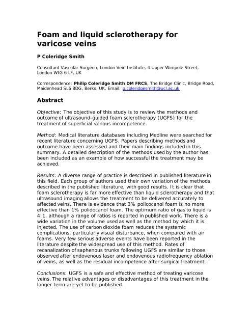
The most widely used method is that of Tessari, which is readily achieved<br />
using materials available in most clinics (Figure 1). Two syringes are<br />
connected using a three-way tap. A 5 µm intravenous filter can be<br />
inserted between the syringes <strong>and</strong> this greatly improves the quality of the<br />
foam. Either 2 mL or 5 mL syringes may be used, or a combination. A<br />
mixture of sclerosant <strong>and</strong> air is drawn into one syringe at a ratio of one<br />
part of sclerosant to four parts of gas. The sclerosant can be sodium<br />
tetradecyl sulphate (STS) 1–3% (Fibrovein STD Pharmaceuticals,<br />
Here<strong>for</strong>d, UK) or POL 0.5–3% (Sclerovein, Resinag AG, Zurich, CH). Low<br />
concentrations of POL (0.5%) make better foam when mixed in the ratio<br />
of 1:1 with air. The mixture is oscillated vigorously between the two<br />
syringes about 10 or 20 times. The tap can be turned slightly to reduce<br />
the aperture <strong>and</strong> increase the smoothness of the foam. The foam<br />
produced in this way is stable <strong>for</strong> about two minutes so it should be<br />
injected as soon as it has been made.<br />
View larger version<br />
(105K):<br />
[in this window]<br />
[in a new window]<br />
Figure 1 Tessari method <strong>for</strong> creating<br />
sclerosant foam. Gas <strong>and</strong> sclerosant are<br />
oscillated between two syringes via a<br />
three-way tap approximately 20 times<br />
The patient with stroke reported by Forlee et al. 18 highlights a potential<br />
hazard of injecting air/foam into <strong>varicose</strong> <strong>veins</strong>, although such events are<br />
rare considering the tens of millions of patients who have been treated<br />
worldwide. A recent letter to the New Engl<strong>and</strong> Journal of Medicine 19
described how air/foam injected in the leg <strong>veins</strong> rapidly finds its way to<br />
the right side of the heart <strong>and</strong> may be a factor in producing visual<br />
disturbance. The problem may lie with the nitrogen in air which is<br />
relatively insoluble, <strong>and</strong> replacing this with carbon dioxide can limit the<br />
reported side-effects of treatment. 20 A mixture of 70% CO2 <strong>and</strong> 30% O2<br />
can be obtained from BOC Special Gases (see<br />
www.bocspecialgases.co.uk) <strong>and</strong> used to make foam. Pure CO2 makes<br />
short lived, poor quality foam.<br />
Patient selection – indications <strong>and</strong> contraindications<br />
Suitability of a patient <strong>for</strong> foam <strong>sclerotherapy</strong> depends on the aims of<br />
treatment as well as the venous anatomy. <strong>Foam</strong> <strong>sclerotherapy</strong> can be<br />
carried out successfully in almost any patient with clinically significant<br />
venous disease no matter how elderly, frail, obese or ill they are. In<br />
patients with leg ulcers more aggressive treatment may be indicated or<br />
more ambitious treatment (very large varices) than in those seeking<br />
cosmetic improvement. The only absolute contraindications are severe<br />
allergy <strong>and</strong> obliterated deep <strong>veins</strong>. Table 1 lists some relative <strong>and</strong><br />
absolute contraindications to treatment.<br />
View this<br />
table:<br />
[in this<br />
window]<br />
[in a new<br />
window]<br />
Table 1 Some relative <strong>and</strong> absolute<br />
contraindications to treatment by foam<br />
<strong>sclerotherapy</strong><br />
For those learning to use ultrasound-guided foam <strong>sclerotherapy</strong>, the<br />
easiest varices to manage are those in patients with moderate size great<br />
or small saphenous trunks (5–8 mm diameter) that have never been<br />
treated previously. In general, the saphenous trunks are straight <strong>and</strong> easy<br />
to cannulate, <strong>and</strong> the varices are not very extensive. Patients with<br />
recurrent varices following previous surgery are more difficult. Residual<br />
non-tortuous saphenous trunks are relatively easy to cannulate, but very<br />
tortuous recurrent vessels near the saphenofemoral junction (SFJ) <strong>and</strong><br />
saphenopopliteal junction (SPJ) need significant previous experience.<br />
Patient consent
Patients should be fully in<strong>for</strong>med of the method of treatment as well as<br />
the complications which may arise. They should be warned about the<br />
lumps caused by thrombophlebitis <strong>and</strong> skin pigmentation. Injection site<br />
ulceration is rare <strong>and</strong> need not be mentioned. Visual disturbance, chest<br />
tightness <strong>and</strong> coughing occur occasionally <strong>and</strong> are included. DVT is also<br />
mentioned as is severe allergy, although this is very uncommon. This<br />
in<strong>for</strong>mation should be supported by written consent <strong>and</strong> an in<strong>for</strong>mation<br />
sheet. Some clinicians may also have access to a website reference <strong>for</strong><br />
further in<strong>for</strong>mation.<br />
Position of the patient<br />
The patient is positioned supine be<strong>for</strong>e placing any needle or cannula to<br />
minimize the risk of syncope. The small saphenous <strong>veins</strong> (SSVs) are most<br />
easily accessed with the patient lying on his left side, facing away from<br />
the surgeon. The leg to be treated is moved towards the operator as<br />
shown in Figure 2. For the left GSV, the same position works well. The left<br />
leg is drawn towards the operator giving easy access to the medial aspect<br />
of the thigh <strong>and</strong> calf. For the right GSV, the patient lies on their right side<br />
(Figure 3).<br />
View larger version<br />
(102K):<br />
[in this window]<br />
[in a new window]<br />
Figure 2 The patient lies in the left<br />
lateral position <strong>for</strong> treatment of the<br />
right small saphenous vein. The left<br />
small <strong>and</strong> long saphenous <strong>veins</strong> are<br />
also accessible in this position
View larger version<br />
(103K):<br />
[in this window]<br />
[in a new window]<br />
Ultrasound-guided cannulation<br />
Figure 3 The patient lies in the right<br />
lateral position to permit cannulation of<br />
the right great saphenous vein<br />
Both saphenous trunks <strong>and</strong> major tributaries are treated through an<br />
intravenous cannula or Butterfly needle. All cannulae <strong>and</strong> butterflies are<br />
positioned be<strong>for</strong>e any foam is injected. This allows confirmation that the<br />
cannula is in the vein at all times, by flushing with saline. Once foam is<br />
injected it spreads rapidly to many superficial <strong>veins</strong>, which makes further<br />
ultrasound-guided injection more difficult. Superficial varices do not need<br />
ultrasound guidance <strong>and</strong> can be treated by direct needle injection without<br />
prejudicing safety or efficacy.<br />
Strategy <strong>for</strong> primary GSV varices<br />
In those cases where the GSV lies in the saphenous compartment<br />
throughout the thigh, an 18 g cannula is placed in the GSV in the lower<br />
third of the thigh. This will treat the vein between the injection point <strong>and</strong><br />
the SFJ. If the GSV lies more superficially in part of its track, this is<br />
strictly speaking an accessory vein <strong>and</strong> not the main saphenous trunk. It<br />
is better to inject less strong sclerosants here. Is there an 18 g cannula<br />
positioned in the saphenous trunk where it lies within the saphenous<br />
fascia? A Butterfly needle is then placed in the superficial accessory vein,<br />
usually in the distal third of the thigh. Any further accessory <strong>veins</strong> in the<br />
region of the GSV are treated by placing further Butterflies in them. These<br />
may be the source of recurrence if left untreated.<br />
Next, any major tributaries of the GSV in the thigh are identified,<br />
especially those above the level of cannulation of the GSV. Larger<br />
tributaries (3 mm diameter <strong>and</strong> above) may supply enough venous flow to<br />
the GSV to keep it open above the level of the junction of the GSV with its<br />
tributaries. They can be managed by direct needle injection with foam.
In the calf, a 23 g Butterfly needle is used to cannulate the saphenous<br />
trunk <strong>and</strong> any varices, again inserting all cannulae <strong>and</strong> Butterflies be<strong>for</strong>e<br />
any treatment is commenced. In patients with primary <strong>varicose</strong> <strong>veins</strong>,<br />
between two <strong>and</strong> six cannulations are required to treat GSV reflux <strong>and</strong><br />
associated varices.<br />
Primary SSV varices<br />
SSV varices are usually much less complex to treat than GSV varices,<br />
since they have fewer tributaries <strong>and</strong> accessory <strong>veins</strong>. With the patient<br />
lying in the left lateral position the SSV is punctured in the mid-calf region<br />
using an 18 g cannula. This avoids making an injection in the popliteal<br />
fossa <strong>and</strong> inserting foam close to the popliteal vein. A 23 g Butterfly is<br />
used to treat the distal SSV. Varices can be managed using a further<br />
Butterfly or by direct needle injection; two or three cannulations are<br />
usually needed <strong>for</strong> the management of SSV varices.<br />
Combined GSV <strong>and</strong> SSV reflux<br />
In patients where both the GSV <strong>and</strong> SSV are incompetent, both <strong>veins</strong> are<br />
cannulated in order to treat the two saphenous trunks in one session.<br />
More limited treatment of superficial varices may be required to minimize<br />
the number of cannulations <strong>and</strong> volume of foam injected during the first<br />
treatment session.<br />
Incompetent per<strong>for</strong>ating <strong>veins</strong><br />
In the calf, most incompetent per<strong>for</strong>ating <strong>veins</strong> can be managed by<br />
cannulation of a superficial vein immediately fed from the per<strong>for</strong>ator. This<br />
works <strong>for</strong> ankle (Cockett's) per<strong>for</strong>ators as well as more proximal paratibial<br />
per<strong>for</strong>ating <strong>veins</strong>. STS 3% in <strong>liquid</strong> <strong>for</strong>m is an alternative <strong>for</strong> medial calf<br />
per<strong>for</strong>ators, which may help to avoid producing thrombosis in the<br />
posterior tibial <strong>veins</strong>. <strong>Foam</strong> is used <strong>for</strong> proximal calf per<strong>for</strong>ating <strong>veins</strong>,<br />
popliteal fossa per<strong>for</strong>ators <strong>and</strong> per<strong>for</strong>ating <strong>veins</strong> in the thigh. In the<br />
popliteal fossa, the cannula is positioned within the per<strong>for</strong>ator so that its<br />
tip lies at or near the fascial opening. Medial thigh per<strong>for</strong>ating <strong>veins</strong> are<br />
cannulated with an 18 g cannula to ensure reliable injection of the source<br />
of varices.<br />
Treatment – injecting the foam<br />
Superficial varices are injected first with 1% sclerosant foam using a<br />
syringe <strong>and</strong> 30 or 25 g needle; 1 mL is given per injection <strong>and</strong> the foam is<br />
distributed to varices near the point of injection by massaging the leg with<br />
the ultrasound probe or by h<strong>and</strong>. Next, all the previously placed<br />
Butterflies <strong>and</strong> cannulae are injected with foam, working from the calf
towards the groin. The leg should be elevated well above the heart to<br />
minimize the diameter of the <strong>veins</strong> – this achieves Fegan's ‘empty vein’<br />
although it is not universally used by sclerotherapists. No more than 2 mL<br />
of foam should be injected at a time, even into the largest <strong>veins</strong>. In small<br />
<strong>veins</strong> in the calf injection of 1 mL at a time may be adequate.<br />
After each injection of foam, the patients are asked to per<strong>for</strong>m a series of<br />
ankle dorsiflexions to clear any foam which has reached the deep <strong>veins</strong>.<br />
<strong>Foam</strong> always reaches the deep <strong>veins</strong> within a few moments of injection.<br />
There is significant difference of opinion among Phlebologists over this<br />
point, some recommend that the patient lies still after injection of foam.<br />
Further injections of 1–2 mL of foam into each needle <strong>and</strong> cannula<br />
rein<strong>for</strong>ce the treatment already given. This strategy results in the <strong>veins</strong><br />
being treated <strong>and</strong> then retreated. Retreatment has been suggested by<br />
French <strong>and</strong> Italian phlebologists as a way of achieving more effective<br />
sclerosis than a single injection. The first injection produces spasm of the<br />
treated vein <strong>and</strong> facilitates the passage of foam to more proximal <strong>veins</strong><br />
with the subsequent injections.<br />
The last vein to be treated is the GSV in the thigh <strong>for</strong> which 2 ml of 3%<br />
STS foam is injected, repeating this twice more making sure the patient<br />
dorsiflexes between each one. The extent of spread of the foam is<br />
monitored using duplex ultrasonography <strong>and</strong> injection is discontinued at<br />
any site where extravasation of foam is seen. Butterflies work well but<br />
sometimes cut through the side of the vein The development of spasm in<br />
treated <strong>veins</strong> is widely regarded as a measure of success following<br />
<strong>sclerotherapy</strong> of a vein.<br />
The total amount of foam required to treat the GSV in the thigh is usually<br />
6 mL or 8 mL if it is a large vein. In the calf section of the GSV 2–4 mL of<br />
1% STS foam is sufficient. In the SSV 4–6 mL of 3% STS foam is required<br />
depending on the size of the vein. Additional foam is required <strong>for</strong><br />
accessory <strong>veins</strong> <strong>and</strong> varices. In per<strong>for</strong>ating <strong>veins</strong> other than the medial<br />
ankle (Cockett's) per<strong>for</strong>ators, 2–4 mL of 3% foam is used. Robust<br />
treatment of per<strong>for</strong>ators is required to achieve a permanent result. For<br />
medial calf per<strong>for</strong>ators connecting directly to the posterior tibial vein 1 mL<br />
of 3% <strong>liquid</strong> STS is needed.<br />
Compression following <strong>sclerotherapy</strong><br />
The treated leg is b<strong>and</strong>aged <strong>for</strong> 7–14 days using a short stretch cohesive<br />
b<strong>and</strong>age (PehaHaft, Hartmann, Germany). A compression roll is placed<br />
between the layers of the b<strong>and</strong>age to increase compression over the<br />
treated <strong>veins</strong>. Traditional crêpe <strong>and</strong> elastocrêpe b<strong>and</strong>ages are similar in<br />
price, but much thicker <strong>and</strong> the compression they apply diminishes
apidly. The b<strong>and</strong>age is secured with Medipore (3M Company, USA) 100<br />
mm wide adhesive tape <strong>and</strong> covered with a Class 2 compression stocking.<br />
The compression regimen should be repeated after each treatment<br />
session. Using elastic compression stockings without b<strong>and</strong>aging may lead<br />
to excessive retained thrombus, thrombophlebitis <strong>and</strong> skin pigmentation<br />
over treated <strong>veins</strong>.<br />
As with earlier practices in <strong>sclerotherapy</strong>, immediate ambulation <strong>and</strong><br />
return to normal activity are encouraged. There is little need <strong>for</strong> time<br />
away from work. Patients are encouraged to bring a friend or relative to<br />
their treatment sessions to ensure that they can travel home safely.<br />
Provided that patients can flex their knee sufficiently, it is acceptable <strong>for</strong><br />
them to drive since the leg is not normally painful or weak following<br />
treatment.<br />
Follow-up sessions<br />
Patients should be reviewed two weeks following their first treatment, as<br />
this is the optimum time <strong>for</strong> managing any adverse consequences such as<br />
thrombophlebitis. Patients usually return having removed b<strong>and</strong>ages<br />
themselves. The treated leg may be checked using duplex<br />
ultrasonography including visualization of the femoral <strong>and</strong> popliteal <strong>veins</strong><br />
<strong>for</strong> any thrombus, although this is rarely seen. Veins <strong>and</strong> saphenous<br />
trunks containing excessive amounts of thrombus can readily be managed<br />
by aspiration. The patient is positioned supine <strong>and</strong> a 5 mL syringe <strong>and</strong> a<br />
19 g needle (with local anaesthetic if necessary) can be inserted into<br />
saphenous trunks or varices under ultrasound guidance or by palpation.<br />
Aspiration rapidly resolves painful <strong>veins</strong> <strong>and</strong> lumps <strong>and</strong> minimizes the risk<br />
of skin pigmentation. Any residual segment of vein can be managed by<br />
further foam <strong>sclerotherapy</strong>, as described above. Re-b<strong>and</strong>aging the leg in<br />
the region of further <strong>sclerotherapy</strong> is usually appropriate.<br />
Management of problems following foam <strong>sclerotherapy</strong><br />
The most frequent problem is thrombophlebitis. This can be managed by<br />
compression <strong>and</strong> analgesia combined with aspiration of thrombus as<br />
described above.<br />
Occasionally, STS foam is injected outside a vein due to a technical<br />
problem with a cannula. This results in an inflammatory lump at the<br />
injection site, which resolves completely over two weeks. Injection site<br />
ulcers are rare since ultrasound monitoring of treatment allows<br />
discontinuation of injection where extravasation has occurred. POL is<br />
much safer in this respect. It causes no significant problem if injected at<br />
low concentrations <strong>and</strong> amounts outside the vein. Extravascular injection
of sclerosant foam usually causes pain during injection, <strong>and</strong> treatment<br />
should then be stopped while the position of the cannula is checked.<br />
DVT has been seen occasionally after this treatment, most frequently<br />
affecting the calf <strong>veins</strong>. This should be managed conventionally with<br />
compression <strong>and</strong> exercise or heparin/warfarin anticoagulation if it extends<br />
to major <strong>veins</strong> using local hospital protocols.<br />
Systemic complications<br />
Visual disturbance occurs in about 2% of patients <strong>and</strong> is probably doserelated.<br />
This occurs following both <strong>liquid</strong> <strong>and</strong> foam <strong>sclerotherapy</strong>, but is<br />
more frequent after foam. It often occurs in patients who have a previous<br />
history of migraine, but may occur in anyone. A scotoma develops<br />
associated with other visual phenomena such as a ground glass<br />
appearance in part of the visual field <strong>and</strong> irregular coloured patterns. This<br />
resolves within 30 minutes in most patients. It is highly likely to return in<br />
subsequent sessions of treatment; it is suggested that affected patients lie<br />
supine <strong>for</strong> up to 30 minutes following injection of foam to try <strong>and</strong> prevent<br />
this problem.<br />
Some patients may develop tightness in the chest or coughing after foam.<br />
This is probably a direct effect of the foam on the lungs <strong>and</strong> can also<br />
occur following injections of <strong>liquid</strong> sclerosant. This also resolves in about<br />
30 minutes. Again, lying supine <strong>for</strong> some time after treatment may be<br />
useful. The incidence of visual disturbance <strong>and</strong> chest symptoms has been<br />
reported to be reduced by using CO2 foams. 20<br />
Severe allergic reactions may follow injection of either of the sclerosants<br />
mentioned here. Appropriate drugs <strong>and</strong> equipment must be available to<br />
manage this problem.<br />
Cost of treatment<br />
<strong>Foam</strong> <strong>sclerotherapy</strong> is relatively inexpensive. With the exception of the<br />
ultrasound machine, no complex or specialist equipment is required. The<br />
consumable items require a cost of about £30 per session <strong>and</strong> the use of<br />
expensive facilities, such as a fully staffed operating theatre, is avoided.<br />
The staff required to complete the treatment include a surgeon or other<br />
expert practitioner <strong>and</strong> a vascular scientist or nurse assistant. These<br />
advantages are mitigated by the need <strong>for</strong> additional sessions of treatment<br />
to monitor the outcome, treat residual varices <strong>and</strong> manage retained<br />
thrombus. This is the least complex <strong>and</strong> most economical of all treatments<br />
<strong>for</strong> <strong>varicose</strong> <strong>veins</strong>.<br />
Sclerotherapy <strong>for</strong> superficial varices <strong>and</strong> telangiectases
Conventional <strong>liquid</strong> <strong>sclerotherapy</strong> is useful in the management of isolated<br />
small superficial varices not associated with truncal saphenous<br />
incompetence. <strong>Foam</strong> treatment has little advantage here <strong>and</strong> ultrasound<br />
guidance is unnecessary.<br />
A frequent problem is patients who present with reticular varices <strong>and</strong><br />
telangiectases. Reticular varices can be managed surgically, but better<br />
outcomes are usually obtained by <strong>sclerotherapy</strong>. All patients should be<br />
checked <strong>for</strong> the presence of saphenous truncal incompetence or other<br />
possible sources of varices by duplex ultrasonography. This commonly<br />
reveals an underlying problem which may lead to an unsatisfactory<br />
outcome if it is ignored. Most saphenous trunks <strong>and</strong> varices can be<br />
managed by foam <strong>sclerotherapy</strong> where they play a significant part in the<br />
development of reticular varices <strong>and</strong> telangiectases.<br />
The detailed management of telangiectases is beyond the scope of this<br />
article. In summary, small <strong>veins</strong> are probably best managed by <strong>liquid</strong>,<br />
rather than foam <strong>sclerotherapy</strong>. <strong>Foam</strong> sclerosants tend to be very strong<br />
<strong>and</strong> can cause even more telangiectases to develop if injected overenthusiastically.<br />
Most publications mention the injection of 0.25–0.5%<br />
POL as being the most appropriate. STS 0.2% may also be used with<br />
similar outcomes. 21 In Europe chromated glycerine is commonly used <strong>and</strong><br />
a r<strong>and</strong>omized trial has been published comparing this to POL <strong>and</strong> POL<br />
foam. 22 Allergy to metals is fairly common among adults; so methods to<br />
manage allergies should be available.<br />
A wide range of practitioners, including surgeons, general practitioners,<br />
dentists, nurse specialists <strong>and</strong> beauticians, treat telangiectases in the UK.<br />
Very few adverse events occur. However, a good result can only be<br />
expected when the methods employed by European phlebologists are<br />
used. 23 The essence is to treat the ‘feeding <strong>veins</strong>’ as well as the<br />
telangiectases. The feeding <strong>veins</strong> comprise the reticular <strong>veins</strong> that drain<br />
the telangiectases, plus any incompetent saphenous <strong>veins</strong>. A region of<br />
telangiectases is selected <strong>and</strong> reticular <strong>veins</strong> in this area are injected with<br />
0.5% POL <strong>liquid</strong>, injecting 0.25–0.5 mL at each location. Sclerosant will<br />
often enter telangiectases following this, demonstrating that the valves in<br />
the reticular <strong>veins</strong> are incompetent. Then all telangiectases in the affected<br />
area should be injected with small amounts of 0.5% POL using 0.1–0.2<br />
mL at each site. This treatment is a skill best learnt from an experienced<br />
practitioner.<br />
If the reticular varices are not treated, a reasonable outcome will be<br />
obtained in about half of patients, but may not be sustained. The other<br />
half of patients may obtain a disappointing outcome or more<br />
telangiectases may develop.
The use of compression following <strong>sclerotherapy</strong> <strong>for</strong> small <strong>veins</strong> varies<br />
widely. A recent r<strong>and</strong>omized trial shows that it is of value 24 <strong>and</strong> it is<br />
advised that patients wear Class 2 compression stockings <strong>for</strong> at least<br />
three days after injecting telangiectases <strong>and</strong> reticular varices.<br />
Outcomes<br />
A detailed review of the outcomes of ultrasound-guided foam<br />
<strong>sclerotherapy</strong> has been published recently. 25 Some important publications<br />
<strong>and</strong> more recent data are discussed below.<br />
Cabrera et al. 26 has published a clinical series of 500 legs treated by foam<br />
<strong>sclerotherapy</strong>. He reported that after three or more years 81% of treated<br />
great saphenous trunks remained occluded <strong>and</strong> 97% of superficial varices<br />
had disappeared. This required one session of <strong>sclerotherapy</strong> in 86% of<br />
patients, two in 11% <strong>and</strong> three sessions in 3% of patients. No DVT or<br />
pulmonary embolism was encountered in this series. Subsequently, a<br />
number of authors have published clinical series based on this technique<br />
including Frullini <strong>and</strong> Cavezzi, 27 who reported a series of 453 patients, <strong>and</strong><br />
Barrett et al., 28 who reported a series of 100 limbs. Cavezzi et al. 29 has<br />
subsequently published a detailed analysis of the efficacy of foam<br />
<strong>sclerotherapy</strong> in 194 patients, reporting a good outcome in 93% of<br />
patients. In fact, this technique has become widely used in southern<br />
Europe, Australia, New Zeal<strong>and</strong>, South America <strong>and</strong> the USA. 11 However,<br />
few surgeons in the UK use this method perhaps because of limited<br />
evidence of efficacy. One series has been recently reported from the UK<br />
involving 60 patients comparing surgical treatment with foam<br />
<strong>sclerotherapy</strong> combined with saphenofemoral ligation. 30<br />
One r<strong>and</strong>omized study of foam <strong>sclerotherapy</strong> in comparison with surgery<br />
has been published. This was a multicentre European study. 31 This<br />
r<strong>and</strong>omized controlled trial included two separate studies: a surgical part<br />
undertaken by surgeons who r<strong>and</strong>omized patients to saphenous stripping<br />
or ultrasound-guided foam <strong>sclerotherapy</strong>. In addition, sclerotherapists<br />
r<strong>and</strong>omized patients to ultrasound-guided <strong>sclerotherapy</strong> conducted using<br />
either foam or <strong>liquid</strong> sclerosant. In all, 654 patients were treated during<br />
this study. Up to four sessions of ultrasound-guided foam <strong>sclerotherapy</strong><br />
were allowed over three months to obliterate the saphenous trunks. After<br />
12 months, the surgeons had eliminated truncal saphenous reflux in 130<br />
of 176 patients (74%) by foam <strong>sclerotherapy</strong> <strong>and</strong> in 84/94 (88%) by<br />
surgery. In comparison, sclerotherapists had eliminated reflux in 239 of<br />
254 patients (91%) by foam <strong>and</strong> 104/125 (83%) by <strong>liquid</strong> <strong>sclerotherapy</strong>.<br />
Post-treatment pain was assessed by a visual analogue scale, which<br />
showed that surgery was much more painful during the first week. Normal<br />
activities were resumed after a median of 13 days in the surgery group
<strong>and</strong> two days in the foam group. DVT was seen in 10 patients treated by<br />
foam <strong>and</strong> one patient treated by <strong>liquid</strong> <strong>sclerotherapy</strong>.<br />
A personal experience of the use of ultrasound-guided foam <strong>sclerotherapy</strong><br />
based on an analysis of all patients treated <strong>for</strong> <strong>varicose</strong> <strong>veins</strong> between<br />
January 2002 <strong>and</strong> August 2005 has been published. 32 A total of 808<br />
patients (666 women, 142 men) were managed by ultrasound-guided<br />
foam <strong>sclerotherapy</strong> <strong>for</strong> truncal saphenous incompetence.<br />
Thrombophlebitis occurred in a small number of patients (5%) <strong>and</strong> was<br />
managed by analgesia, compression <strong>and</strong> aspiration of thrombus. Calf vein<br />
thrombosis was confined to isolated gastrocnemius <strong>veins</strong> or to part of the<br />
posterior tibial vein (10 cases). All resolved with compression by stocking<br />
or b<strong>and</strong>age <strong>and</strong> exercise without use of anticoagulants. In one case, a<br />
short occlusive thrombus arose in the common femoral vein two weeks<br />
following treatment of the GSV. The mechanism appeared to be due to<br />
direct extension of thrombus from the GSV into the femoral vein. This was<br />
managed by anticoagulation using low-molecular weight heparin <strong>and</strong><br />
warfarin continued <strong>for</strong> six months. The occluded femoral vein recanalized<br />
within four weeks <strong>and</strong> at six months follow-up no residual scarring or<br />
valve damage could be demonstrated on duplex ultrasonography. In two<br />
further cases thrombus extended from the SFJ <strong>and</strong> SPJ (one case each)<br />
into the femoral <strong>and</strong> popliteal vein. The extent of the thrombus was<br />
limited <strong>and</strong> firmly adherent to the vein wall. This was managed by<br />
compression stockings <strong>and</strong> exercise while monitoring the extent of the<br />
thrombus by serial duplex ultrasonography.<br />
In all, 457 legs have been reviewed at six months or more following<br />
treatment (average 11 months, range 6–46 months). This includes 367 of<br />
886 GSV <strong>and</strong> 145 of 263 SSV which had been treated. A substantial<br />
improvement in clinical venous disease was obtained. Duplex examination<br />
of the GSV showed that occlusion had been obtained in 322 of 364 (88%).<br />
In the SSV occlusion was present in 118 of 143 (83%). The median<br />
diameter of the GSV <strong>and</strong> SSV fell from 5 mm be<strong>for</strong>e treatment to GSV: 2<br />
mm <strong>and</strong> SSV: 1 mm at follow-up of six months or more.<br />
The only residual adverse events still present at six months or more of<br />
follow-up were skin pigmentation <strong>and</strong> palpable lumps. Skin pigmentation<br />
was seen in 115 of 457 legs reviewed after six months <strong>and</strong> palpable lumps<br />
were present in 21 legs. The skin pigmentation was almost always minor<br />
<strong>and</strong> continued to fade with the passage of time. In those patients<br />
reviewed one year or more following treatment, skin pigmentation was<br />
present in 11 of 115 legs. Small palpable lumps were sometimes<br />
detectable in the calf <strong>and</strong> comprised residual elements of treated <strong>veins</strong>.<br />
These could be identified by ultrasound imaging, but were not otherwise<br />
visible. In contrast to surgery, no scars, neurological damage or lymphatic<br />
injuries were encountered.
The relative efficacy of foam <strong>and</strong> <strong>liquid</strong> <strong>sclerotherapy</strong> has been<br />
investigated in a detailed study. 33 Patients with truncal saphenous<br />
incompetence were injected with either 2–2.5 mL of 3% POL <strong>liquid</strong> or<br />
foam into the GSV under ultrasound guidance, on only one occasion.<br />
Obliteration of saphenous incompetence was obtained in 35% of <strong>liquid</strong>treated<br />
patients <strong>and</strong> 85% of foam-treated patients after three weeks. At<br />
two years 53% of foam-treated <strong>and</strong> 12% of <strong>liquid</strong>-treated patients had<br />
successful obliteration of the GSV. A further study was per<strong>for</strong>med to<br />
assess the relative efficacy of 1% <strong>and</strong> 3% sclerosant foam. 34 Patients with<br />
truncal saphenous reflux were r<strong>and</strong>omized to treatment with either 1% or<br />
3% POL foam, in a single session. An average of 4.5 mL of foam was<br />
injected in both groups. Immediate occlusion rates were 96% (3% foam)<br />
<strong>and</strong> 86% (1%) foam. After two years saphenous occlusion was seen in<br />
69% of the 3% group <strong>and</strong> 68% of the 1% group. In both of these studies,<br />
a rather small volume of foam was used compared with the maximum of<br />
10 mL recommended in the Tegernsee document 12 <strong>and</strong> 20 mL that I have<br />
suggested above was used. This probably prejudiced the outcome but<br />
allowed the authors to demonstrate clearly the advantages of foam <strong>and</strong><br />
lack of increased efficacy with 3% POL.<br />
A number of papers have addressed particular problems in the<br />
management of <strong>varicose</strong> <strong>veins</strong>. Recurrent varices managed surgically<br />
have a poor outcome with further recurrence from neovascularization as a<br />
common feature. 35 The clinical series mentioned above found similar<br />
outcomes in primary <strong>and</strong> recurrent varices managed by foam<br />
<strong>sclerotherapy</strong>, with 88% of SFJs <strong>and</strong> saphenous trunks remaining<br />
obliterated at 11 months of follow-up. 32 Creton <strong>and</strong> Uhl 36 have reported a<br />
combined surgical <strong>and</strong> foam <strong>sclerotherapy</strong> technique (in one session) <strong>for</strong><br />
patients with recurrent varices, with obliteration of 93% of varices <strong>and</strong><br />
saphenous trunks at 40 days of follow-up. Perrin <strong>and</strong> Gillet 37 have<br />
reviewed the available literature on recurrent varices of the popliteal fossa<br />
<strong>and</strong> concluded that, unless a grossly incompetent SPJ stump is present,<br />
ultrasound-guided foam <strong>sclerotherapy</strong> is the most appropriate treatment.<br />
They acknowledge that this conclusion is supported by reports of clinical<br />
series <strong>and</strong> not r<strong>and</strong>omized clinical trials.<br />
A number of papers have discussed the outcome of foam <strong>sclerotherapy</strong> in<br />
patients with venous ulceration <strong>and</strong> severe venous disease. In one study,<br />
185 limbs with venous disease (CEAP C4–6: 109 limbs, CEAP C1–3: 76<br />
limbs). 38 In about three-quarters of patients in both groups, the<br />
saphenous trunk remained obliterated at six months. The authors<br />
conclude that foam <strong>sclerotherapy</strong> is equally applicable in complicated <strong>and</strong><br />
uncomplicated venous disease. The clinical outcome of treatment has<br />
been reported in a number of papers. 39–41 In general, rapid healing of<br />
ulcers is reported following foam <strong>sclerotherapy</strong> confirming that this
treatment can probably achieve the same outcomes that result from<br />
saphenous obliteration in leg ulcer patients.<br />
Systemic complications of foam <strong>sclerotherapy</strong> have been examined in<br />
some detail in view of occasional visual disturbance reported by some<br />
patients following both foam <strong>and</strong> <strong>liquid</strong> <strong>sclerotherapy</strong>. 42 Visual disturbance<br />
has been reported following the injection of a range of <strong>liquid</strong> sclerosants<br />
although it is clear that it is far more common following foam<br />
<strong>sclerotherapy</strong>. Some serious neurological adverse events, from which<br />
recovery was eventually complete have been reported. 43 This has led to<br />
attention focussing on the effects of a patent <strong>for</strong>amen ovale. This was the<br />
subject of a study by Morrison, in which 20 patients with visual or<br />
respiratory symptoms following foam <strong>sclerotherapy</strong> were studied by transthoracic<br />
echocardiography during treatment <strong>and</strong> 65% were found to have<br />
echoes in the left atrium after injection of foam sclerosant. Five of nine<br />
patients who then underwent transcranial Doppler investigation during<br />
foam <strong>sclerotherapy</strong> were found to have high-intensity events in the<br />
middle cerebral artery during treatment. Clearly, gas bubbles injected in<br />
the lower limb may reach the cerebral circulation, but the relationship to<br />
this phenomenon to the visual disturbance reported in some patients is<br />
yet to be established. Morrison has also reported the outcome of treating<br />
patients with foam made with carbon dioxide in comparison with air foam.<br />
This resulted in a substantial reduction in visual <strong>and</strong> other adverse events<br />
following treatment. It seems logical to consider moving to CO2 foams to<br />
minimize these side-effects, even if they are usually benign <strong>and</strong> resolve<br />
swiftly. In general, the number of serious adverse events is very small<br />
compared with the number of treatments which have been done. These<br />
remind us that even minimally invasive treatments may have significant<br />
side-effects <strong>and</strong> that we shall take all reasonable precautions to prevent<br />
these.<br />
One study has examined the relative efficacy of minimally invasive means<br />
of treating incompetent saphenous trunks in comparison with surgical<br />
management. 44 A meta-analysis of 119 studies including 12,320 limbs<br />
was undertaken. The main outcome measure was obliteration of the<br />
saphenous trunk as assessed by duplex ultrasonography at an average of<br />
30 months following treatment. A small advantage in favour of<br />
endovenous laser ablation was found, but the authors concluded that<br />
surgery <strong>and</strong> endovenous laser ablation, endovenous RF ablation <strong>and</strong><br />
ultrasound-guided foam <strong>sclerotherapy</strong> are equally effective.<br />
Conclusions<br />
There is sufficient evidence to conclude that ultrasound-guided foam<br />
<strong>sclerotherapy</strong> is a safe <strong>and</strong> effective treatment <strong>for</strong> superficial truncal<br />
saphenous incompetence as well as <strong>for</strong> varices. This treatment is suitable
<strong>for</strong> the management of primary uncomplicated varices, recurrent <strong>varicose</strong><br />
<strong>veins</strong> <strong>and</strong> patients with lipodermatosclerosis. The majority of clinical<br />
series published so far have limited follow-up (3 years at most), so the<br />
five-year clinical outcome remains uncertain at present.<br />
Conflict of interest<br />
The author hereby declares no conflict of interests.<br />
Accepted January 5, 2009<br />
References<br />
1. Hobbs JT. Surgery or <strong>sclerotherapy</strong> <strong>for</strong> <strong>varicose</strong> <strong>veins</strong>: 10-year<br />
results of a r<strong>and</strong>om trial. In: Tesi M, Dorm<strong>and</strong>y JA, eds. Superficial<br />
<strong>and</strong> Deep Venous Diseases of the Lower Limbs. Turin, Italy:<br />
Panminerva Medica, 1984:243–8<br />
2. Fegan WG. Injection with compression as a treatment <strong>for</strong> <strong>varicose</strong><br />
<strong>veins</strong>. Proc R Soc Med 1965;58:874–6[Medline]<br />
3. Schadeck M, Allaert F. Echotomographie de la sclérose. Phlébologie<br />
1991;44:111–30[Medline]<br />
4. Vin F. Echo-sclérothérapie de la veine saphène externe. Phlébologie<br />
1991;44:79–84[Medline]<br />
5. Bishop CC, Fronek HS, Fronek A, Dilley RB, Bernstein EF. Real-time<br />
color duplex scanning after <strong>sclerotherapy</strong> of the greater saphenous<br />
vein. J Vasc Surg 1991;14:505–8[Medline]<br />
6. Kanter A, Thibault P. Dermatol saphenofemoral incompetence<br />
treated by ultrasound-guided <strong>sclerotherapy</strong>. Surgery 1996;22:648–<br />
52<br />
7. Cabrera Garido JR, Cabrera Garcia Olmedo JR, Garcia Olmedo<br />
Dominguez. Nuevo método de esclerosis en las varices tronculares.<br />
Patologia Vasculares 1995;1:55–72<br />
8. Foote RR. Varicose Veins. London: Butterworth & Co., 1949:1–225
9. Orbach EJ. The thrombogenic activity of foam of a synthetic anionic<br />
detergent (sodium tetradecyl sulfate NNR). Angiology 1950;1:237–<br />
43[Medline]<br />
10. Cabrera Garrido JR, Cabrera Garcia-Olmedo JR, Garcia-<br />
Olmedo Dominguez MA. Elargissement des limites de la<br />
schlérothérapie:noveaux produits sclérosants Phlébologie<br />
1997;50:181–8<br />
11. Breu FX, Guggenbichler S, Wollmann JC. 2nd European<br />
Consensus Meeting on <strong>Foam</strong> Sclerotherapy, 2006. Tegernsee,<br />
Germany, VASA 2008;3 Suppl 1:1–29<br />
12. Breu FX, Guggenbichler S, Wollmann JC. Second European<br />
Consensus Meeting on <strong>Foam</strong> Sclerotherapy. Duplex ultrasound <strong>and</strong><br />
efficacy criteria in foam <strong>sclerotherapy</strong> from the Second European<br />
Consensus Meeting on <strong>Foam</strong> Sclerotherapy 2006, Tegernsee,<br />
Germany. VASA 2008;37:90–5[Medline]<br />
13. Monfreux A. Traitement sclérosant des troncs saphènies et<br />
leurs collatérales de gros calibre par le méthode mus. Phlébologie<br />
1997;50:351–3<br />
14. Sadoun S, Benigni JP. The treatment of varicosities <strong>and</strong><br />
telangiectases with TDS <strong>and</strong> Lauromacrogol foam. XIII World<br />
Congress of Phlebology, 1998. Conrad P, Myers K, eds. Abstract<br />
book. International Union of Phlebology, Sydney, 1998<br />
15. Tessari L. Nouvelle technique d'obtention de la scléromousse.<br />
Phlébologie 2000;53:129<br />
16. Frullini A. New technique in producing sclerosing foam in a<br />
disposable syringe. Derm Surg 2000;26:705–6<br />
17. Flückinger P. Nicht-operative retrograde Varizenverödung mit<br />
Varsylschaum. Schweizer Med Wochenschrift 1956;86:1368–70<br />
18. Forlee MV, Grouden M, Moore DJ, Shanik G. Stroke after<br />
<strong>varicose</strong> vein foam injection <strong>sclerotherapy</strong>. J Vasc Surg<br />
2006;43:162–4[Medline]<br />
19. Ceulen RP, Sommer A, Vernooy K. Microembolism during<br />
foam <strong>sclerotherapy</strong> of <strong>varicose</strong> <strong>veins</strong>. N Engl J Med<br />
2008;358:1525–6[Free Full Text]
20. Morrison N, Neuhardt DL, Rogers CR, et al. Comparisons of<br />
side effects using air <strong>and</strong> carbon dioxide foam <strong>for</strong> endovenous<br />
chemical ablation. J Vasc Surg 2008;47:830–6[Medline]<br />
21. Goldman MP. Treatment of <strong>varicose</strong> <strong>and</strong> telangiectatic leg<br />
<strong>veins</strong>: double-blind prospective comparative trial between<br />
aethoxyskerol <strong>and</strong> sotradecol. Dermatol Surg 2002;28:52–<br />
5[Medline]<br />
22. Kern P, Ramelet AA, Wutschert R, Bounameaux H, Hayoz D.<br />
Single-blind, r<strong>and</strong>omized study comparing chromated glycerin,<br />
polidocanol solution, <strong>and</strong> polidocanol foam <strong>for</strong> treatment of<br />
telangiectatic leg <strong>veins</strong>. Dermatol Surg 2004;30:367–72[Medline]<br />
23. Guex JJ. Micro<strong>sclerotherapy</strong>. Semin Dermatol 1993;12:129–<br />
34[Medline]<br />
24. Kern P, Ramelet AA, Wütschert R, Hayoz D. Compression<br />
after <strong>sclerotherapy</strong> <strong>for</strong> telangiectasias <strong>and</strong> reticular leg <strong>veins</strong>: a<br />
r<strong>and</strong>omized controlled study. J Vasc Surg 2007;45:1212–<br />
6[Medline]<br />
25. Jia X, Mowatt G, Burr JM, Cassar K, Cook J, Fraser C.<br />
Systematic review of foam <strong>sclerotherapy</strong> <strong>for</strong> <strong>varicose</strong> <strong>veins</strong>. Br J<br />
Surg 2007;94:925–36[Medline]<br />
26. Cabrera J, Cabrera JJr, Garcia-Olmedo MA. Treatment of<br />
<strong>varicose</strong> long saphenous <strong>veins</strong> with sclerosant in microfoam <strong>for</strong>m:<br />
long-term outcomes. Phlebology 2000;15:19–23<br />
27. Frullini A, Cavezzi A. Sclerosing foam in the treatment of<br />
<strong>varicose</strong> <strong>veins</strong> <strong>and</strong> telangiectases: history <strong>and</strong> analysis of safety<br />
<strong>and</strong> complications. Dermatol Surg 2002;28:11–15[Medline]<br />
28. Barrett JM, Allen B, Ockel<strong>for</strong>d A, Goldman MP. Microfoam<br />
ultrasound-guided <strong>sclerotherapy</strong> of <strong>varicose</strong> <strong>veins</strong> in 100 legs.<br />
Dermatol Surg 2004;30:6–12[Medline]<br />
29. Cavezzi A, Frullini A, Ricci S, Tessari L. Treatment of <strong>varicose</strong><br />
<strong>veins</strong> by foam <strong>sclerotherapy</strong>: two clinical series. Phlebology<br />
2002;17:13–18<br />
30. Bountouroglou DG, Azzam M, Kakkos SK, Pathmarajah M,<br />
Young P, Geroulakos G. Ultrasound-guided foam <strong>sclerotherapy</strong><br />
combined with sapheno-femoral ligation compared to surgical<br />
treatment of <strong>varicose</strong> <strong>veins</strong>: early results of a r<strong>and</strong>omised
controlled trial. Eur J Vasc Endovasc Surg 2006;31:93–<br />
100[Medline]<br />
31. Wright D, Gobin JP, Bradbury AW, et al.; the Varisolve ®<br />
European Phase III Investigators Group. Varisolve ® polidocanol<br />
microfoam compared with surgery or <strong>sclerotherapy</strong> in the<br />
management of <strong>varicose</strong> <strong>veins</strong> in the presence of trunk vein<br />
incompetence: European r<strong>and</strong>omized controlled trial. Phlebology<br />
2006;21:180–90[Abstract/Free Full Text]<br />
32. Coleridge Smith P. Chronic venous disease treated by<br />
ultrasound guided foam <strong>sclerotherapy</strong>. Eur J Vasc Endovasc Surg<br />
2006;32:577–83[Medline]<br />
33. Ouvry P, Allaert FA, Desnos P, Hamel-Desnos C. Efficacy of<br />
polidocanol foam versus <strong>liquid</strong> in <strong>sclerotherapy</strong> of the great<br />
saphenous vein: a multicentre r<strong>and</strong>omised controlled trial with a 2year<br />
follow-up. Eur J Vasc Endovasc Surg 2008;36:366–<br />
70[Medline]<br />
34. Hamel-Desnos C, Ouvry P, Benigni JP, et al. Comparison of<br />
1% <strong>and</strong> 3% polidocanol foam in ultrasound guided <strong>sclerotherapy</strong> of<br />
the great saphenous vein: a r<strong>and</strong>omised, double-blind trial with 2<br />
year-follow-up. ‘The 3/1 Study’. Eur J Vasc Endovasc Surg<br />
2007;34:723–9[Medline]<br />
35. Winterborn RJ, Foy C, Heather BP, Earnshaw JJ. R<strong>and</strong>omised<br />
trial of flush saphenofemoral ligation <strong>for</strong> primary great saphenous<br />
<strong>varicose</strong> <strong>veins</strong>. Eur J Vasc Endovasc Surg 2008;36:477–<br />
84[Medline]<br />
36. Creton D, Uhl JF. <strong>Foam</strong> <strong>sclerotherapy</strong> combined with surgical<br />
treatment <strong>for</strong> recurrent <strong>varicose</strong> <strong>veins</strong>: short term results. Eur J<br />
Vasc Endovasc Surg 2007;33:619–24[Medline]<br />
37. Perrin M, Gillet JL. Management of recurrent varices at the<br />
popliteal fossa after surgical treatment. Phlebology 2008;23:64–<br />
8[Abstract/Free Full Text]<br />
38. O'Hare JL, Parkin D, V<strong>and</strong>enbroeck CP, Earnshaw JJ. Mid term<br />
results of ultrasound guided foam <strong>sclerotherapy</strong> <strong>for</strong> complicated<br />
<strong>and</strong> uncomplicated <strong>varicose</strong> <strong>veins</strong>. Eur J Vasc Endovasc Surg<br />
2008;36:109–13[Medline]
39. Hertzman PA, Owens R. Rapid healing of chronic venous<br />
ulcers following ultrasound-guided foam <strong>sclerotherapy</strong>. Phlebology<br />
2007;22:34–9[Abstract/Free Full Text]<br />
40. Ergan J, Pascarella L, Mekenas L. Venous disorders:<br />
treatment with sclerosant foam. J Cardiovasc Surg (Torino)<br />
2006;47:9–18[Medline]<br />
41. Pascarella L, Bergan JJ, Mekenas LV. Severe chronic venous<br />
insufficiency treated by foamed sclerosant. Ann Vasc Surg<br />
2006;20:83–91[Medline]<br />
42. Guex JJ, Allaert FA, Gillet JL, Chleir F. Immediate <strong>and</strong><br />
midterm complications of <strong>sclerotherapy</strong>: report of a prospective<br />
multicenter registry of 12,173 <strong>sclerotherapy</strong> sessions. Dermatol<br />
Surg 2005;31:123–8[Medline]<br />
43. Bush RG, Derrick M, Manjoney D. Major neurological events<br />
following foam <strong>sclerotherapy</strong>. Phlebology 2008;23:189–<br />
92[Abstract/Free Full Text]<br />
44. van den Bos R, Arends L, Kockaert M, Neumann M, Nijsten T.<br />
Endovenous therapies of lower extremity varicosities: a metaanalysis.<br />
J Vasc Surg 2009;49:230–9[Medline]