38 Foot and Ankle Amputations: Ray Resections - Sarcoma.org
38 Foot and Ankle Amputations: Ray Resections - Sarcoma.org
38 Foot and Ankle Amputations: Ray Resections - Sarcoma.org

You also want an ePaper? Increase the reach of your titles
YUMPU automatically turns print PDFs into web optimized ePapers that Google loves.
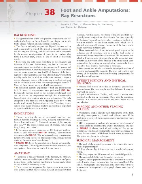
13282_ON-<strong>38</strong>.qxd 4/10/09 12:35 PM Page 1Chapter <strong>38</strong><strong>Foot</strong> <strong>and</strong> <strong>Ankle</strong> <strong>Amputations</strong>:<strong>Ray</strong> <strong>Resections</strong>Loretta B. Chou, H. Thomas Temple, Yvette Ho,<strong>and</strong> Martin M. MalawerBACKGROUND■ Malignant tumors of the foot present a significant <strong>and</strong> formidablechallenge to the orthopaedic oncologist due to thefoot’s unique function <strong>and</strong> anatomic peculiarities.■ The foot is uniquely adapted for bipedal motion <strong>and</strong> assuch is essentially a tripod. The tripod is basically formed bythe first ray, the fifth ray, <strong>and</strong> the calcaneus <strong>and</strong> is supportedby the osseous configuration of bones in the midfoot thatform a Roman arch, which in <strong>and</strong> of itself is inherentlystable.■ Both bone <strong>and</strong> soft tissue contribute to the structure <strong>and</strong>function of the foot. Furthermore, the foot is composed ofcompact compartments that are interconnected by nerves <strong>and</strong>vascular structures. Complete resection (wide local excision) ofosseous structures of the foot is difficult because of the interruptionof these complex anatomic relationships, which affordstability to the foot, in addition to the interconnected compartments.Malignant tumors of bone are rare in the foot <strong>and</strong> rarerstill in locations distal to the metatarsophalangeal joints. 3,5–9Many of these tumors are treated with amputations.■ In the senior author’s experience of foot <strong>and</strong> ankle tumorsin 153 cases, 31 amputations were performed (FIG 1A).Fortunately, tumors distal to the metatarsophalangeal jointcan be treated by amputation through the metatarsophalangealjoint with minimal disruption to function. The onlyexception is the first ray, which typically bears 50% of theweight with toe-off during each gait cycle. Therefore, preservationof as much proximal phalanx as possible is importantto optimize this important structure.INDICATIONS■ Tumors involving the toe or metatarsal bone are rare.Primary tumors affecting the foot, including osteosarcomas,have a low incidence. 1,2,7 Metastatic tumors of the foot arealso rare. 5,11 The most common sites of primary tumors arelung, kidney, <strong>and</strong> colon. 5■ In the senior author’s experience of 153 foot <strong>and</strong> ankle tumors,73 cases were bony (FIG 1B); of these, 7 cases involvedthe metatarsals (FIG 1C). The indications for ray resections aretumors involving the toe or metatarsal bone. Benign tumorsmake up the majority of these lesions. 6■ FIGURE 1D shows the distribution of benign versus malignantdiagnoses. The malignant lesions include metastatic disease,primary bony tumors, <strong>and</strong> soft tissue tumors.ANATOMY■ The tripod is basically formed by the first ray, the fifth ray,<strong>and</strong> the calcaneus <strong>and</strong> is supported by the osseous configurationof bones in the midfoot that forms a Roman arch, whichin <strong>and</strong> of itself is inherently stable.■ These bones are supported by ligamentous structures aswell as tendons that help preserve the arch <strong>and</strong> support footfunction. For this reason, resections of the first <strong>and</strong> fifth raystypically result in significant alterations in function, especiallythe first ray. The resulting forces after resection of the first rayresult in transfer to the lesser metatarsals, which are illadapted to structurally support the weight of the body, resultingin transverse metatarsalgia.■ These structural alterations can be mitigated in part by thejudicious use of orthotics, such as a medial heel wedge totransfer the forces farther lateral or the use of a metatarsal bar,which will more evenly distribute the weight across the lessermetatarsals. Resection of the fifth ray is relatively easily compensatedfor by creating an orthosis that transfers the forcesmedially; thus, a lateral heel wedge is beneficial.■ Resection of the middle rays results in insignificant loss offunction <strong>and</strong> acceptable cosmesis. The only net result is narrowingof the forefoot, which can be easily compensated forwith shoe modifications.PATIENT HISTORY AND PHYSICALFINDINGS■ Patients with tumors of the toe or metatarsal present withpain <strong>and</strong> mass. The mass may be small <strong>and</strong> chronic. It may appearwith an injury.■ Physical examination (Table 1) will reveal a tender masslocalized to the toe or metatarsal. There may be associatedswelling. If a sensory nerve overlies the mass, there may beparesthesias.IMAGING AND OTHER STAGINGSTUDIES■ Preoperative studies include plain radiographs of the foot,including anteroposterior, lateral, <strong>and</strong> oblique views. If theankle joint is involved, then anteroposterior <strong>and</strong> mortise viewsare obtained.■ MRI is important to evaluate the amount of involvement ofthe metatarsals to determine the level of amputation. The casein FIGURE 2 shows cortical thickening of the secondmetatarsal. The clinical photographs show increased space betweenthe metatarsals. MRI shows the soft tissue involvement.The lesion was found to be benign.SURGICAL MANAGEMENT■ The goal of the surgical procedure is to remove the tumorwith adequate margins.■ A long plantar flap is important for a sturdy end-bearingstump.■ The bony edges should be smooth <strong>and</strong> beveled if possible.■ Myodesis is helpful to pad the end of the stump.■ Figure 2 shows pigmented villonodular synovitis (PVNS) localizedto the plantar aspect of the first metatarsal head. Theclinical photographs show the mass. MRI shows the soft tissueinvolvement. Intraoperative photograph shows resection ofthe tumor.1
13282_ON-<strong>38</strong>.qxd 4/10/09 12:35 PM Page 22 Part 4 ONCOLOGY • Section IV LOWER EXTREMITIESABCDFIG 1 • A. Distribution of types of amputations for 153 tumors of the foot <strong>and</strong> ankle (n 31). B. All diagnoses of bony tumors of thefoot <strong>and</strong> ankle (n 73). C. Diagnoses of metatarsal tumors (n 7). D. Distribution of bony tumors of the foot <strong>and</strong> ankle by diagnosis<strong>and</strong> site (n 73).Table 1Physical Examination MethodsExamination Technique SignificanceInspection Evaluate the lower extremity with Examination of both extremities allows forthe patient undressed from the knees.comparison to identify abnormalities.Palpation Gentle <strong>and</strong> deep palpation of Evaluates tenderness of the mass; evaluatesthe mass or area of painwhether the mass is mobile or fixedRange of motion Observe passive <strong>and</strong> active range Identifies joint involvement with tumorof motion of the foot <strong>and</strong> ankle.Vascular Palpate dorsalis pedis <strong>and</strong> Evaluates vascularity of the extremity <strong>and</strong>examination posterior tibia arteries. whether there is tumor involvementNeurologic Evaluate motor strength <strong>and</strong> sensation Evaluates involvement of muscles <strong>and</strong>examination to light touch. sensory nerves
13282_ON-<strong>38</strong>.qxd 4/10/09 12:35 PM Page 3Chapter <strong>38</strong> FOOT AND ANKLE AMPUTATIONS: RAY RESECTIONS 3ABCDEFGFIG 2 • A. Benign tumors canshow cortical thickening, mimickinga stress fracture. In this case, it wasa benign lesion of the secondmetatarsal. B. Clinical photographshows increased space between themetatarsals from the mass. C. MRIshows the amount of soft tissueinvolvement. D,E. Pigmentedvillonodular synovitis (PVNS) isusually treated with simpleexcision. This case of PVNS waslocalized to the plantar aspect ofthe first metatarsal head. Clinicalphotographs show the mass. F. MRIshows soft tissue involvement.G. Intraoperative photographshows tumor resection.Preoperative Planning■ Preoperative planning is crucial for a good outcome. Thepreoperative radiographs <strong>and</strong> CT <strong>and</strong> MRI studies are importantto determine the amount of tumor involvement. They alsoshow the extent of soft tissue tumors <strong>and</strong> may be helpful indistinguishing benign from malignant tumors. 10 The biopsyresults will determine the level of amputation.■ The level of amputation is an important part of preoperativeplanning. The length of the residual stump is as important asthe quality of soft tissue. There must be adequate padding ofskin, subcutaneous fat, muscle, <strong>and</strong> tendons to cover the endof the bones.Positioning■ The patient is placed supine on the operating table. A thightourniquet is placed over adequate cotton padding. A bumpmay be placed proximal to the sciatic notch on the ipsilateralhip to limit external rotation of the extremity during theprocedure.Approach■ The surgical approach is planned preoperatively. It is importantto maintain as much plantar skin as possible because thisskin is thicker <strong>and</strong> has specialized columns of plantar fat forweight bearing.
13282_ON-<strong>38</strong>.qxd 4/10/09 12:35 PM Page 44 Part 4 ONCOLOGY • Section IV LOWER EXTREMITIESTECHNIQUESRAY RESECTION■■■■In performing a ray resection, an incision is made longitudinally<strong>and</strong> dorsally in line with the involved metatarsal(TECH FIG 1A,B). At the metatarsophalangeal joint, theincision is carried plantarly in a curvilinear fashion aroundthe joint. This tissue is then used to reconstruct the resultingweb space between the adjacent digits.The sensory nerves are identified just beneath the skin<strong>and</strong> are pulled distally <strong>and</strong> transected sharply with ascalpel. The extensor tendon is also transected sharply at<strong>and</strong> proximally near the tarsometatarsal joint.The common digital nerve is identified along with thevascular bundle. If they are involved or closely adherentto the tumor pseudocapsule, they are ligated proximally.The lumbrical <strong>and</strong> interosseous muscles are transectedproximally, exposing the base of the metatarsal. It ispreferable to preserve the base of the metatarsal if possiblebecause this does not cause any disruption in thearch formed by the tarsal–metatarsal articulation.An oscillating saw is used to transect the metatarsal, orthe metatarsal is disarticulated at the tarsometatarsaljoint <strong>and</strong> elevated. The resection then is performed fromproximal to distal. The flexor tendon is identified <strong>and</strong>transected. The entire metatarsal is then excised alongwith the adjacent soft tissue (lumbricals, intrinsics, <strong>and</strong>■■flexor extensor tendons). The dissection is then carrieddistally <strong>and</strong> plantarward. The capsular structures of themetatarsophalangeal joint are then separated from theunderlying dermis, <strong>and</strong> the ray is removed.A suture is placed through the capsular structures of theadjacent metatarsal heads. Pressure is then applied to thetibial <strong>and</strong> fibular aspect of the foot to close the defect betweenthe adjacent metatarsals, <strong>and</strong> a 0 nonabsorbablesuture is used to anchor the capsular structures betweenthe adjacent metatarsals to bring the metatarsals together<strong>and</strong> narrow the defect between the adjacent rays.A small drain is placed in the defect <strong>and</strong> brought outthrough a separate puncture wound distally. The subcutaneoustissue is then closed with 3-0 absorbable suturesplaced in interrupted fashion. The skin is closed with 4-0nylon sutures placed in interrupted fashion.A bulky dressing is applied, maintaining even compressionacross the foot <strong>and</strong> compressing the adjacent metatarsalsto decrease tension on the capsular stitch maintaining themetatarsal heads in close proximity. If the flexor or extensortendons are not involved with tumor, they can bewoven through the metatarsal heads to create a sling toanchor the metatarsal heads in close proximity <strong>and</strong> maintainthe close space between the metatarsal heads.A B CDTECH FIG 1 • A. Skin incision location in a metatarsalray resection. B. Site of osteotomy <strong>and</strong> skin closure.C. This example shows a recurrent giant cell tumor ofthe first metatarsal that was treated successfully witha fibular strut graft. The lateral radiograph showsthe healed graft 7 years postoperatively. D. Surgicaltechnique for first metatarsophalangeal joint toe amputation.The plantar skin flap is longer. It is broughtdorsally to cover the bony end of the stump.
13282_ON-<strong>38</strong>.qxd 4/10/09 12:35 PM Page 5Chapter <strong>38</strong> FOOT AND ANKLE AMPUTATIONS: RAY RESECTIONS 5First <strong>Ray</strong> Resection withReconstruction with AutogenousFibular Graft■■■■Techniques Figure 1C shows a recurrent giant cell tumorof the first metatarsal that was treated successfully witha fibular strut graft. The lateral radiograph shows thehealed graft 7 years postoperatively.Under epidural anesthesia an incision is made over thefirst metatarsal. Anterior <strong>and</strong> posterior fasciocutaneousflaps are made extending to the level of the secondmetatarsal anterior <strong>and</strong> posterior. The proximal limb ofthe incision extends beyond the metatarsal cuneiformjoint, <strong>and</strong> the distal incision extends beyond the metacarpophalangealjoint.Resection consists of the following planes between thefirst <strong>and</strong> second metatarsals following the bone in thesecond metatarsal through the metatarsal cuneiformjoint <strong>and</strong> posteriorly the flexor longus muscle tendon.The distal metatarsal is osteomized at the condyles <strong>and</strong>proximally just distal to the joint. The tumor mass is removed;the anterior tibial tendon, peroneal tendons,<strong>and</strong> anterior tibial vessels are preserved.Reconstruction is performed with a fibular graft from themidshaft of the fibula of the ipsilateral limb. This isplaced within the defect, measuring about 8 cm betweenthe cuneiform <strong>and</strong> the metatarsal head. Fixation is■achieved with two small cortical interfragmentary screwsproximally <strong>and</strong> distally. The hallux is maintained in neutralposition.Corticocancellous bone graft is placed around the base<strong>and</strong> the distal osteotomy sites with fibrin glue for hemostasis<strong>and</strong> to promote healing. The fibular bone graft istaken through a separate incision with separate instrumentsin the midportion of the fibula on the same sidebetween the peroneus longus <strong>and</strong> the soleus interval.The muscles are retracted, <strong>and</strong> the intermuscular septumis released with the cutting electrocautery. An oscillatingsaw is used to remove 8 to 10 cm of the fibula. Thewound is then irrigated, <strong>and</strong> the peroneal muscles arerepaired. A drain is used.Metatarsophalangeal JointAmputation■■■A longer plantar flap is planned (TECH FIG 1D). Theincision is carried out at the level of the metatarsophalangealjoint on the dorsum of the foot.Sharp dissection is used to expose the joint, <strong>and</strong> the capsule,ligaments, <strong>and</strong> tendons are cut at that level. Thevessels are cauterized.The plantar flap is brought over the dorsal aspect <strong>and</strong>nylon suture is used to repair the skin.TECHNIQUESPEARLS AND PITFALLSIndicationsSurgical incisionWound healingcomplicationsDeep infectionContracturesPainful stump■ A careful <strong>and</strong> complete history <strong>and</strong> physical examination are essential. Preoperative studies arenecessary to plan the resection <strong>and</strong> reconstruction.■ A long plantar flap results in a better end-bearing stump. The natural cascade of metatarsal lengthsshould be maintained.■ Local wound care with dressings <strong>and</strong> oral antibiotics are usually sufficient for healing.■ Parenteral antibiotics <strong>and</strong> surgical débridement may be necessary to treat deep infections. Earlydiagnosis <strong>and</strong> treatment can affect outcome.■ Postoperative splinting can help prevent contractures. Once a contracture occurs, it is treated withstretching if mild. Serial casting may be needed.■ The end of the bones should be contoured to be smooth. A rasp is effective. Adequate soft tissuecoverage will help prevent bony prominence, which may become symptomatic.POSTOPERATIVE CARE■After a ray resection, the patient is placed in a well-paddedsplint. Crutches are used until the sutures are removed. Thepatient may begin range-of-motion exercises <strong>and</strong> weight bearingas tolerated.■Toe amputation patients may ambulate in a postoperativeshoe immediately. The sutures are removed at 2 to 3 weeks.A wide comfort shoe is worn <strong>and</strong> activities are progressed astolerated.OUTCOMES■ After a toe amputation at the level of the metatarsophalangealjoint, full preoperative function is regained.■ A cosmetic toe prosthesis may be used if the patient desiresone, but it has no functional purpose.■ <strong>Ray</strong> resection outcomes depend on the number of rays amputated<strong>and</strong> whether the first ray is involved. A ray resectionof the second, third, fourth, or fifth metatarsal does not needshoe modification with a molded insert or filler. Generally,ordinary store-bought shoes that are wide <strong>and</strong> have adequatecushioning are worn.■ A first ray resection affects the remaining foot functionbecause of loss of the windlass mechanism. Reconstruction ofthe first ray would prevent this problem.■ Removal of two central rays will leave a narrower foot <strong>and</strong>shoewear problems.
13282_ON-<strong>38</strong>.qxd 4/10/09 12:35 PM Page 66 Part 4 ONCOLOGY • Section IV LOWER EXTREMITIES■ Patients with a metatarsal ray or transmetatarsal amputationhave better outcomes than patients with a Syme amputation. 4COMPLICATIONS■ Wound healing complications are treated with local woundcare, elevation, <strong>and</strong> non-weight bearing. Oral antibiotics maybe indicated. If there is skin flap necrosis, débridement with askin graft may be necessary.■ Superficial infection can be treated with a short course oforal antibiotics.■ Deep infection may require immediate operative débridementof devitalized tissue. Parenteral antibiotics are importantto treat this limb-threatening problem. At the time of thedébridement, a bone culture should be ordered to help determinethe appropriate antibiotic.REFERENCES1. Bugnone AN, Temple HT, Pitcher JD. Low-grade central osteosarcomaof the foot <strong>and</strong> ankle: radiographic <strong>and</strong> pathologic features intwo patients: case report <strong>and</strong> literature review. <strong>Foot</strong> <strong>Ankle</strong> Int 2005;26:494–500.2. Choong PF, Qureshi AA, Sim FH, et al. Osteosarcoma of the foot: areview of 52 patients at the Mayo Clinic. Acta Orthop Sc<strong>and</strong> 1999;70:361–364.3. Chou LB, Malawer MM. Analysis of surgical treatment of 33 foot<strong>and</strong> ankle tumors. <strong>Foot</strong> <strong>Ankle</strong> Int 1994;15:175–181.4. Greene WB, Cary JM. Partial foot amputations in children: a comparisonof the several types with the Syme amputation. J Bone JointSurg Am 1982;64A:4<strong>38</strong>–443.5. Hattrup SJ, Amadio PC, Sim FH, et al. Metastatic tumors of the foot<strong>and</strong> ankle. <strong>Foot</strong> <strong>Ankle</strong> 1988;8:243–247.6. Kirby EJ, Shereff MJ, Lewis MM. Soft-tissue tumors <strong>and</strong> tumor-likelesions of the foot: an analysis of eighty-three cases. J Bone Joint SurgAm 1989;71A:621–626.7. Murari TM, Callaghan JJ, Berrey BH Jr, et al. Primary benign <strong>and</strong>malignant osseous neoplasms of the foot. <strong>Foot</strong> <strong>Ankle</strong> 1989;10:68–80.8. Seale KS, Lange TA, Monson D, et al. Soft tissue tumors of the foot<strong>and</strong> ankle. <strong>Foot</strong> <strong>Ankle</strong> 1988;9:19–27.9. Sundberg SB, Carlson WO, Johnson KA. Metastatic lesions of thefoot <strong>and</strong> ankle. <strong>Foot</strong> <strong>Ankle</strong> 1982;3:167–169.10. Wetzel LH, Levine E. Soft-tissue tumors of the foot: value of MRimaging for specific diagnosis. AJR Am J Roentgenol 1990;155:1025–1030.11. Wu KK, Guise ER. Metastatic tumors of the foot. South Med J 1978;71:807–812.