Morphological subtypes of ovarian carcinoma: a ... - BPA Pathology
Morphological subtypes of ovarian carcinoma: a ... - BPA Pathology
Morphological subtypes of ovarian carcinoma: a ... - BPA Pathology

You also want an ePaper? Increase the reach of your titles
YUMPU automatically turns print PDFs into web optimized ePapers that Google loves.
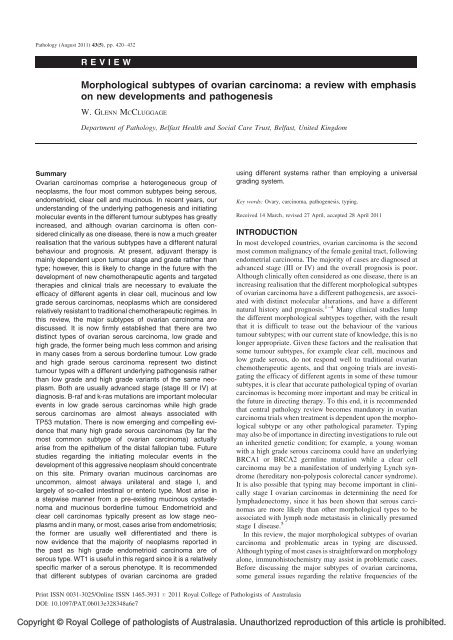
<strong>Pathology</strong> (August 2011) 43(5), pp. 420–432REVIEW<strong>Morphological</strong> <strong>subtypes</strong> <strong>of</strong> <strong>ovarian</strong> <strong>carcinoma</strong>: a review with emphasison new developments and pathogenesisW. GLENN MCCLUGGAGEDepartment <strong>of</strong> <strong>Pathology</strong>, Belfast Health and Social Care Trust, Belfast, United KingdomSummaryOvarian <strong>carcinoma</strong>s comprise a heterogeneous group <strong>of</strong>neoplasms, the four most common <strong>subtypes</strong> being serous,endometrioid, clear cell and mucinous. In recent years, ourunderstanding <strong>of</strong> the underlying pathogenesis and initiatingmolecular events in the different tumour <strong>subtypes</strong> has greatlyincreased, and although <strong>ovarian</strong> <strong>carcinoma</strong> is <strong>of</strong>ten consideredclinically as one disease, there is now a much greaterrealisation that the various <strong>subtypes</strong> have a different naturalbehaviour and prognosis. At present, adjuvant therapy ismainly dependent upon tumour stage and grade rather thantype; however, this is likely to change in the future with thedevelopment <strong>of</strong> new chemotherapeutic agents and targetedtherapies and clinical trials are necessary to evaluate theefficacy <strong>of</strong> different agents in clear cell, mucinous and lowgrade serous <strong>carcinoma</strong>s, neoplasms which are consideredrelatively resistant to traditional chemotherapeutic regimes. Inthis review, the major <strong>subtypes</strong> <strong>of</strong> <strong>ovarian</strong> <strong>carcinoma</strong> arediscussed. It is now firmly established that there are twodistinct types <strong>of</strong> <strong>ovarian</strong> serous <strong>carcinoma</strong>, low grade andhigh grade, the former being much less common and arisingin many cases from a serous borderline tumour. Low gradeand high grade serous <strong>carcinoma</strong> represent two distincttumour types with a different underlying pathogenesis ratherthan low grade and high grade variants <strong>of</strong> the same neoplasm.Both are usually advanced stage (stage III or IV) atdiagnosis. B-raf and k-ras mutations are important molecularevents in low grade serous <strong>carcinoma</strong>s while high gradeserous <strong>carcinoma</strong>s are almost always associated withTP53 mutation. There is now emerging and compelling evidencethat many high grade serous <strong>carcinoma</strong>s (by far themost common subtype <strong>of</strong> <strong>ovarian</strong> <strong>carcinoma</strong>) actuallyarise from the epithelium <strong>of</strong> the distal fallopian tube. Futurestudies regarding the initiating molecular events in thedevelopment <strong>of</strong> this aggressive neoplasm should concentrateon this site. Primary <strong>ovarian</strong> mucinous <strong>carcinoma</strong>s areuncommon, almost always unilateral and stage I, andlargely <strong>of</strong> so-called intestinal or enteric type. Most arise ina stepwise manner from a pre-existing mucinous cystadenomaand mucinous borderline tumour. Endometrioid andclear cell <strong>carcinoma</strong>s typically present as low stage neoplasmsand in many, or most, cases arise from endometriosis;the former are usually well differentiated and there isnow evidence that the majority <strong>of</strong> neoplasms reported inthe past as high grade endometrioid <strong>carcinoma</strong> are <strong>of</strong>serous type. WT1 is useful in this regard since it is a relativelyspecific marker <strong>of</strong> a serous phenotype. It is recommendedthat different <strong>subtypes</strong> <strong>of</strong> <strong>ovarian</strong> <strong>carcinoma</strong> are gradedusing different systems rather than employing a universalgrading system.Key words: Ovary, <strong>carcinoma</strong>, pathogenesis, typing.Received 14 March, revised 27 April, accepted 28 April 2011INTRODUCTIONIn most developed countries, <strong>ovarian</strong> <strong>carcinoma</strong> is the secondmost common malignancy <strong>of</strong> the female genital tract, followingendometrial <strong>carcinoma</strong>. The majority <strong>of</strong> cases are diagnosed atadvanced stage (III or IV) and the overall prognosis is poor.Although clinically <strong>of</strong>ten considered as one disease, there is anincreasing realisation that the different morphological <strong>subtypes</strong><strong>of</strong> <strong>ovarian</strong> <strong>carcinoma</strong> have a different pathogenesis, are associatedwith distinct molecular alterations, and have a differentnatural history and prognosis. 1–4 Many clinical studies lumpthe different morphological <strong>subtypes</strong> together, with the resultthat it is difficult to tease out the behaviour <strong>of</strong> the varioustumour <strong>subtypes</strong>; with our current state <strong>of</strong> knowledge, this is nolonger appropriate. Given these factors and the realisation thatsome tumour <strong>subtypes</strong>, for example clear cell, mucinous andlow grade serous, do not respond well to traditional <strong>ovarian</strong>chemotherapeutic agents, and that ongoing trials are investigatingthe efficacy <strong>of</strong> different agents in some <strong>of</strong> these tumour<strong>subtypes</strong>, it is clear that accurate pathological typing <strong>of</strong> <strong>ovarian</strong><strong>carcinoma</strong>s is becoming more important and may be critical inthe future in directing therapy. To this end, it is recommendedthat central pathology review becomes mandatory in <strong>ovarian</strong><strong>carcinoma</strong> trials when treatment is dependent upon the morphologicalsubtype or any other pathological parameter. Typingmay also be <strong>of</strong> importance in directing investigations to rule outan inherited genetic condition; for example, a young womanwith a high grade serous <strong>carcinoma</strong> could have an underlyingBRCA1 or BRCA2 germline mutation while a clear cell<strong>carcinoma</strong> may be a manifestation <strong>of</strong> underlying Lynch syndrome(hereditary non-polyposis colorectal cancer syndrome).It is also possible that typing may become important in clinicallystage I <strong>ovarian</strong> <strong>carcinoma</strong>s in determining the need forlymphadenectomy, since it has been shown that serous <strong>carcinoma</strong>sare more likely than other morphological types to beassociated with lymph node metastasis in clinically presumedstage I disease. 5In this review, the major morphological <strong>subtypes</strong> <strong>of</strong> <strong>ovarian</strong><strong>carcinoma</strong> and problematic areas in typing are discussed.Although typing <strong>of</strong> most cases is straightforward on morphologyalone, immunohistochemistry may assist in problematic cases.Before discussing the major <strong>subtypes</strong> <strong>of</strong> <strong>ovarian</strong> <strong>carcinoma</strong>,some general issues regarding the relative frequencies <strong>of</strong> thePrint ISSN 0031-3025/Online ISSN 1465-3931 # 2011 Royal College <strong>of</strong> Pathologists <strong>of</strong> AustralasiaDOI: 10.1097/PAT.0b013e328348a6e7Copyright © Royal College <strong>of</strong> pathologists <strong>of</strong> Australasia. Unauthorized reproduction <strong>of</strong> this article is prohibited.
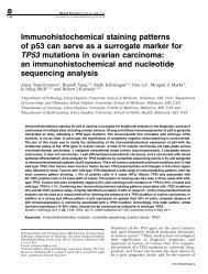
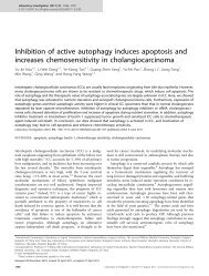
SUBTYPES OF OVARIAN CARCINOMA 421Table 1 Relative frequencies <strong>of</strong> <strong>subtypes</strong> <strong>of</strong> <strong>ovarian</strong> <strong>carcinoma</strong> based on tworecent population based studies68–71% serous3% mucinous9–11% endometrioid12–13% clear cell1% transitional6% mixedvarious tumour types, morphological changes secondary tochemotherapy and tumour grading are covered.RELATIVE FREQUENCIES OF MAJORMORPHOLOGICAL SUBTYPES OF OVARIANCARCINOMAThe major morphological <strong>subtypes</strong> <strong>of</strong> <strong>ovarian</strong> <strong>carcinoma</strong> areserous, endometrioid, clear cell and mucinous. 6 Other <strong>subtypes</strong>include transitional, undifferentiated and mixed. Primary <strong>ovarian</strong>squamous <strong>carcinoma</strong>s also occur; these are rare, usuallyarise in a dermoid cyst or more uncommonly in association withendometriosis or a Brenner tumour, and will not be discussedfurther. Two relatively recent population based studies whichhave included central pathology review using modern diagnosticcriteria have provided updated information regarding therelative frequencies <strong>of</strong> the major <strong>subtypes</strong> <strong>of</strong> <strong>ovarian</strong> <strong>carcinoma</strong>7,8 (Table 1). It can be seen that serous is the mostcommon, followed by clear cell and endometrioid which occurwith approximately equal frequency. Mucinous <strong>carcinoma</strong>s areless common. This represents a change from most older studieswhere mucinous <strong>carcinoma</strong> was the second most commonsubtype and accounted for approximately 12% <strong>of</strong> primary<strong>ovarian</strong> <strong>carcinoma</strong>s, 9 a much higher frequency than in thetwo recent studies; reasons for this decline in the frequency<strong>of</strong> mucinous <strong>carcinoma</strong> are discussed later. The two studies alsoindicate an increase and decrease in the frequency <strong>of</strong> serous andendometrioid <strong>carcinoma</strong>, respectively; this is likely a reflection<strong>of</strong> the fact that the distinction between a high grade serous andhigh grade endometrioid <strong>carcinoma</strong> was previously poorlyreproducible 10–14 and there is now a realisation that manyneoplasms which were previously diagnosed as advanced stage,high grade endometrioid <strong>carcinoma</strong> were, in fact, <strong>of</strong> serous type(discussed later). When divided into early stage (stage I–II) andlate stage (stage III–IV), it can be seen that serous, clear celland endometrioid <strong>carcinoma</strong>s are approximately equallyrepresented in early stage, nearly all mucinous <strong>carcinoma</strong>sare early stage and a very high percentage <strong>of</strong> advanced stageneoplasms are serous in type (Table 2). Put another way, a highpercentage <strong>of</strong> clear cell, endometrioid and mucinous <strong>carcinoma</strong>sare diagnosed at early stage and in fact these tumourtypes (especially endometrioid and mucinous) are usuallyconfined to the ovary at diagnosis (stage I).Table 2 Early stage (I/II) versus late stage (III/IV) distribution <strong>of</strong> <strong>subtypes</strong> <strong>of</strong><strong>ovarian</strong> <strong>carcinoma</strong> based on two recent population based studiesStage I/IIStage III/IVSerous 36% 88%Clear cell 26% 5%Endometrioid 27% 3%Mucinous 8% 1%MORPHOLOGICAL CHANGES IN OVARIANCARCINOMAS SECONDARY TOCHEMOTHERAPYFor many years, the traditional management <strong>of</strong> advanced stage<strong>ovarian</strong> <strong>carcinoma</strong> has been surgical debulking followed byadjuvant chemotherapy. However, in an increasing number <strong>of</strong>cases, upfront chemotherapy is administered, especially inpatients who are a poor operative risk or patients with miliarydisease or widespread metastasis where optimal debulking isnot considered feasible; chemotherapy may or may not befollowed by surgery. The morphological features <strong>of</strong> <strong>ovarian</strong><strong>carcinoma</strong>s treated by chemotherapy <strong>of</strong>ten differ markedlyfrom native tumour. 15,16 Post-chemotherapy, many <strong>ovarian</strong><strong>carcinoma</strong>s (the vast majority will represent serous <strong>carcinoma</strong>ssince almost all advanced stage <strong>ovarian</strong> malignancies are <strong>of</strong> thismorphological subtype) have abundant clear or eosinophiliccytoplasm and the nuclear features are <strong>of</strong>ten bizarre withmultinucleate tumour giant cells 15,16 (Fig. 1). There may beno residual tumour or it may be difficult to identify residualneoplastic cells due to pronounced chemotherapy effect withmarked fibrosis, necrosis, inflammation, cholesterol cleft formation,haemosiderin deposition and dystrophic calcification.Unless there is no or minimal response, it can be very difficultto type an <strong>ovarian</strong> <strong>carcinoma</strong> following chemotherapy and thereis a tendency to misdiagnose some as clear cell <strong>carcinoma</strong> dueto the abundant clear cytoplasm. 15,16 If upfront chemotherapy isbeing administered, a pre-chemotherapy tissue biopsy (usuallya percutaneous radiologically guided biopsy or a biopsy takenat laparoscopy) should be obtained for definitive typing, apartfrom in exceptional circumstances, rather than relying oncytology <strong>of</strong> the ascitic fluid in combination with serumCA125 levels and imaging. The procurement <strong>of</strong> a tissue biopsy,as well as facilitating tumour typing and excluding a metastasisfrom other sites, means that material is available for futurestudies and research; for example, targeted therapies may bedeveloped against specific proteins and if tissue is available thiswill be useful in assessing whether the target protein is presentin the tumour cells. Tissue obtained at different stages intreatment may also be useful in assessing tumour progressionand response to therapy.GRADING OF OVARIAN CARCINOMASSeveral grading systems are used for <strong>ovarian</strong> <strong>carcinoma</strong> butgrading is <strong>of</strong>ten poorly performed by pathologists and in manystudies the grading system used has not been specified. 17–19Most <strong>of</strong> the grading systems are universal in that they can beapplied to all the major morphological <strong>subtypes</strong> <strong>of</strong> <strong>ovarian</strong><strong>carcinoma</strong>; 17–19 for example, the Shimizu-Silverberg system isbased on the Nottingham grading system for breast <strong>carcinoma</strong>and uses three parameters, namely the degree <strong>of</strong> nuclear atypia,the mitotic count and the architectural features, specifically theamount <strong>of</strong> glandular, papillary or solid growth. 17 Eachparameter is given a score <strong>of</strong> 1–3 and a grade is derived basedon the summation <strong>of</strong> the scores. However, although manypathologists use this or other universal grading systems, suchas FIGO or World Health Organization (WHO), there is anincreasing tendency to employ different grading systems fordifferent morphological <strong>subtypes</strong> <strong>of</strong> <strong>ovarian</strong> <strong>carcinoma</strong>; thispractice has been recommended in the Royal College <strong>of</strong>Pathologists Ovarian Cancer Datasets in the United Kingdom. 20Grading <strong>of</strong> the different <strong>subtypes</strong> <strong>of</strong> <strong>ovarian</strong> <strong>carcinoma</strong> isdiscussed with each specific tumour type.Copyright © Royal College <strong>of</strong> pathologists <strong>of</strong> Australasia. Unauthorized reproduction <strong>of</strong> this article is prohibited.
422 MCCLUGGAGE <strong>Pathology</strong> (2011), 43(5), AugustFig. 1 (A) Ovarian serous <strong>carcinoma</strong> with clear cytoplasm and (B) bizarre nuclear features. These are examples <strong>of</strong> serous <strong>carcinoma</strong>s with morphological featuressecondary to chemotherapy.SEROUS CARCINOMAThe perceived relationship between benign, borderline andmalignant <strong>ovarian</strong> serous neoplasms was controversial and asource <strong>of</strong> confusion for many years. It has always been temptingto speculate that a continuum <strong>of</strong> <strong>ovarian</strong> serous neoplasiaexists from benign to borderline to malignant. However, pathologicalevidence for this is lacking and until recently it wasgenerally assumed that there was no firm relationship betweenborderline and malignant serous neoplasms, although occasionallythe two were found to coexist. Recent studies have shedsignificant light on this issue and have convincingly demonstratedthat there are two distinct types <strong>of</strong> <strong>ovarian</strong> serous<strong>carcinoma</strong>, low grade and high grade. 1–4,21–27 Although termedlow grade and high grade serous <strong>carcinoma</strong>, it is important toemphasise that these are not two grades <strong>of</strong> the same neoplasmbut rather two distinct tumour types with different underlyingpathogenesis, molecular events, behaviour and prognosis. Highgrade serous <strong>carcinoma</strong> is much more common than low grade.Low grade serous <strong>carcinoma</strong> is thought to arise in many cases ina stepwise fashion from a benign serous cystadenoma through aserous borderline tumour to an invasive low grade serous<strong>carcinoma</strong>. Thus, there is a well defined adenoma-<strong>carcinoma</strong>sequence. It has been suggested that a micropapillary architecturein a serous borderline tumour is an intermediate stepbetween a usual serous borderline tumour and an invasive lowgrade serous <strong>carcinoma</strong>. 1,27–29 However, a micropapillaryarchitecture is not present in many serous borderline tumourswith coexistent low grade serous <strong>carcinoma</strong>. I also make thepoint that it is relatively uncommon to see areas <strong>of</strong> invasive lowgrade serous <strong>carcinoma</strong> within a serous borderline tumour andconversely in many low grade serous <strong>carcinoma</strong>s a borderlinecomponent is not seen. Therefore, it is not proven that all lowgrade serous <strong>carcinoma</strong>s arise from a pre-existing serousborderline tumour and it is possible that some or many donot. In contrast, high grade serous <strong>carcinoma</strong> is not related toserous borderline tumour and was thought until recently to arisedirectly from the <strong>ovarian</strong> surface epithelium or the epithelium<strong>of</strong> cortical inclusion cysts with no well defined precursor lesion.There is now emerging and quite compelling evidence (discussedlater) that many high grade <strong>ovarian</strong> serous <strong>carcinoma</strong>sactually originate from the epithelium <strong>of</strong> the distal fimbrialportion <strong>of</strong> the fallopian tube. 30–35 Instead <strong>of</strong> grading <strong>ovarian</strong>serous <strong>carcinoma</strong> using a three-tiered system (well, moderate,poor; grade 1, 2 or 3), there is now a growing tendency amongstpathologists and oncologists to classify these as high grade orlow grade and this practice has been adopted in the UnitedKingdom; 20 anecdotally many gynaecological pathologistsnow use this two tiered grading system which was originallyproposed by the MD Anderson Group (Houston, Texas, USA).The classification <strong>of</strong> a serous <strong>carcinoma</strong> as low grade or highgrade has been shown to be reproducible amongst pathologists.36,37 Almost all serous <strong>carcinoma</strong>s which would havepreviously been classified as moderately or poorly differentiatedrepresent high grade neoplasms while those which wouldhave been classified as well differentiated may be either lowgrade or high grade using the two tier classification. Forexample, some architecturally well differentiated serous <strong>carcinoma</strong>shave high nuclear grade and represent examples <strong>of</strong>high grade serous <strong>carcinoma</strong>. Although representing two distincttumour types, on rare occasions a low grade serous<strong>carcinoma</strong> component may coexist with and probably transforminto a high grade serous <strong>carcinoma</strong> or even an undifferentiated<strong>carcinoma</strong> or a high grade serous <strong>carcinoma</strong> may arisedirectly from a serous borderline tumour (Dehari et al. 38 andpersonal observations). However, most low grade serous<strong>carcinoma</strong>s when they recur do so as low grade neoplasms.Some advocate the term invasive micropapillary serous <strong>carcinoma</strong>as an alternative to low grade serous <strong>carcinoma</strong> but thisterminology is not recommended since some low grade serous<strong>carcinoma</strong>s do not exhibit a micropapillary architecture andconversely many high grade serous <strong>carcinoma</strong>s do so. Figure 2illustrates the postulated pathways <strong>of</strong> development <strong>of</strong> low gradeand high grade serous <strong>carcinoma</strong>.<strong>Morphological</strong> and immunohistochemical features <strong>of</strong> lowgrade and high grade serous <strong>carcinoma</strong>The distinction between low and high grade serous <strong>carcinoma</strong> isbased on morphology, the chief discriminator being the degree<strong>of</strong> nuclear atypia in the worst area <strong>of</strong> the tumour. The amount <strong>of</strong>mitotic activity is also taken into account. 36,37 In low gradeserous <strong>carcinoma</strong>, the nuclei are uniform with mild or at themost moderate atypia and less than or equal to 12 mitoses per 10high power fields (the mitotic count is usually approximately 2per 10 high power fields or less than this) (Fig. 3). There is nonecrosis or multinucleation. High grade serous <strong>carcinoma</strong>exhibits moderate to marked nuclear atypia and greater than12 mitoses per 10 high power fields (Fig. 4). Necrosis andmultinucleate cells are <strong>of</strong>ten present. It is generally not necessaryto count mitotic figures since these are typically abundantin high grade serous <strong>carcinoma</strong>s and difficult to find in lowgrade neoplasms. In both low grade and high grade serous<strong>carcinoma</strong>s, there may be a variety <strong>of</strong> architectural patterns,Copyright © Royal College <strong>of</strong> pathologists <strong>of</strong> Australasia. Unauthorized reproduction <strong>of</strong> this article is prohibited.
SUBTYPES OF OVARIAN CARCINOMA 423Postulated development <strong>of</strong> low-grade and high-grade<strong>ovarian</strong> serous <strong>carcinoma</strong>Fimbria <strong>of</strong> fallopian tubeOvarian surface epitheliumBenign serous cystadenomaSerous tubalintraepithelial<strong>carcinoma</strong> (serousTIC)OvarianintraepithelialneoplasiaCorticalinclusioncystsUsual serous borderlineneoplasmMicropapillary variant <strong>of</strong> serousborderline neoplasmHigh gradeserous <strong>carcinoma</strong>Low gradeserous <strong>carcinoma</strong>Fig. 2Postulated developmental pathways <strong>of</strong> low grade and high grade serous <strong>carcinoma</strong>.including nested, papillary (micropapillary or macropapillary),glandular (slit-like or round spaces), cribriform, solid and singlecells. The glands and papillae in low grade serous <strong>carcinoma</strong>sare <strong>of</strong>ten surrounded by clefts or non-epithelial lined spaces andintracytoplasmic mucin is <strong>of</strong>ten a feature. 39 Psammoma bodiesare common in both tumour types. Psammo<strong>carcinoma</strong> is a termused for a tumour with extensive psammoma body formationand is usually morphologically a low grade serous <strong>carcinoma</strong>,although occasional examples have high grade nuclear cytology,in keeping with high grade serous <strong>carcinoma</strong>.Both low grade and high grade serous <strong>carcinoma</strong>s typicallyexpress WT1. High grade serous <strong>carcinoma</strong> exhibits significantlygreater expression <strong>of</strong> p53, MIB1, bcl-2, c-kit, Her-2 neu,HLA-G and p16 than low grade serous <strong>carcinoma</strong>. 40,41 p16 istypically diffusely positive in high grade serous <strong>carcinoma</strong> andfocally positive or negative in low grade. Low grade serous<strong>carcinoma</strong>s are almost always diffusely positive with oestrogenreceptor (ER), as are a high percentage <strong>of</strong> high grade serous<strong>carcinoma</strong>s (70–80%). High grade serous <strong>carcinoma</strong>s mostcommonly exhibit diffuse intense nuclear immunoreactivitywith p53 but totally absent staining (‘all or nothing’) is also anaberrant pattern <strong>of</strong> p53 staining and suggestive <strong>of</strong> an underlyingTP53 abnormality (see next section). So-called ‘wild-type’ p53staining, with a focal, weak and heterogenous pattern, is notindicative <strong>of</strong> a TP53 abnormality and is characteristic <strong>of</strong> lowgrade serous <strong>carcinoma</strong>. 42Molecular events in low grade and high grade serous<strong>carcinoma</strong>The underlying molecular events differ between low grade andhigh grade serous <strong>carcinoma</strong>. Low grade serous <strong>carcinoma</strong> isassociated with k-ras or b-raf mutation in approximately twothirds<strong>of</strong> cases. 21–27 These mutations occur early in the evolution<strong>of</strong> low grade serous <strong>carcinoma</strong> since they are also found inborderline and benign areas within the same neoplasm. K-rasand b-raf mutations appear mutually exclusive; in other wordsone, but not both, may be present in a particular neoplasm. TheERBB2 gene is also frequently mutated. 27 Low grade serous<strong>carcinoma</strong> is not associated with TP53 mutations. 21–27 Incontrast, high grade serous <strong>carcinoma</strong>s harbour, in most cases,a TP53 mutation or exhibit p53 dysfunction and this appears tooccur early in neoplastic development; 20–26 they are not, exceptin occasional cases, associated with k-ras, b-raf or ERBB2mutation. A recent study using stringent techniques identifiedTP53 mutations in 97% <strong>of</strong> <strong>ovarian</strong> high grade serous <strong>carcinoma</strong>s;most <strong>of</strong> the mutation negative cases exhibited TP53Fig. 3 (A) Low grade serous <strong>carcinoma</strong> with a micropapillary architecture and clefts surrounding the groups <strong>of</strong> tumour cells. (B) On high power <strong>of</strong> another low gradeserous <strong>carcinoma</strong>, the tumour cells are bland with little mitotic activity.Copyright © Royal College <strong>of</strong> pathologists <strong>of</strong> Australasia. Unauthorized reproduction <strong>of</strong> this article is prohibited.
424 MCCLUGGAGE <strong>Pathology</strong> (2011), 43(5), Augustserous <strong>carcinoma</strong> was 9% with a median survival <strong>of</strong> 1.7 years 36while low grade serous <strong>carcinoma</strong>s had a median survival <strong>of</strong>4.2 years and a 5 year survival <strong>of</strong> 40%. 36 In another study <strong>of</strong> lowgrade serous <strong>carcinoma</strong>s, the median overall survival with stageIII or IV disease was 6.8 years. 44 Low grade serous <strong>carcinoma</strong>s<strong>of</strong>ten prove fatal but in some cases the course is indolent and ina few cases the patient survives for a considerable period <strong>of</strong>time, for example in excess <strong>of</strong> 10 or 20 years.Fig. 4 High grade serous <strong>carcinoma</strong> with slit-like spaces and abundant mitoticactivity.dysfunction, illustrating that p53 is abnormal in almost 100% <strong>of</strong>high grade serous <strong>carcinoma</strong>s. 43 High grade serous <strong>carcinoma</strong>sare also associated with BRCA1 and BRCA2 abnormalities,including germline mutations and somatic abnormalities.Behaviour <strong>of</strong> low grade and high grade serous <strong>carcinoma</strong>Most low grade and high grade serous <strong>carcinoma</strong>s are stage IIIor IV when diagnosed. At present, management is similarbetween these two tumour types and consists, in most cases,<strong>of</strong> total surgical debulking if possible followed by adjuvantchemotherapy. In cases where surgical debulking is not consideredfeasible, upfront chemotherapy may be the method <strong>of</strong>treatment and this may or may not be followed by surgicaldebulking. However, adjuvant chemotherapy is usually notadministered for a stage I low grade serous <strong>carcinoma</strong>.Additionally, there is a growing realisation amongst oncologiststhat low grade serous <strong>carcinoma</strong>s do not respond well totraditional chemotherapeutic agents and if the tumour is optimallydebulked with no gross residual disease, some oncologistsdo not administer adjuvant chemotherapy. High gradeserous <strong>carcinoma</strong> is an aggressive neoplasm which usuallyresponds well initially to chemotherapy but which commonlyrecurs. Few studies have directly compared the outcomebetween low grade and high grade serous <strong>carcinoma</strong>. In onestudy, the survival <strong>of</strong> patients with low grade neoplasms wassignificantly better than that <strong>of</strong> patients with high gradetumours. 36 In this study, the 5 year survival for high gradeEVIDENCE FOR ORIGIN OF HIGH GRADEPELVIC SEROUS CARCINOMA FROM DISTALFALLOPIAN TUBEThere is recent convincing and accumulating evidence thatmany currently classified high grade serous <strong>carcinoma</strong>s <strong>of</strong> theovary, fallopian tube and peritoneum (collectively referred to ashigh grade pelvic serous <strong>carcinoma</strong>) are derived from thefimbria <strong>of</strong> the fallopian tube. 30–35 Most <strong>of</strong> the work hasemanated from Christopher Crum and colleagues in the Brighamand Women’s Hospital, Boston, USA. The initial evidencefor this came from prophylactic salpingo-oophorectomy specimensin patients with germline BRCA1 or BRCA2 mutation,these patients having a high risk <strong>of</strong> developing <strong>ovarian</strong> highgrade serous <strong>carcinoma</strong>. In initial studies, small high gradeserous <strong>carcinoma</strong>s were occasionally identified in the ovary butthis was rare. However, when pathologists began examining thefallopian tubes in their entirety, it was found that there wasmore likely to be a small in situ or invasive high grade serous<strong>carcinoma</strong> involving the mucosa <strong>of</strong> the fimbria <strong>of</strong> the fallopiantube; the in situ lesions are referred to as serous tubal intraepithelial<strong>carcinoma</strong> (serous TIC). This is relatively uncommonbut is occasionally seen in prophylactic salpingo-oophorectomyspecimens in patients with BRCA1 or BRCA2 germlinemutation. Further studies systematically examined the fallopiantubes in patients with sporadic high grade <strong>ovarian</strong> serous<strong>carcinoma</strong> and found similar lesions involving the fimbria ina significant percentage <strong>of</strong> cases (Fig. 5). 30,33 Furthermore,identical TP53 mutations were demonstrated within the <strong>ovarian</strong>and tubal lesions. While this does not unequivocally prove thatthe origin is within the fallopian tube (this could represent atumour originating within the ovary and spreading to thefallopian tube), there is accumulating evidence that many highgrade pelvic serous <strong>carcinoma</strong>s arise from the tubal fimbriafrom serous TIC. A p53 signature has also been demonstrated inthe fallopian tube. 45 This takes the form <strong>of</strong> small foci <strong>of</strong> intensep53 nuclear staining involving consecutive secretory cells,most commonly <strong>of</strong> the fimbria, in the absence <strong>of</strong> morphologicalFig. 5 (A) Serous tubal intraepithelial <strong>carcinoma</strong> (serous TIC) involving fimbria <strong>of</strong> fallopian tube and exhibiting diffuse nuclear p53 positivity; (B) the adjacent tubalepithelium is p53 negative.Copyright © Royal College <strong>of</strong> pathologists <strong>of</strong> Australasia. Unauthorized reproduction <strong>of</strong> this article is prohibited.
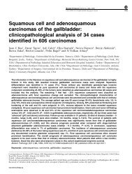
SUBTYPES OF OVARIAN CARCINOMA 425changes. TP53 mutations have been demonstrated in some p53signatures and these may represent the earliest stage <strong>of</strong> development<strong>of</strong> high grade pelvic serous <strong>carcinoma</strong>. However, p53signatures are extremely common in the fallopian tube, even inpatients with benign disease and no hereditary predisposition todeveloping <strong>ovarian</strong> cancer, and it is clear that only a smallproportion will ever develop into a serous TIC. Serous TIC isnot diagnosed on the basis <strong>of</strong> p53 staining unless associatedmorphological alterations are present associated with a highMIB1 proliferation index.It is probable that not all high grade pelvic serous <strong>carcinoma</strong>sare derived from the fallopian tube. A recent study whichsystematically examined all <strong>of</strong> the fallopian tubes in a consecutiveseries <strong>of</strong> <strong>ovarian</strong> <strong>carcinoma</strong>s identified serous TIC innearly 60% <strong>of</strong> high grade serous <strong>carcinoma</strong>s but not in othermorphological <strong>subtypes</strong> <strong>of</strong> <strong>ovarian</strong> <strong>carcinoma</strong>. 33 It is possiblethat some high grade serous <strong>carcinoma</strong>s do arise from the<strong>ovarian</strong> surface epithelium or the epithelium <strong>of</strong> corticalinclusion cysts, the latter being lined by ciliated epitheliumidentical to that lining the fallopian tube. Another possibility inthose cases in which no premalignant or malignant lesion isfound in the fallopian tube is that tubal epithelium mayexfoliate and become incorporated into the ovary and subsequentlygive rise to a high grade serous <strong>carcinoma</strong>. 33It can be summarised that there is accumulating evidence thatmany, or even most, high grade pelvic serous <strong>carcinoma</strong>s (highgrade serous <strong>carcinoma</strong>s which are currently classified as<strong>ovarian</strong>, tubal or peritoneal in origin) arise from the fimbria<strong>of</strong> the fallopian tube. In most cases, the malignant cellsexfoliate from the fimbria into the pelvis and abdomen andresult in the formation <strong>of</strong> an <strong>ovarian</strong> mass or masses usually, butnot always, with disease elsewhere in the pelvis and abdomen;this is conventionally referred to as <strong>ovarian</strong> high grade serous<strong>carcinoma</strong>. In other cases, the neoplasm remains localised tothe fallopian tube, resulting in a fallopian tube high gradeserous <strong>carcinoma</strong>, or gives rise to extensive peritoneal diseasein the absence <strong>of</strong> significant <strong>ovarian</strong> or tubal involvement; thisis conventionally referred to as primary peritoneal high gradeserous <strong>carcinoma</strong>. It is likely that neoplasms which are currentlyclassified as high grade serous <strong>carcinoma</strong>s <strong>of</strong> the ovary,fallopian tube and peritoneum are all different manifestations<strong>of</strong> the same disease and the designation high grade pelvicserous <strong>carcinoma</strong> may be more appropriate. A consequence<strong>of</strong> these observations is that screening programmes for <strong>ovarian</strong><strong>carcinoma</strong> may be relatively ineffective in downstaging highgrade serous <strong>carcinoma</strong>s since these are most likely disseminatedfrom the outset. However, screening may be <strong>of</strong> value inpicking up serous <strong>carcinoma</strong>s when the burden <strong>of</strong> disease islower and will also identify other morphological <strong>subtypes</strong> <strong>of</strong><strong>carcinoma</strong> which probably do arise within the ovary. Futurestudies investigating the underlying molecular events in thedevelopment <strong>of</strong> high grade pelvic serous <strong>carcinoma</strong> shouldconcentrate on the distal fallopian tube.MUCINOUS CARCINOMAPrimary <strong>ovarian</strong> mucinous <strong>carcinoma</strong>s affect a wide age range,including occasionally children and adolescents. As discussed,they are relatively uncommon, the two studies referred toearlier suggesting that these account for approximately 3%<strong>of</strong> primary <strong>ovarian</strong> <strong>carcinoma</strong>s, 7,8 a significantly lower percentagethan in older studies. The reasons behind the apparentmarked decline in primary <strong>ovarian</strong> mucinous <strong>carcinoma</strong>s arewell known. In older studies, it is likely that many presumedprimary <strong>ovarian</strong> mucinous <strong>carcinoma</strong>s, especially <strong>of</strong> advancedstage, were metastases from extra<strong>ovarian</strong> sites. Advances inimaging, serum markers and preoperative workup have resultedin better recognition <strong>of</strong> metastatic <strong>ovarian</strong> neoplasms with theresult that many <strong>of</strong> these are not surgically removed. Moreover,pathologists are now better at recognising the morphologicalfeatures <strong>of</strong> metastatic mucinous <strong>carcinoma</strong> in the ovary, 1,46–52including the tendency to cystic change and the well knownmaturation phenomenon resulting in areas resembling benignand borderline mucinous cystadenoma. The use <strong>of</strong> differentialcytokeratin staining and other immunohistochemical markers53–60 has also improved the situation, although problemsstill exist. It is now clear that <strong>ovarian</strong> mucinous neoplasmsassociated with pseudomyxoma peritonei are almost always <strong>of</strong>appendiceal origin, 61–63 with the very rare exception <strong>of</strong> primary<strong>ovarian</strong> mucinous neoplasms <strong>of</strong> overtly large intestinal typearising in a dermoid cyst. 64 It seems to be overtly large intestinaltype mucinous epithelium which has the potential to result inpseudomyxoma peritonei. There has also been a redefinition <strong>of</strong>the criteria for diagnosis <strong>of</strong> a well differentiated mucinous<strong>carcinoma</strong> with so-called expansile (non-destructive, confluentglandular) invasion; 1,46–52,65,66 the distinction <strong>of</strong> this from amucinous borderline tumour at the upper end <strong>of</strong> the spectrum stillrepresents a somewhat poorly reproducible area amongst pathologists,resulting in some variation in the reported prevalence<strong>of</strong> primary <strong>ovarian</strong> mucinous <strong>carcinoma</strong> between centres.Most primary <strong>ovarian</strong> mucinous <strong>carcinoma</strong>s are unilateraland stage I and advanced stage neoplasms (stage III or IV) areextremely uncommon. In this scenario, a secondary shouldalways be strongly considered. Although metastatic mucinous<strong>carcinoma</strong>s in the ovary are still sometimes misdiagnosed as aprimary <strong>ovarian</strong> mucinous <strong>carcinoma</strong> or even a mucinousborderline tumour due to the prounced maturation effect seenwith some secondary mucinous <strong>carcinoma</strong>s in the ovary, wehave to some extent come full circle in that, in my opinion,there is now a tendency to overplay the possibility <strong>of</strong> ametastasis even when the pathological features are obviouslythose <strong>of</strong> a primary <strong>ovarian</strong> neoplasm. I feel that in a largemajority <strong>of</strong> cases the distinction between a primary and secondarymucinous <strong>carcinoma</strong> in the ovary can be achieved bycareful pathological examination encompassing both the grossand microscopic findings and taking into account the distribution<strong>of</strong> the disease. It has been stated that when a mucinous<strong>carcinoma</strong> is diagnosed in the ovary, further investigations,such as colonoscopy and detailed imaging <strong>of</strong> the upperabdomen, should be undertaken to exclude a primary neoplasmelsewhere but I feel this is unnecessary. Features suggesting ametastatic mucinous <strong>carcinoma</strong> in the ovary have been extensivelydiscussed elsewhere 46–52 and are listed in Table 3.Table 3Features favouring metastasis in <strong>ovarian</strong> mucinous <strong>carcinoma</strong>Bilateral tumoursSmall tumoursNodular pattern <strong>of</strong> <strong>ovarian</strong> involvementMicroscopic surface deposits <strong>of</strong> tumourMarked lymphovascular space invasion (especially outside ovary and in hilum)Marked variation in growth pattern from one nodule to anotherDestructive stromal invasionSingle cell infiltration and signet ring cellsCells ‘floating’ in mucinExtra<strong>ovarian</strong> spreadNone <strong>of</strong> these features are pathognomonic but are <strong>of</strong>ten seen in combination.Copyright © Royal College <strong>of</strong> pathologists <strong>of</strong> Australasia. Unauthorized reproduction <strong>of</strong> this article is prohibited.
426 MCCLUGGAGE <strong>Pathology</strong> (2011), 43(5), AugustPathological features <strong>of</strong> primary <strong>ovarian</strong> mucinous<strong>carcinoma</strong>sMost primary <strong>ovarian</strong> mucinous <strong>carcinoma</strong>s (and borderlinetumours) are <strong>of</strong> so-called intestinal (enteric or non-specific)type. While many <strong>of</strong> these contain goblet cells, and evenoccasionally Paneth or neuroendocrine cells, the presence <strong>of</strong>goblet cells is not a prerequisite for an intestinal type mucinoustumour. In fact, with regard to their mucin histochemicalpr<strong>of</strong>ile, many <strong>of</strong> these more closely resemble gastric or pancreaticobiliary(upper gastrointestinal) mucinous neoplasms. 67A much more uncommon müllerian (endocervical) type <strong>of</strong><strong>ovarian</strong> mucinous <strong>carcinoma</strong> and borderline tumour alsoexists. 68,69 While borderline mucinous neoplasms <strong>of</strong> mülleriantype are well described, malignant müllerian mucinous tumoursare extremely uncommon and may, in the main, representendometrioid <strong>carcinoma</strong>s with marked accumulation <strong>of</strong> intracytoplasmicmucin; 69 given their rarity, they will not be discussedfurther in this review.Primary <strong>ovarian</strong> mucinous <strong>carcinoma</strong>s <strong>of</strong> intestinal type areusually large unilateral neoplasms with a smooth capsule andconfined to the ovary at diagnosis (stage I). They are usuallymultiloculated, <strong>of</strong>ten with thick tenacious mucus. On sectioning,solid areas and foci <strong>of</strong> necrosis are commonly present; thesolid and necrotic areas may histologically be borderline ormalignant. Ovarian mucinous neoplasms <strong>of</strong> intestinal typecomprise a spectrum or continuum from benign through borderlineto malignant. In other words, intestinal type <strong>ovarian</strong>mucinous <strong>carcinoma</strong>s, like low grade serous <strong>carcinoma</strong>s, arethought to arise through a well defined adenoma-<strong>carcinoma</strong>sequence from a benign cystadenoma through a borderlinetumour to a mucinous <strong>carcinoma</strong>. 1,46–52 Categories <strong>of</strong> intraepithelial<strong>carcinoma</strong> and microinvasion are also described. Thedesignation intraepithelial <strong>carcinoma</strong> should be reserved forborderline tumours in which there is severe atypia <strong>of</strong> theepithelial lining. 48 Microinvasion is not uncommon in mucinousborderline tumours. The upper size limit has variedbetween studies but most use 5 mm (others use 3 mm or10 mm 2 ); multiple separate areas <strong>of</strong> microinvasion may occur.It is recommended that microinvasive areas are classified asborderline with microinvasion or microinvasive <strong>carcinoma</strong>. 48The former is more common and is characterised by smallgroups or single morphologically bland cells, <strong>of</strong>ten with abundanteosinophilic cytoplasm, within the stroma. Cytokeratinstains sometimes highlight more invasive cells than can beappreciated on examination <strong>of</strong> the haematoxylin and eosinstained slides. Microinvasive <strong>carcinoma</strong> is characterised bythe presence <strong>of</strong> a small invasive <strong>carcinoma</strong> with high gradenuclear features, sometimes representing anaplastic <strong>carcinoma</strong>.The first pattern <strong>of</strong> microinvasion is not thought to have anyinfluence on prognosis while microinvasive <strong>carcinoma</strong> maybehave aggressively. Because <strong>of</strong> the continuum from benign tomalignant, it is not uncommon to see an admixture <strong>of</strong> differentmorphological patterns (benign, borderline, borderline withintraepithelial <strong>carcinoma</strong>, microinvasion, <strong>carcinoma</strong>) side byside within an individual neoplasm. Given this heterogeneity,and the fact that primary <strong>ovarian</strong> intestinal type mucinousneoplasms are typically very large, extensive pathologicalsampling is mandatory to rule out a small focus <strong>of</strong> invasionwhich may potentially result in adverse behaviour. The degree<strong>of</strong> sampling necessary has been discussed previously 1 and it isgenerally recommended that one block per cm should be takenfrom tumours 10 cm or smaller and two blocks per cm <strong>of</strong>neoplasms larger than this. However, a degree <strong>of</strong> commonsense should also be applied and areas which are solid ordifferent in appearance should be preferentially sampled andif there are worrisome features in the initial sections, such asintraepithelial <strong>carcinoma</strong> or areas suspicious <strong>of</strong> invasion ormicroinvasion, additional sections should be examined.Invasion in primary <strong>ovarian</strong> mucinous <strong>carcinoma</strong>s can beeither expansile (non-destructive or confluent glandular) orinfiltrative (destructive) in type (Fig. 6); 1,46–52,65,66 the formeris more common and is associated with a relatively goodprognosis. The pattern <strong>of</strong> invasion should be detailed in thepathology report and sometimes the two types <strong>of</strong> invasioncoexist. It may be difficult to diagnose a mucinous <strong>carcinoma</strong>with expansile invasion due to the orderly growth pattern andabsence <strong>of</strong> a stromal reaction and, as discussed, this is an areawhere there is significant interobserver variability betweenpathologists. My criteria for the distinction between a borderlinemucinous tumour and a <strong>carcinoma</strong> exhibiting expansileinvasion are that the latter contain closely packed small tointermediate sized glands with a confluent back to backarrangement and no or minimal intervening stroma. A labyrinthineor cribriform growth pattern is also common. 1 This isanalogous to the criteria used to diagnose a grade 1 endometrioidadeno<strong>carcinoma</strong> in the uterus where the diagnosis is madeon the basis <strong>of</strong> a back to back architecture with stromal exclusion.Establishing a diagnosis <strong>of</strong> a mucinous <strong>carcinoma</strong> with infiltrative(destructive) stromal invasion is straightforward. Althoughthis may occur, usually in association with expansile invasion, asecondary neoplasm should always be considered. Althoughthere is no evidence base, the Royal College <strong>of</strong> PathologistsOvarian Cancer Datasets in the United Kingdom recommendthat primary <strong>ovarian</strong> mucinous <strong>carcinoma</strong>s are graded in theFig. 6(A) Primary <strong>ovarian</strong> mucinous <strong>carcinoma</strong>s exhibiting expansile and (B) a mixture <strong>of</strong> expansile (top) and infiltrative (bottom) stromal invasion.Copyright © Royal College <strong>of</strong> pathologists <strong>of</strong> Australasia. Unauthorized reproduction <strong>of</strong> this article is prohibited.
SUBTYPES OF OVARIAN CARCINOMA 427same way as endometrioid <strong>carcinoma</strong>s 20 (see later). Mostprimary <strong>ovarian</strong> mucinous <strong>carcinoma</strong>s are well or moderatelydifferentiated (grade 1 or 2) and confined to the ovary.Occasionally, especially in the context <strong>of</strong> a borderline mucinousneoplasm, there is a sharp demarcation between a borderlinearea and a solid cellular area, a so-called mural nodule. 70,71The mural nodules may be benign (either sarcoma-like composed<strong>of</strong> a mixture <strong>of</strong> osteoclast-like giant cells, inflammatorycells and mononuclear cells or corresponding to some specificbenign neoplasm such as leiomyoma, rhabdomyoma or haemangioma)or malignant. The malignancy is most commonlyanaplastic <strong>carcinoma</strong> but may be sarcoma or carcinosarcoma. 70The anaplastic <strong>carcinoma</strong> may be composed <strong>of</strong> spindled,pleomorphic or rhabdoid cells. 70 It is likely that some previouslyreported examples <strong>of</strong> sarcoma-like mural nodulesactually represent anaplastic spindle cell <strong>carcinoma</strong> and cytokeratinsstains may be useful in confirming this.Immunohistochemistry <strong>of</strong> primary <strong>ovarian</strong> mucinous<strong>carcinoma</strong>sAs discussed, most primary <strong>ovarian</strong> mucinous <strong>carcinoma</strong>s (andborderline tumours) are <strong>of</strong> so-called intestinal type. Intestinaltype <strong>ovarian</strong> mucinous neoplasms are typically diffuselypositive with CK7 but also commonly express, focally ordiffusely, enteric markers such as CK20, CDX2, CEA andCA19.9 and are negative with hormone receptors, CA125 andWT1. 72,73 CA19.9 especially is <strong>of</strong>ten diffusely positive andthere may be elevation <strong>of</strong> the serum level <strong>of</strong> this marker. 74Serum CA19.9 levels may be extremely high and are <strong>of</strong> novalue in predicting preoperatively whether an <strong>ovarian</strong> mucinousneoplasm is benign, borderline or malignant. 74Molecular events in primary <strong>ovarian</strong> mucinous <strong>carcinoma</strong>sSimilar to low grade serous <strong>carcinoma</strong>s, <strong>ovarian</strong> mucinoustumours <strong>of</strong> intestinal type commonly exhibit k-ras mutationsand identical mutations have been demonstrated in benign,borderline and malignant areas within the same neoplasm,suggesting that k-ras mutation is an early event in the evolution<strong>of</strong> these tumours. 75–77 Unlike low grade serous <strong>carcinoma</strong>s,b-raf mutations are not a feature <strong>of</strong> <strong>ovarian</strong> mucinousneoplasms <strong>of</strong> intestinal type.Behaviour <strong>of</strong> primary <strong>ovarian</strong> mucinous <strong>carcinoma</strong>sAs discussed previously, most primary <strong>ovarian</strong> mucinous <strong>carcinoma</strong>s<strong>of</strong> intestinal type are unilateral and stage I. Advancedstage (stage III or IV) primary <strong>ovarian</strong> mucinous <strong>carcinoma</strong>sare very rare and a secondary should always be excluded. Theprognosis <strong>of</strong> stage I primary <strong>ovarian</strong> mucinous <strong>carcinoma</strong> isrelatively good, although some cases recur, usually in the pelvisor abdomen; recurrence is associated with a poor prognosis. Inone recent population-based study <strong>of</strong> 31 primary <strong>ovarian</strong>mucinous <strong>carcinoma</strong>s, eight <strong>of</strong> 31 recurred. 65 Infiltrative stromalinvasion is associated with a worse prognosis than expansileinvasion. Advanced stage primary <strong>ovarian</strong> mucinous <strong>carcinoma</strong>shave a dismal prognosis and respond poorly to thetraditional chemotherapeutic agents used to treat <strong>ovarian</strong> <strong>carcinoma</strong>s.However, it is again stressed that these are extremelyuncommon. Malignant mural nodules are usually associatedwith a poor prognosis, although a recent study has shown thatstage 1A tumours with malignant mural nodules may beassociated with a relatively favourable outcome. 70ENDOMETRIOID ADENOCARCINOMAMost, but not all, <strong>ovarian</strong> endometrioid adeno<strong>carcinoma</strong>s arelow grade and low stage (usually confined to the ovary, stage I).They are usually unilateral but approximately 10% are bilateral.They <strong>of</strong>ten, although not always, arise from endometriosis(especially an endometriotic cyst) or a pre-existing borderlineaden<strong>of</strong>ibroma. 78,79 The prevalence <strong>of</strong> primary <strong>ovarian</strong> endometrioidadeno<strong>carcinoma</strong> is lower in recent than in olderstudies. 7,8 This is almost certainly due to the recognition thatmany neoplasms which were previously diagnosed as highgrade and high stage endometrioid <strong>carcinoma</strong>s are, in fact,serous in type. This is an area where previously there was poorreproducibility amongst pathologists and where WT1 stainingmay be useful (discussed later). With an endometrioid adeno<strong>carcinoma</strong>involving the ovary, there is not uncommonly asynchronous endometrioid proliferation, either premalignant ormalignant, within the uterine corpus. 80 If conservative management(unilateral salpingo-oophorectomy) is undertaken for astage I <strong>ovarian</strong> endometrioid adeno<strong>carcinoma</strong>, the endometriumshould be sampled to exclude significant pathology here.Diagnosing a low grade endometrioid adeno<strong>carcinoma</strong> isusually straightforward, although problems may arise in thedistinction between a grade 1 endometrioid adeno<strong>carcinoma</strong>and a borderline endometrioid aden<strong>of</strong>ibroma. 79 Some lowgrade endometrioid adeno<strong>carcinoma</strong>s exhibit obvious stromalinvasion but in many (probably the majority) the diagnosis ismade on the basis <strong>of</strong> a back-to-back glandular architecture withexclusion <strong>of</strong> stroma, similar to the criteria used to diagnose anendometrioid adeno<strong>carcinoma</strong> <strong>of</strong> the endometrium (Fig. 7).The presence <strong>of</strong> glandular confluence and a back-to-backarchitecture with stromal exclusion should result in a diagnosis<strong>of</strong> endometrioid adeno<strong>carcinoma</strong> even when the cytologicalfeatures are low grade. The presence <strong>of</strong> one or more <strong>of</strong> theimportant triad <strong>of</strong> aden<strong>of</strong>ibromatous areas, squamous elements(morular or non-morular) and endometriosis may be a usefulpointer towards an endometrioid neoplasm. Unusual morphologicalfeatures occasionally seen in <strong>ovarian</strong> endometrioidadeno<strong>carcinoma</strong>s, either focally or diffusely, include sexcord-like formations and spindle cell differentiation; thesefeatures also occur more uncommonly in the correspondinguterine neoplasms. 81 The other morphological variants <strong>of</strong>uterine endometrioid adeno<strong>carcinoma</strong>, such as secretory andoxyphilic, may also occur in the ovary. High grade <strong>ovarian</strong>Fig. 7 Low grade endometrioid adeno<strong>carcinoma</strong> <strong>of</strong> ovary with back to backglandular arrangement.Copyright © Royal College <strong>of</strong> pathologists <strong>of</strong> Australasia. Unauthorized reproduction <strong>of</strong> this article is prohibited.
428 MCCLUGGAGE <strong>Pathology</strong> (2011), 43(5), AugustTable 4 Markers <strong>of</strong> use in distinction between <strong>ovarian</strong> endometrioid adeno<strong>carcinoma</strong>and colorectal adeno<strong>carcinoma</strong>Ovarian endometrioid adeno<strong>carcinoma</strong>Colorectal adeno<strong>carcinoma</strong>CK7 Diffusely positive NegativeCK20 Negative Diffusely positiveER Diffusely positive NegativeCA125 Diffusely positive NegativeCEA Negative Diffusely positiveCDX2 Negative or focally positive(squamous morules are positive)Diffusely positiveendometrioid adeno<strong>carcinoma</strong>s exist but are relatively uncommon.The distinction between a high grade endometrioid and ahigh grade serous <strong>carcinoma</strong> is discussed later. Endometrioidadeno<strong>carcinoma</strong>s <strong>of</strong> the ovary are usually ER positive, negativewith WT1, negative or patchily positive with p16 and exhibit‘wild-type’ staining with p53. Occasionally there is an admixture<strong>of</strong> low grade (grade 1 or 2) endometrioid adeno<strong>carcinoma</strong>and undifferentiated <strong>carcinoma</strong>, so-called dedifferentiatedendometrioid <strong>carcinoma</strong> (this is discussed later in the sectionon undifferentiated <strong>carcinoma</strong>).Metastatic adeno<strong>carcinoma</strong>s from several sites, especiallythe colorectum, may mimic a primary <strong>ovarian</strong> endometrioidadeno<strong>carcinoma</strong>. 49–52 Careful morphological examination(squamous elements, aden<strong>of</strong>ibromatous areas or endometriosissuggesting an endometrioid adeno<strong>carcinoma</strong>; segmental ordirty necrosis or a garland-like growth pattern suggesting acolorectal adeno<strong>carcinoma</strong>) combined, if necessary, withimmunohistochemistry facilitates the distinction. Differentialcytokeratin (CK7 and CK20) staining is virtually diagnostic inthe distinction between an endometrioid adeno<strong>carcinoma</strong> and ametastatic colorectal adeno<strong>carcinoma</strong> with a pseudoendometrioidappearance. Other markers which may assist include ER,CA125, CEA and CDX2 (Table 4). 53–60 The latter is an entericmarker which is diffusely positive in most colorectal adeno<strong>carcinoma</strong>sbut which on occasions is focally positive in<strong>ovarian</strong> endometrioid <strong>carcinoma</strong>s (personal observations);squamous morules, which are common in endometrioid proliferationswithin the ovary and endometrium, are almostalways CDX2 positive. 82In the Royal College <strong>of</strong> Pathologists Ovarian Cancer Dataset,20 it is recommended that <strong>ovarian</strong> endometrioid adeno<strong>carcinoma</strong>sare graded using the FIGO system used to gradeuterine endometrioid adeno<strong>carcinoma</strong>s. Although less extensivelystudied, endometrioid adeno<strong>carcinoma</strong>s <strong>of</strong> the ovaryexhibit similar molecular events to those seen in uterineendometrioid adeno<strong>carcinoma</strong>s; these include PTEN, b-catenin,k-ras and PIK 3CA mutations and microsatellite instability.83 b-catenin mutations are especially characteristic <strong>of</strong> lowgrade endometrioid adeno<strong>carcinoma</strong>s with squamous morules.84CLEAR CELL CARCINOMARecent studies suggest that primary <strong>ovarian</strong> clear cell <strong>carcinoma</strong>soccur with approximate equal frequency to endometrioidadeno<strong>carcinoma</strong>s. Clear cell <strong>carcinoma</strong>s are typicallycomposed <strong>of</strong> cells with abundant clear cytoplasm, <strong>of</strong>ten withprominent cell membranes; an oxyphilic variant also exists. 85The latter is usually a focal finding within an otherwise typicalclear cell <strong>carcinoma</strong> but occasionally is the predominant orexclusive element. Clear cell <strong>carcinoma</strong>s have a characteristicmorphological appearance typically consisting <strong>of</strong> an admixture<strong>of</strong> architectural arrangements including tubulocystic, glandular,solid and papillary (Fig. 8). Hobnail cells and eosinophilicstromal hyalinisation are common. 85 A plasmacytic or neutrophilicinfiltrate may be present. Some have a low powerarchitecture which closely mimics a serous borderlinetumour. 86 On occasions, a component <strong>of</strong> benign or borderlineclear cell aden<strong>of</strong>ibroma is identified, although it may bedifficult to ascertain whether this represents a precursor lesionor merely a growth pattern in a clear cell <strong>carcinoma</strong>. It is theadmixture <strong>of</strong> architectural patterns which is more characteristic<strong>of</strong> clear cell <strong>carcinoma</strong> than the presence <strong>of</strong> clear cells per sesince clear cells are sometimes a feature <strong>of</strong> both serous andendometrioid <strong>carcinoma</strong>s. In the absence <strong>of</strong> the architecturalfeatures listed, a diagnosis <strong>of</strong> clear cell <strong>carcinoma</strong> should bedoubted. 85 Most clear cell <strong>carcinoma</strong>s are diagnosed at earlystage (stage I or II) and the majority arise in endometriosis.Careful pathological sampling, especially concentrating oncystic areas, will usually reveal background endometriosis,<strong>of</strong>ten in the form <strong>of</strong> an endometriotic cyst.Most authorities recommend that <strong>ovarian</strong> clear cell <strong>carcinoma</strong>sare automatically graded as grade 3. 20 Since theseneoplasms are <strong>of</strong>ten architecturally well differentiated, sometimeshave a relatively low cytological grade and are typicallymitotically quite inactive (average 3–4 mitoses per 10 highpower fields 85 ), formal grading using one <strong>of</strong> the universalsystems may result in these being categorised as grade 1 or2. Clear cell <strong>carcinoma</strong>s are usually negative with ER, WT1 andp53 (so-called ‘triple negative’). They are usually negative orfocally positive with p16.It is widely assumed that clear cell <strong>carcinoma</strong>s have arelatively poor prognosis. However, the prognosis <strong>of</strong> stage I<strong>ovarian</strong> clear cell <strong>carcinoma</strong>s is relatively good. Advancedstage clear cell <strong>carcinoma</strong>s have a poor prognosis and appearrelatively resistant to the traditional chemotherapeutic agentsused in the treatment <strong>of</strong> <strong>ovarian</strong> <strong>carcinoma</strong>; it is possible thatthis is because these neoplasms exhibit a low proliferationindex.The underlying molecular events in <strong>ovarian</strong> clear cell <strong>carcinoma</strong>have not been extensively investigated 87 but a recentstudy identified ARID1A mutations in a significant percentage<strong>of</strong> cases. 88TRANSITIONAL CARCINOMAIn my opinion, primary <strong>ovarian</strong> transitional <strong>carcinoma</strong>s arerare, although they do exist. 89 I feel most tumours which arediagnosed as transitional <strong>carcinoma</strong> represent variants <strong>of</strong> highgrade serous <strong>carcinoma</strong> and that transitional <strong>carcinoma</strong> is apoorly reproducible diagnosis. Other tumours diagnosed astransitional <strong>carcinoma</strong> may represent endometrioid adeno<strong>carcinoma</strong>swith a transitional-like growth pattern. Transitional<strong>carcinoma</strong>s <strong>of</strong> the ovary express müllerian and not urothelialmarkers and are usually positive with WT1, 90 a point in favour<strong>of</strong> many being variants <strong>of</strong> high grade serous <strong>carcinoma</strong>. Somerecur or metastasise as high grade serous <strong>carcinoma</strong> and this isfurther evidence that many are variants <strong>of</strong> the latter.UNDIFFERENTIATED CARCINOMAThe WHO definition <strong>of</strong> an undifferentiated <strong>ovarian</strong> <strong>carcinoma</strong>is a primary <strong>ovarian</strong> <strong>carcinoma</strong> with no differentiation or onlysmall foci <strong>of</strong> differentiation. 6 These are uncommon but not rareCopyright © Royal College <strong>of</strong> pathologists <strong>of</strong> Australasia. Unauthorized reproduction <strong>of</strong> this article is prohibited.
SUBTYPES OF OVARIAN CARCINOMA 429Fig. 8Ovarian clear cell <strong>carcinoma</strong> with (A) papillary, (B) tubulocystic and (C) solid growth patterns and (D) hobnail cells.and most probably represent the extreme end <strong>of</strong> the spectrum <strong>of</strong>high grade serous <strong>carcinoma</strong> since undifferentiated areas arenot uncommonly seen in the latter. With a seemingly undifferentiated<strong>ovarian</strong> <strong>carcinoma</strong>, further sampling may reveal areasmore diagnostic <strong>of</strong> high grade serous <strong>carcinoma</strong>, such as vaguepapillary formations, slit-like spaces or psammoma bodies(Fig. 9). Global gene expression studies support the hypothesisthat most undifferentiated <strong>ovarian</strong> <strong>carcinoma</strong>s are related tohigh grade serous <strong>carcinoma</strong>. 3 WT1 staining also supports thissince many, but not all, undifferentiated <strong>ovarian</strong> <strong>carcinoma</strong>sexhibit nuclear staining with this marker which is commonlyexpressed in <strong>ovarian</strong> serous <strong>carcinoma</strong>. 1,3 A smaller number <strong>of</strong>undifferentiated <strong>ovarian</strong> <strong>carcinoma</strong>s may represent dedifferentiationwithin a low grade endometrioid adeno<strong>carcinoma</strong>;the concept <strong>of</strong> dedifferentiation within uterine and <strong>ovarian</strong>Fig. 9 Largely undifferentiated <strong>ovarian</strong> <strong>carcinoma</strong> (top) where foci with apapillary architecture are present in keeping with high grade serous <strong>carcinoma</strong>(bottom).endometrioid adeno<strong>carcinoma</strong>s, especially the former, has beenhighlighted in recent years. 91MIXED CARCINOMASAccording to the WHO, a mixed <strong>carcinoma</strong> should only bediagnosed when the minor component makes up at least 10% <strong>of</strong>the neoplasm. 6 However, all morphological <strong>subtypes</strong> within an<strong>ovarian</strong> <strong>carcinoma</strong> should be documented and the percentageslisted, even if the minor component accounts for less than 10%.True mixed <strong>carcinoma</strong>s <strong>of</strong> the ovary (unlike in the uterus) arerelatively uncommon, although they do occur. 1–4 A combination<strong>of</strong> endometrioid and clear cell <strong>carcinoma</strong> occasionallyoccurs since both tumour types commonly arise in endometriosis.Neoplasms which are diagnosed as mixed serous andendometrioid or mixed serous and clear cell <strong>carcinoma</strong> mostlyrepresent high grade serous <strong>carcinoma</strong>s with areas which mimicendometrioid or clear cell <strong>carcinoma</strong>; the combination <strong>of</strong>serous and endometrioid or serous and clear cell <strong>carcinoma</strong>is uncommon. This is discussed further in the next section. Thecombinations <strong>of</strong> serous and undifferentiated or endometrioidand undifferentiated <strong>carcinoma</strong> have already been discussed.The former should be reported as a high grade serous <strong>carcinoma</strong>with a comment that undifferentiated areas are present and thatthis is in keeping with the spectrum <strong>of</strong> high grade serous<strong>carcinoma</strong>.PROBLEMATIC AREAS IN TYPING OF OVARIANCARCINOMASThere are problems with regard to typing some poorly differentiated<strong>ovarian</strong> <strong>carcinoma</strong>s. The situation has improved overthe past few years and a recent study has shown that when usingmodern diagnostic criteria, excellent interobserver agreementcan be achieved amongst specialist gynaecological and generalpathologists in the typing <strong>of</strong> <strong>ovarian</strong> <strong>carcinoma</strong>s. 92 In fact, theCopyright © Royal College <strong>of</strong> pathologists <strong>of</strong> Australasia. Unauthorized reproduction <strong>of</strong> this article is prohibited.
430 MCCLUGGAGE <strong>Pathology</strong> (2011), 43(5), Augustsituation is much better with <strong>ovarian</strong> than uterine <strong>carcinoma</strong>swhere considerable problems still exist in the categorisation <strong>of</strong>high grade <strong>carcinoma</strong>s. Many <strong>of</strong> the difficulties in classification<strong>of</strong> <strong>ovarian</strong> <strong>carcinoma</strong>s relate to the categories <strong>of</strong> high gradeserous, high grade endometrioid and undifferentiated <strong>carcinoma</strong>in which there is morphological overlap. Another problematicarea is the categorisation <strong>of</strong> clear cell areas within an <strong>ovarian</strong><strong>carcinoma</strong>, specifically whether these represent a clear cell<strong>carcinoma</strong> or component <strong>of</strong> clear cell <strong>carcinoma</strong> or clear cellswithin a serous, endometrioid or undifferentiated <strong>carcinoma</strong>.Previously some pathologists tended to diagnose manypoorly differentiated <strong>ovarian</strong> <strong>carcinoma</strong>s as serous in type,while others classified them as endometrioid or mixed serousand endometrioid. The prevailing view, and my personalopinion, is that the vast majority represent high grade serous<strong>carcinoma</strong>s. In this distinction, WT1 immunohistochemicalstaining may be <strong>of</strong> value. 93–96 Most (80–90%) primary <strong>ovarian</strong>(as well as primary peritoneal and tubal) serous <strong>carcinoma</strong>sexhibit diffuse nuclear positivity with WT1 while most endometrioidadeno<strong>carcinoma</strong>s are negative or at the most focallypositive. 93–96 In problematic cases, WT1 staining is recommendedas an adjunct to help distinguish between a high gradeserous and a high grade endometrioid adeno<strong>carcinoma</strong>. Asdiscussed, most undifferentiated <strong>ovarian</strong> <strong>carcinoma</strong>s representthe extreme end <strong>of</strong> the spectrum <strong>of</strong> high grade serous <strong>carcinoma</strong>and WT1 staining may be useful in this regard. There is now awidely held view that most high grade serous and undifferentiated<strong>carcinoma</strong>s, and what some report as high gradeendometrioid <strong>carcinoma</strong>s, represent variants <strong>of</strong> high gradeserous <strong>carcinoma</strong>; this is supported by global gene expressionstudies which cannot distinguish these at a molecular level. 3Characteristically in <strong>ovarian</strong> clear cell <strong>carcinoma</strong>, an admixture<strong>of</strong> growth patterns is present, as discussed previously. 85Most clear cell <strong>carcinoma</strong>s are diagnosed without difficulty butthere is a tendency to overdiagnose clear cell <strong>carcinoma</strong> or aclear cell <strong>carcinoma</strong> component due to the presence <strong>of</strong> clearcells within serous (Fig. 10) and to a lesser extent endometrioidadeno<strong>carcinoma</strong>s. The presence <strong>of</strong> areas <strong>of</strong> more typical serousor endometrioid adeno<strong>carcinoma</strong> are useful pointers in diagnosis(as discussed, sometimes a combination <strong>of</strong> clear cell andendometrioid <strong>carcinoma</strong> occurs) and it is stressed that themere presence <strong>of</strong> clear cells does not constitute a clear cell<strong>carcinoma</strong>. WT1, ER and p53 are usually negative in <strong>ovarian</strong>clear cell <strong>carcinoma</strong>; as discussed, most high grade serous<strong>carcinoma</strong>s are positive with these markers while most endometrioid<strong>carcinoma</strong>s are ER positive. Recently, hepatocytenuclear factor 1 beta has been shown to be a promising nuclearmarker <strong>of</strong> <strong>ovarian</strong> (and uterine) clear cell <strong>carcinoma</strong>s, 97,98although further studies are needed to more fully evaluatethe full range <strong>of</strong> immunoreactivity with this marker.CONCEPT OF TYPE I AND TYPE II OVARIANCARCINOMAGiven the major recent advances in our knowledge regardingthe pathogenesis, natural history and behaviour <strong>of</strong> the major<strong>subtypes</strong> <strong>of</strong> <strong>ovarian</strong> <strong>carcinoma</strong>s, we can now think <strong>of</strong> a broaddualistic pathway <strong>of</strong> <strong>ovarian</strong> epithelial carcinogenesis; similarto the uterus, the terms type I and type II <strong>ovarian</strong> <strong>carcinoma</strong>shave been proposed. 2 This does not imply that the terms type Iand type II <strong>carcinoma</strong> should replace the specific histological<strong>subtypes</strong> but, like the dualistic pathway relating to the pathogenesis<strong>of</strong> uterine <strong>carcinoma</strong>s, this terminology is concernedwith broad mechanisms <strong>of</strong> tumour development. Type Itumours are considered to arise via a well defined adenoma<strong>carcinoma</strong>sequence from a benign precursor lesion, such as aborderline tumour or endometriosis, and to evolve in a stepwisefashion. They are associated with a variety <strong>of</strong> molecular events,as discussed earlier. Type I tumours are, in general, slowgrowing and indolent neoplasms and include low grade serous,endometrioid, mucinous and clear cell <strong>carcinoma</strong> and malignantBrenner tumour. There are obvious parallels with type Iendometrial cancers which are also, in general, indolent andarise from a well defined precursor, atypical hyperplasia. Incontrast, type II <strong>ovarian</strong> <strong>carcinoma</strong>s are high grade clinicallyaggressive neoplasms. Most represent high grade serous <strong>carcinoma</strong>.Carcinosarcoma (similar to the situation in the uterinecorpus, most <strong>ovarian</strong> examples represent high grade serous<strong>carcinoma</strong>s with sarcomatous elements which derive from theepithelial malignancy 99,100 ) and undifferentiated <strong>carcinoma</strong>,which are both predominantly variants <strong>of</strong> high grade serous<strong>carcinoma</strong>, are also included in this category. Type II <strong>carcinoma</strong>sare <strong>of</strong>ten associated with TP53 mutations and there isemerging evidence that many arise from the epithelium <strong>of</strong> thedistal fallopian tube. Again there are obvious parallels with typeII endometrial cancers. In rare cases, a type I <strong>carcinoma</strong> cantransform into a type II, as discussed earlier.Address for correspondence: Pr<strong>of</strong>essor W. G. McCluggage, Department <strong>of</strong><strong>Pathology</strong>, Royal Group <strong>of</strong> Hospitals Trust, Grosvenor Road, Belfast BT126BA, Northern Ireland. E-mail: glenn.mccluggage@belfasttrust.hscni.netFig. 10 High grade serous <strong>carcinoma</strong> <strong>of</strong> ovary containing cells with clearcytoplasm resulting in potential for misdiagnosis as clear cell <strong>carcinoma</strong>.References1. McCluggage WG. My approach to and thoughts on typing <strong>of</strong> <strong>ovarian</strong><strong>carcinoma</strong>s. J Clin Pathol 2008; 61: 152–63.2. Shih IM, Kurman RJ. Ovarian tumorigenesis. A proposed model based onmorphological and molecular genetic analysis. Am J Pathol 2004; 164:1511–8.3. Gilks CB. Subclassification <strong>of</strong> <strong>ovarian</strong> surface epithelial tumors based oncorrelation <strong>of</strong> histologic and molecular pathologic data. Int J GynecolPathol 2004; 23: 200–2005.4. Soslow RA. Histologic <strong>subtypes</strong> <strong>of</strong> <strong>ovarian</strong> <strong>carcinoma</strong>: an overview. Int JGynecol Pathol 2008; 27: 161–74.5. Nomura H, Tsuda H, Susumu N, et al. Lymph node metastasis in grosslyapparent stages I and II epithelial <strong>ovarian</strong> cancer. Int J Gynecol Cancer2010; 20: 341–5.6. Tavassoli FA, Devilee P, editors. World Health Organisation Classification<strong>of</strong> Tumours. <strong>Pathology</strong> and Genetics. Tumours <strong>of</strong> the Breast and FemaleGenital Organs. Lyon: IARC Press, 2003.Copyright © Royal College <strong>of</strong> pathologists <strong>of</strong> Australasia. Unauthorized reproduction <strong>of</strong> this article is prohibited.
SUBTYPES OF OVARIAN CARCINOMA 4317. Seidman JD, Horkayne-Szakaly I, Haiba M, Boice CR, Kurman RJ,Ronnett BM. The histologic type and stage distribution <strong>of</strong> <strong>ovarian</strong><strong>carcinoma</strong>s <strong>of</strong> surface epithelial origin. Int J Gynecol Pathol 2004; 23:41–4.8. Köbel M, Kalloger SE, Huntsman DG, et al. Differences in tumor type inlow-stage versus high-stage <strong>ovarian</strong> <strong>carcinoma</strong>s. Int J Gynecol Pathol2010; 29: 203–11.9. Koonings PP, Campbell K, Mishell Dr, et al. Relative frequency <strong>of</strong>primary <strong>ovarian</strong> neoplasms: a ten year review. Obstet Gynecol 1989;74: 921–6.10. Bertelsen K, Holund B, Andersen E. Reproducibility and prognostic value<strong>of</strong> histologic type and grade in early epithelial <strong>ovarian</strong> cancer. Int JGynecol Cancer 1993; 3: 72–9.11. Baak JP, Langley FA, Talerman A, Delemarre JF. Interpathologist andintrapathologist disagreement in <strong>ovarian</strong> tumor grading and typing. AnalQuant Cytol Histol 1986; 8: 354–7.12. Cramer SF, Roth LM, Ulbright TM, et al. Evaluation <strong>of</strong> the reproducibility<strong>of</strong> the World Health Organization classification <strong>of</strong> common <strong>ovarian</strong>cancers. With emphasis on methodology. Arch Pathol Lab Med 1987;111: 819–29.13. Lund B, Thomsen HK, Olsen J. Reproducibility <strong>of</strong> histopathologicalevaluation in epithelial <strong>ovarian</strong> <strong>carcinoma</strong>. Clinical implications. APMIS1991; 99: 353–8.14. Sakamoto A, Sasaki H, Furusato M, et al. Observer disagreement inhistological classification <strong>of</strong> <strong>ovarian</strong> tumors in Japan. Gynecol Oncol1996; 54: 54–8.15. McCluggage WG, Lyness RW, Atkinson RJ, et al. <strong>Morphological</strong>effects <strong>of</strong> chemotherapy on <strong>ovarian</strong> <strong>carcinoma</strong>. J Clin Pathol 2002; 55:27–31.16. Miller K, Price JH, Dobbs SP, et al. An immunohistochemical andmorphological analysis <strong>of</strong> post-chemotherapy <strong>ovarian</strong> <strong>carcinoma</strong>. J ClinPathol 2008; 61: 652–7.17. Silverberg SG. Histopathologic grading <strong>of</strong> <strong>ovarian</strong> <strong>carcinoma</strong>: a reviewand proposal. Int J Gynecol Pathol 2000; 19: 7–15.18. Shimizu Y, Kamoi S, Amada S, et al. Toward the development <strong>of</strong> auniversal grading system for <strong>ovarian</strong> epithelial <strong>carcinoma</strong>. I. Prognosticsignificance <strong>of</strong> histopathologic features – problems involved in thearchitectural grading system. Gynecol Oncol 1998; 70: 2–12.19. Shimizu Y, Kamoi S, Amada S, et al. Toward the development <strong>of</strong> auniversal grading system for <strong>ovarian</strong> epithelial <strong>carcinoma</strong>: testing <strong>of</strong> aproponed system in a series <strong>of</strong> 461 patients with uniform treatment andfollow-up. Cancer 1998; 82: 893–901.20. Wilkinson N, McCluggage WG. Datasets for the HistopathologicalReporting <strong>of</strong> Neoplasms <strong>of</strong> the Ovaries and Fallopian Tubes and PrimaryCarcinomas <strong>of</strong> the Peritoneum. London: Royal College <strong>of</strong> Pathologists,July 2008.21. Russell HE, McCluggage WG. A multistep model for <strong>ovarian</strong> tumorigenesis:the value <strong>of</strong> mutation analysis in the KRAS and BRAF genes.J Pathol 2004; 203: 617–9.22. Singer G, Kurman RJ, Chang HW, et al. Diverse tumorigenic pathways in<strong>ovarian</strong> serous <strong>carcinoma</strong>. Am J Pathol 2002; 160: 1223–8.23. Singer G, Shih IM, Truskinovsky A, et al. Mutational analysis <strong>of</strong> K-rassegregates <strong>ovarian</strong> serous <strong>carcinoma</strong>s into two types: invasive MPSC(low-grade tumor) and conventional serous <strong>carcinoma</strong>. Int J GynecolPathol 2003; 22: 37–41.24. Singer G, Stohr R, Cope L, et al. Patterns <strong>of</strong> p53 mutations separate<strong>ovarian</strong> serous borderline tumors and low and high grade <strong>carcinoma</strong>s andprovide support for a new model <strong>of</strong> <strong>ovarian</strong> carcinogenesis: a mutationalanalysis with immunohistochemical correlation. Am J Surg Pathol 2005;29: 218–24.25. Ho C-L, Kurman RJ, Dehari R, et al. Mutations <strong>of</strong> BRAF and KRASprecede the development <strong>of</strong> <strong>ovarian</strong> serous borderline tumors. Can Res2004; 64: 6915–8.26. Sieben NLG, Macropoulos P, Roemen GMJM, et al. In <strong>ovarian</strong> neoplasms,BRAF, but not KRAS, mutation are restricted to low-grade seroustumours. J Pathol 2004; 202: 336–40.27. Vang R, Shih IeM, Kurman RJ. Ovarian low-grade and high-gradeserous <strong>carcinoma</strong>: pathogenesis, clinicopathologic and molecular biologicfeatures, and diagnostic problems. Adv Anat Pathol 2009; 16: 267–82.28. McCluggage WG. The pathology <strong>of</strong> and controversial aspects <strong>of</strong> <strong>ovarian</strong>borderline tumours. Curr Opin Oncol 2010; 22: 462–72.29. Seidman JD, Kurman RJ. Subclassification <strong>of</strong> serous borderline tumors <strong>of</strong>the ovary into benign and malignant types. A clinicopathologic study <strong>of</strong> 65advanced stage cases. Am J Surg Pathol 1996; 20: 1331–45.30. Kindelberger DW, Lee Y, Miron A, et al. Intraepithelial <strong>carcinoma</strong> <strong>of</strong> thefimbria and pelvic serous <strong>carcinoma</strong>: Evidence for a causal relationship.Am J Surg Pathol 2007; 31: 161–9.31. Lee Y, Medeiros F, Mindelberger D, et al. Advances in the recognition <strong>of</strong>tubal intraepithelial <strong>carcinoma</strong>: applications to cancer screening and thepathogenesis <strong>of</strong> <strong>ovarian</strong> cancer. Adv Anat Pathol 2006; 13: 1–7.32. Medeiros F, Muto MG, Lee Y, et al. The tubal fimbria is a preferred site forearly adeno<strong>carcinoma</strong> in women with familial <strong>ovarian</strong> cancer syndrome.Am J Surg Pathol 2007; 30: 230–6.33. Przybycin CG, Kurman RJ, Ronnett BM, et al. Are all pelvic (nonuterine)serous <strong>carcinoma</strong>s <strong>of</strong> tubal origin? Am J Surg Pathol 2010; 34: 1407–16.34. Lee Y, Miron A, Drapkin R, et al. A candidate precursor to serous<strong>carcinoma</strong> that originates in the distal fallopian tube. J Pathol 2007;211: 26–35.35. Herrington CS, McCluggage WG. The emerging role <strong>of</strong> the distalfallopian tube and p53 in pelvic serous carcinogenesis. J Pathol 2010;220: 5–6.36. Malpica A, Deavers MT, Lu K, et al. Grading <strong>ovarian</strong> serous <strong>carcinoma</strong>using a two-tier system. Am J Surg Pathol 2004; 28: 496–504.37. Malpica A, Deavers MT, Tornos C, et al. Interobserver and intraobservervariability <strong>of</strong> a two-tier system for grading <strong>ovarian</strong> serous <strong>carcinoma</strong>. Am JSurg Pathol 2007; 31: 1168–74.38. Dehari R, Kurman RJ, Logani S, et al. The development <strong>of</strong> high-gradeserous <strong>carcinoma</strong> from atypical proliferative (borderline) serous tumorsand low-grade micropapillary serous <strong>carcinoma</strong>: a morphologic andmolecular genetic analysis. Am J Surg Pathol 2007; 31: 1007–12.39. Silva EG, Deavers MT, Malpica A. Patterns <strong>of</strong> low-grade serous <strong>carcinoma</strong>with emphasis on the nonepitheliallined spaces pattern <strong>of</strong> invasionand the disorganised orphan papillae. Int J Gynecol Pathol 2010; 29: 507–12.40. O’Neill CJ, McBride HA, Connolly LE, et al. High-grade <strong>ovarian</strong> serous<strong>carcinoma</strong> exhibits significantly higher p16 expression than low-gradeserous <strong>carcinoma</strong> and serous borderline tumour. Histopathology 2007; 50:773–9.41. O’Neill CJ, Deavers MT, Malpica A, Foster H, McCluggage WG. Animmunohistochemical comparison between low-grade and high-grade<strong>ovarian</strong> serous <strong>carcinoma</strong>s. Significantly higher expression <strong>of</strong> p53,MIB1, bcl2, her-2/neu and C-KIT in high-grade neoplasms. Am J SurgPathol 2005; 29: 1034–41.42. Kobel M, Reuss A, Du Bois A, et al. The biological and clinical value <strong>of</strong>p53 expression in pelvic high-grade serous <strong>carcinoma</strong>s. J Pathol 2010;222: 191–8.43. Ahmed AA, Etemadmoghadam D, Temple J, et al. Driver mutations inTP53 are ubiquitous in high grade serous <strong>carcinoma</strong>s <strong>of</strong> the ovary. J Pathol2010; 221: 49–56.44. Gershenson DM, Sun CC, Lu KH, et al. Clinical behaviour <strong>of</strong> stage II-IVlow-grade serous <strong>carcinoma</strong> <strong>of</strong> the ovary. Obstet Gynecol 2006; 108: 361–8.45. Folkins A, Jarboe E, Saleemuddin A, et al. A candidate precursor to pelvicserous cancer (p53 signature) and its prevalence in ovaries and fallopiantubes from women with BRCA mutations. Gynecol Oncol 2008; 109:168–73.46. Hart WR. Mucinous tumors <strong>of</strong> the ovary: a review. Int J Gynecol Pathol2005; 24: 4–25.47. Lerwill MF, Young RH. Mucinous tumours <strong>of</strong> the ovary. Diagn Histopathol2008; 14: 366–87.48. Ronnett BM, Kajdacsy-Balla A, Gilks CB, et al. Mucinous borderline<strong>ovarian</strong> tumors: points <strong>of</strong> general agreement and persistent controversiesregarding nomenculature, diagnostic criteria, and behaviour. Hum Pathol2004; 35: 959–60.49. Lee KR, Young RH. The distinction between primary and secondarymucinous <strong>carcinoma</strong>s <strong>of</strong> the ovary: gross and histologic findings in50 cases. Am J Surg Pathol 2003; 27: 281–92.50. Seidman JD, Kurman RJ, Ronnett BM. Primary and metastatic mucinousadeno<strong>carcinoma</strong>s in the ovary: incidence in routine practice with a newapproach to improve intraoperative diagnosis. Am J Surg Pathol 2003; 27:985–93.51. Lewis MR, Deavers MT, Silva EG, Malpica A. Ovarian involvement bymetastatic colorectal adeno<strong>carcinoma</strong>: still a diagnostic challenge. Am JSurg Pathol 2006; 30: 177–84.52. McCluggage WG, Wilkinson N. Metastatic neoplasms involving theovary: a review with an emphasis on morphological and immunohistochemicalfeatures. Histopathology 2005; 47: 231–47.53. Berezowski K, Stasny JF, Kornstein MJ. Cytokeratins 7 and 20 andcarcinoembryonic antigen in <strong>ovarian</strong> and colonic <strong>carcinoma</strong>. Mod Pathol1996; 9: 1040–4.54. Lagendijk JH, Mullink H, van Diest PJ, Meijer GA, Meijer CJ. Immunohistochemicaldifferentiation between primary adeno<strong>carcinoma</strong>s <strong>of</strong> theovary and <strong>ovarian</strong> metastases <strong>of</strong> colon and breast origin. Comparisonbetween a statistical and intuitive approach. J Clin Pathol 1999; 52: 283–90.55. Park SO, Kim HS, Hong EK, et al. Expression <strong>of</strong> cytokeratins 7 and 20 inprimary <strong>carcinoma</strong>s <strong>of</strong> the stomach and colorectum and their value in thedifferential diagnosis <strong>of</strong> metastatic <strong>carcinoma</strong>s to the ovary. Hum Pathol2002; 33: 1078–85.56. Vang R, Gown AM, Wu LS, et al. Immunohistochemical expression <strong>of</strong>CDX2 in primary <strong>ovarian</strong> mucinous tumors and metastatic mucinousCopyright © Royal College <strong>of</strong> pathologists <strong>of</strong> Australasia. Unauthorized reproduction <strong>of</strong> this article is prohibited.
432 MCCLUGGAGE <strong>Pathology</strong> (2011), 43(5), August<strong>carcinoma</strong>s involving the ovary: comparison with CK20 and correlationwith coordinate expression <strong>of</strong> CK7. Mod Pathol 2006; 19: 1421–8.57. Vang R, Gown AM, Barry TS, et al. Cytokeratins 7 and 20 in primary andsecondary mucinous tumors <strong>of</strong> the ovary: analysis <strong>of</strong> coordinate immunohistochemicalexpression pr<strong>of</strong>iles and staining distribution in 179 cases.Am J Surg Pathol 2006; 30: 1130–9.58. Fragetta F, Pelosi G, Cafici A, et al. CDX2 immunoreactivity in primaryand metastatic <strong>ovarian</strong> mucinous tumours. Virchows Arch 2003; 443: 782–6.59. Logani S, Oliva E, Arnell PM, et al. Use <strong>of</strong> novel immunohistochemicalmarkers expressed in colonic adeno<strong>carcinoma</strong> to distinguish primary<strong>ovarian</strong> tumors from metastatic colorectal <strong>carcinoma</strong>. Mod Pathol2005; 18: 19–25.60. Raspollini MR, Amunni G, Villanucci A, et al. Utility <strong>of</strong> CDX-2 indistinguishing between primary and secondary (intestinal) mucinous<strong>ovarian</strong> <strong>carcinoma</strong>: an immunohistochemical comparison <strong>of</strong> 43 cases.Appl Immunohistochem Mol Morphol 2004; 12: 127–31.61. Ronnett BM, Zahn CM, Kurman RJ, et al. Disseminated peritonealadenomucinosis and peritoneal mucinous <strong>carcinoma</strong>tosis: a clinicopathologicanalysis <strong>of</strong> 109 cases with emphasis on distinguishing pathologicfeatures, site <strong>of</strong> origin, prognosis and relationship to ‘pseudomyxomaperitonei’. Am J Surg Pathol 1995; 19: 1390–408.62. Guerrieri C, Franlund B, Boeryd B. Expression <strong>of</strong> cytokeratin 7 insimultaneous mucinous tumors <strong>of</strong> the ovary and appendix. Mod Pathol1995; 8: 573–6.63. Ronnett BM, Shmookler BM, Diener-West M, et al. Immunohistochemicalevidence supporting the appendiceal origin <strong>of</strong> pseudomyxoma peritoneiin women. Int J Gynecol Pathol 1997; 16: 1–9.64. Vang R, Gown AM, Zhao C, et al. Ovarian mucinous tumors associatedwith mature cystic teratomas: morphologic and immunohistochemicalanalysis identifies a subset <strong>of</strong> potential teratomatous origin that sharesfeatures <strong>of</strong> lower gastrointestinal tract mucinous tumors more commonlyencountered as secondary tumors in the ovary. Am J Surg Pathol 2007; 31:854–69.65. Tabrizi AD, Kalloger SE, Kobel M, et al. Primary <strong>ovarian</strong> mucinous<strong>carcinoma</strong> <strong>of</strong> intestinal type: significance <strong>of</strong> pattern <strong>of</strong> invasion andimmunohistochemical expression pr<strong>of</strong>ile in a series <strong>of</strong> 31 cases. Int JGynecol Pathol 2010; 29: 99–107.66. Chiesa AG, Deavers MT, Veras E, et al. Ovarian intestinal type mucinousborderline tumors: are we ready for a nomenculature change? Int JGynecol Pathol 2010; 29: 108–12.67. Tenti P, Aguzzi A, Riva C, et al. Ovarian mucinous tumors frequentlyexpress markers <strong>of</strong> gastric, intestinal and pancreaticobiliary epithelialcells. Cancer 1992; 69: 2131–42.68. Rodriguez IM, Irving JA, Prat J. Endocervical-like mucinous borderlinetumors <strong>of</strong> the ovary: a clinicopathologic analysis <strong>of</strong> 31 cases. Am J SurgPathol 2004; 28: 1311–8.69. Lee KR, Nucci MR. Ovarian mucinous and mixed epithelial <strong>carcinoma</strong>s <strong>of</strong>mullerian (endocervical-like) type: a clinicopathologic analysis <strong>of</strong> fourcases <strong>of</strong> an uncommon variant associated with endometriosis. Int JGynecol Pathol 2003; 22: 42–51.70. Provenza C, Young RH, Prat J. Anaplastic <strong>carcinoma</strong> in mucinous <strong>ovarian</strong>tumors: a clinicopathologic study <strong>of</strong> 34 cases emphasizing the crucialimpact <strong>of</strong> stage on prognosis, their histologic spectrum, and overlap withsarcomalike mural nodules. Am J Surg Pathol 2008; 32: 383–9.71. Baergen RN, Rutgers J. Mural nodules in common epithelial tumors <strong>of</strong> theovary. Int J Gynecol Pathol 1994; 13: 62–72.72. Vang R, Gown AM, Barry TS, et al. Ovarian atypical proliferative(borderline) mucinous tumors: gastrointestinal and seromucinous (endocervical-like)types are immunophenotypically distinctive. Int J GynecolPathol 2006; 25: 83–9.73. Vang R, Gown AM, Barry TS, Wheeler DT, Ronnett BM. Immunohistochemistryfor estrogen and progesterone receptors in the distinction <strong>of</strong>primary and metastatic mucinous tumors in the ovary: an analysis <strong>of</strong> 124cases. Mod Pathol 2006; 19: 97–105.74. Kelly P, Archbold P, Price JH, Cardwell C, McCluggage WG. SerumCA19.9 levels are commonly elevated in primary <strong>ovarian</strong> mucinoustumours but cannot be used to predict the histological subtype. J ClinPathol 2010; 63: 169–73.75. Scott M, McCluggage WG. Current concepts in <strong>ovarian</strong> epithelial tumorigenesis:correlation between morphological and molecular data. HistolHistopathol 2006; 21: 81–92.76. Cantrecasas M, Villaneuva A, Matias-Guiu X, Prat J. K-ras mutations inmucinous <strong>ovarian</strong> tumors: a clinicopathologic and molecular study <strong>of</strong> 95cases. Cancer 1997; 70: 1581–6.77. Gemignani ML, Schlaerth AC, Bogomolnly F, et al. Role <strong>of</strong> KRAS andBRAF gene mutations in mucinous <strong>ovarian</strong> <strong>carcinoma</strong>. Gynecol Oncol2003; 90: 378–81.78. Stern RC, Dash R, Bentley RC, et al. Malignancy in endometriosis:frequency and comparison <strong>of</strong> <strong>ovarian</strong> and extra<strong>ovarian</strong> types. Int JGynecol Pathol 2001; 20: 133–9.79. Bell KA, Kurman RJ. A clinicopathologic analysis <strong>of</strong> atypical proliferative(borderline) tumors and well-differentiated endometrioid adeno<strong>carcinoma</strong>s<strong>of</strong> the ovary. Am J Surg Pathol 2000; 24: 1465–79.80. Zaino RJ. Synchronous <strong>carcinoma</strong>s <strong>of</strong> the uterine corpus and ovary.Gynecol Oncol 1984; 19: 329–35.81. Tornos C, Silva EG, Ordonez NG, Gershenson DM, Young RH, Scully RE.Endometrioid <strong>carcinoma</strong> <strong>of</strong> the ovary with a prominent spindle-cellcomponent, a source <strong>of</strong> diagnostic confusion. A report <strong>of</strong> 14 cases. AmJ Surg Pathol 1995; 19: 1343–53.82. Houghton O, Connolly LE, McCluggage WG. Morules in endometrioidproliferations <strong>of</strong> the uterus and ovary consistently express the intestinaltranscription factor CDX2. Histopathology 2008; 53: 156–65.83. Catasus L, Bussaglia E, Rodriguez I, et al. Molecular genetic alterations inendometrioid <strong>carcinoma</strong>s <strong>of</strong> the ovary: similar frequency <strong>of</strong> beta-cateninabnormalities but lower rate <strong>of</strong> microsatellite instability and PTENalterations than in uterine endometrioid <strong>carcinoma</strong>s. Hum Pathol 2004;35: 1360–8.84. Brachtel EF, Sánchez-Estevez C, Moreno-Bueno G, Prat J, Palacios J,Oliva E. Distinct molecular alterations in complex endometrial hyperplasia(CEH) with and without immature squamous metaplasia (squamousmorules). Am J Surg Pathol 2009; 29: 1322–9.85. DeLair D, Oliva E, Koble M, et al. Morphologic spectrum <strong>of</strong> immunohistochemicallycharacterized clear cell <strong>carcinoma</strong> <strong>of</strong> the ovary: a study <strong>of</strong>155 cases. Am J Surg Pathol 2011; 35: 36–44.86. Sangoi AR, Soslow RA, Teng NN, Longacre TA. Ovarian clear cell<strong>carcinoma</strong> with papillary features: a potential mimic <strong>of</strong> serous tumor<strong>of</strong> low malignant potential. Am J Surg Pathol 2008; 32: 269–74.87. Tan DS, Kaye S. Ovarian clear cell <strong>carcinoma</strong>: a continuing enigma. J ClinPathol 2007; 60: 355–60.88. Wiegand KC, Shah SP, Al-Agha OM, et al. ARID1A mutations inendometriosis-associated <strong>ovarian</strong> <strong>carcinoma</strong>s. N Eng J Med 2010; 363:1532–43.89. Eichhorn JH, Young RH. Transitional cell <strong>carcinoma</strong> <strong>of</strong> the ovary: amorphologic study with emphasis on differential diagnosis. Am J SurgPathol 2004; 28: 453–63.90. Logani S, Oliva E, Amin MB, et al. Immunopr<strong>of</strong>ile <strong>of</strong> <strong>ovarian</strong> tumors withputative transitional cell (urothelial) differentiation using novel urothelialmarkers: histogenetic and diagnostic implications. Am J Surg Pathol 2003;27: 1434–41.91. Silva EG, Deavers MT, Malpica A. Undifferentiated <strong>carcinoma</strong> <strong>of</strong> theendometrium: a review. <strong>Pathology</strong> 2007; 39: 134–8.92. Kobel M, Kalloger SE, Baker PM, et al. Diagnosis <strong>of</strong> <strong>ovarian</strong> <strong>carcinoma</strong>cell type is highly reproducible: a transcanadian study. Am J Surg Pathol2010; 34: 984–93.93. McCluggage WG. WT1 is <strong>of</strong> value in ascertaining the site <strong>of</strong> origin <strong>of</strong>serous <strong>carcinoma</strong>s within the female genital tract. Int J Gynecol Pathol2004; 23: 97–9.94. Shimizu M, Toki T, Takagi Y, Konishi I, Fujii S. Immunohistochemicaldetection <strong>of</strong> the Wilms’ tumor gene (WT1) in epithelial <strong>ovarian</strong> tumors. IntJ Gynecol Pathol 2000; 19: 158–63.95. Al-Hussaini M, Stockman A, Foster H, McCluggage WG. WT-1 assists indistinguishing <strong>ovarian</strong> from uterine serous <strong>carcinoma</strong> and in distinguishingbetween serous and endometrioid <strong>ovarian</strong> <strong>carcinoma</strong>. Histopathology2004; 44: 109–15.96. Goldstein NS, Uzieblo A. WT-1 immunoreactivity in uterine papillaryserous <strong>carcinoma</strong>s is different from <strong>ovarian</strong> serous <strong>carcinoma</strong>s. Am J ClinPathol 2002; 117: 541–5.97. Köbel M, Kalloger SE, Carrick J, et al. A limited panel <strong>of</strong> immunomarkerscan reliably distinguish between clear cell and high-grade serous <strong>carcinoma</strong><strong>of</strong> the ovary. Am J Surg Pathol 2009; 33: 14–21.98. Yamamoto S, Tsuda H, Aida S, Shimazaki H, Tamai S, Matsubara O.Immunohistochemical detection <strong>of</strong> hepatocyte nuclear factor 1beta in<strong>ovarian</strong> and endometrial clear-cell adeno<strong>carcinoma</strong>s and nonneoplasticendometrium. Hum Pathol 2007; 38: 1074–80.99. McCluggage WG. Malignant biphasic uterine tumours: carcinosarcomasor metaplastic <strong>carcinoma</strong>s? J Clin Pathol 2002; 55: 321–5.100. McCluggage WG. Uterine carcinosarcomas (malignant mixed Mulleriantumors) are metaplastic <strong>carcinoma</strong>s. Int J Gynecol Cancer 2002; 12: 687–90.Copyright © Royal College <strong>of</strong> pathologists <strong>of</strong> Australasia. Unauthorized reproduction <strong>of</strong> this article is prohibited.