<strong>Smear</strong> <strong>layer</strong> <strong>and</strong> <strong>sealing</strong> <strong>ability</strong> <strong>of</strong> <strong>three</strong> <strong>root</strong> <strong>canal</strong> <strong>sealers</strong>The presence <strong>of</strong> smear <strong>layer</strong> has received considerableattention in the last decade. Some researchersrecommend the removal <strong>of</strong> smear <strong>layer</strong> while othersinsist on an intact smear <strong>layer</strong>. According Shahravanet al. <strong>Smear</strong>-free obturated <strong>canal</strong>s leaked significantlyless than groups with intact smear <strong>layer</strong>. 9 HoweverGalvan et al. reported that the presence <strong>of</strong> smear <strong>layer</strong>resulted in reduced apical leakage as compared tothose without smear <strong>layer</strong>. 10, Evans <strong>and</strong> Simon concludedthat the presence or absence <strong>of</strong> smear <strong>layer</strong> hasno significant effect on the apical seal. 11Controversies still exists over the effect <strong>of</strong> smear<strong>layer</strong> on the apical <strong>sealing</strong> <strong>ability</strong> <strong>of</strong> various <strong>root</strong> <strong>canal</strong>s<strong>sealers</strong>. There is no one solution for different situations.More underst<strong>and</strong>ing is required as to the influence<strong>of</strong> smear <strong>layer</strong> when different <strong>sealing</strong> materialsare used in the presence or absence <strong>of</strong> smear <strong>layer</strong>.The objectives <strong>of</strong> this study were to study theinfluence <strong>of</strong> the presence or absence <strong>of</strong> smear <strong>layer</strong> onthe <strong>sealing</strong> <strong>ability</strong> <strong>of</strong> the <strong>three</strong> individual <strong>sealers</strong> <strong>and</strong> tocompare the <strong>sealing</strong> <strong>ability</strong> <strong>of</strong> <strong>three</strong> <strong>sealers</strong> in thepresence or absence <strong>of</strong> smear <strong>layer</strong>.METHODOLOGYSixty freshly extracted m<strong>and</strong>ibular first premolarteeth with single <strong>canal</strong> were selected for the study. Theteeth had no caries or restorations <strong>and</strong> were thoseindicated for extraction for orthodontic treatment. Theteeth were stored in 0.5% Chloramines solution.The pulp tissue from the <strong>root</strong> was extirpated witha barbed broach (xxfine, Maillefer Switzerl<strong>and</strong>). A size15 K file (Colorinox, Dentsply, Maillefer, Switzerl<strong>and</strong>)was inserted into the <strong>canal</strong> to verify the patency untilit was visible at the apical foramen. The teeth weresectioned at cemento-enamel junction, where the rubberstop was adjusted in level with the coronal cut end<strong>of</strong> the <strong>root</strong>. Measurement was taken to obtain the <strong>root</strong>length. The working length was determined by subtracting1 mm from the measurement previously obtained.The same procedure was followed for all thespecimens.Instrumentation was carried out using step-backtechnique. During instrumentation 5.25% NaOCl wasused as irrigant for all specimens.Before instrumentation <strong>and</strong> between each instrument,irrigation with 1ml <strong>of</strong> 5.25% NaOCl was doneusing a 25-gauge needle placed up to two thirds thelength <strong>of</strong> the <strong>root</strong> <strong>canal</strong>. The <strong>canal</strong>s were instrumentedup to master apical file size 35.After completion <strong>of</strong> the instrumentation, thespecimens were divided into two groups withthirty specimens in each group. The groups wereidentified by labeling them as Group A (specimens withPakistan Oral & Dental Journal Vol 31, No. 1 (June 2011)smear <strong>layer</strong>) <strong>and</strong> Group B(specimens with no smear<strong>layer</strong>).Group A specimens were irrigated with a final flush<strong>of</strong> 5.25% NaOCl solution after instrumentation to keepthe smear <strong>layer</strong> intact. While the Group B specimensirrigated with a final flush <strong>of</strong> 10ml <strong>of</strong> 17% EDTAsolution <strong>and</strong> followed by 10ml <strong>of</strong> 5.25% NaOCl solutionafter instrumentation to remove the smear <strong>layer</strong>.The specimens in Group A were divided into <strong>three</strong>sub groups as A1 (AH Plus sub-group), A2 (Ketac-endosub-group), <strong>and</strong> A3 (Roth 801 sub-group) with each subgroupconsisting <strong>of</strong> ten (10) specimens.The specimens in Group B were divided into <strong>three</strong>sub-groups as B1 (AH Plus sub-group), B2 (Ketac-endosub-group), <strong>and</strong> B3 (Roth 801 sub-group) with each subgroupconsisting <strong>of</strong>ten (10) specimens.All the specimens were kept in separate plasticcontainers along with a piece <strong>of</strong> gauze soaked indistilled water until obturation.All the specimens in sub-groups were obturatedwith lateral condensation technique. The <strong>canal</strong> <strong>of</strong> eachspecimen was dried with absorbent paper points(Dentslpy, Maillefer, Switzerl<strong>and</strong>) before obturation.The <strong>sealers</strong> were mixed according to manufacturerrecommendations. A st<strong>and</strong>ardized gutta percha cone(Color coded, Maillefer instruments SA, Switzerl<strong>and</strong>)<strong>of</strong> the same size as the master apical file was placed intothe <strong>root</strong> <strong>canal</strong> up to the working length <strong>and</strong> the tugback was verified, for each specimen. Post obturationradiographs were taken for all specimens in mesiodistal<strong>and</strong> buccolingual views to assess the quality <strong>of</strong>obturation <strong>and</strong> corrections were made where neededthrough reobturation or by addition <strong>of</strong> additional guttapercha cones. After completion <strong>of</strong> obturation, an endodonticplugger was heated <strong>and</strong> 2 to 3 mm <strong>of</strong> guttapercha from the coronal end was removed in all thespecimens, to facilitate the placement <strong>of</strong> a restorationto seal the coronal access. Cavit-w (ESPE) restorationswere done for all specimens.All the specimens were placed in separate containerswith wet gauze to maintain 100% humidity <strong>and</strong>maintained at 37°C in an incubator (Memmert GmbH+Co, Schwabach, W-Germany) for seven (7) days duringthe complete setting <strong>of</strong> the <strong>sealers</strong>.After seven days each specimen was blotted dry <strong>and</strong>coated with nail polish (Express finish, Maybelline,USA), except for the apical 2 mm which was coveredwith college wax (Stretch-toughened Modelling wax,Metrodent limited, Engl<strong>and</strong>). Three coats were givento each specimen.The specimens were then suspended upright inairtight containers containing 10 ml <strong>of</strong> 2% solution <strong>of</strong>179
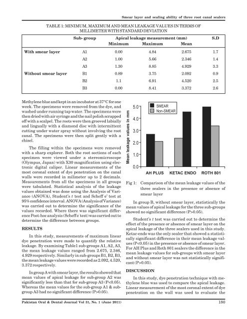
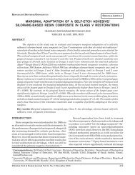
<strong>Smear</strong> <strong>layer</strong> <strong>and</strong> <strong>sealing</strong> <strong>ability</strong> <strong>of</strong> <strong>three</strong> <strong>root</strong> <strong>canal</strong> <strong>sealers</strong>TABLE 1: MINIMUM, MAXIMUM AND MEAN LEAKAGE VALUES IN TERMS OFMILLIMETER WITH STANDARD DEVIATIONSub- group Apical leakage measurement (mm) S.DMinimum Maximum MeanWith smear <strong>layer</strong> A1 0.00 4.84 2.675 1.7A2 1.00 5.66 2.346 1.4A3 1.30 8.85 4.929 3.3Without smear <strong>layer</strong> B1 0.89 3.75 2.092 0.9B2 1.1 6.91 4.520 2.5B3 0.00 8.41 3.372 2.6Methylene blue <strong>and</strong> kept in an incubator at 37°C for oneweek. The specimens were removed from the dye, <strong>and</strong>washed under running tap water. The specimens werethen dried with air syringe <strong>and</strong> the nail polish scrapped<strong>of</strong>f with a scalpel. The <strong>root</strong>s were then grooved labially<strong>and</strong> lingually with a diamond disc with intermittentcutting under water spray without involving the <strong>root</strong><strong>canal</strong>. The specimens were then split gently with achisel.The filling within the specimens were removedwith a sharp explorer. Both the <strong>root</strong> sections <strong>of</strong> eachspecimen were viewed under a stereomicroscope(Olympus, Japan) with X30 magnification using electronicdigital caliper. Linear measurements <strong>of</strong> themost coronal extent <strong>of</strong> dye penetration on the <strong>canal</strong>walls were recorded in milimeter up to 2 decimals.Measurements from all the specimens in all groupswere tabulated. Statistical analysis <strong>of</strong> the leakagevalues obtained was done using the Analysis <strong>of</strong> Variance(ANOVA), Student’s t test <strong>and</strong> Scheff e’ test at95% confidence interval. ANOVA (Analysis <strong>of</strong> Variance)was carried out to determine the significance <strong>of</strong> thevalues recorded. Where there was significant differencePost-hoc analysis (Scheff e ’ test) was carried out todetermine the difference between groups.RESULTSIn this study, measurements <strong>of</strong> maximum lineardye penetration were made to quantify the relativeleakage. By examining Table1 sub-groups A1, A2, A3,the mean leakage values ranged from 2.675, 2.346,4.929 respectively. Similarly in sub-groups B1, B2, B3,the mean leakage values were recorded as 2.092, 4.520,3.372 respectively.In group A with smear <strong>layer</strong>, the results showed thatmean values <strong>of</strong> apical leakage for sub-group A2 wassignificantly less than that for sub-group A3 (P0.05).Pakistan Oral & Dental Journal Vol 31, No. 1 (June 2011)Fig 1:Comparison <strong>of</strong> the mean leakage values <strong>of</strong> the<strong>three</strong> <strong>sealers</strong> in the presence or absence <strong>of</strong>smear <strong>layer</strong>In group B, without smear <strong>layer</strong>, statistically themean values <strong>of</strong> apical leakage for the <strong>three</strong> sub-groupsshowed no significant difference (P>0.05).Student’s t test was carried out to determine theeffect <strong>of</strong> the presence or absence <strong>of</strong> smear <strong>layer</strong> on theapical leakage <strong>of</strong> the <strong>three</strong> <strong>sealers</strong> used in this study.Ketac-endo was the only sealer that showed a statisticallysignificant difference in their mean leakage values(P0.05).DISCUSSIONIn this study, dye penetration technique with methyleneblue was used to compare the apical leakage.Linear measurement <strong>of</strong> the most coronal extent <strong>of</strong> dyepenetration on the wall was used to evaluate the180