June 2012 - Macquarie University Hospital
June 2012 - Macquarie University Hospital
June 2012 - Macquarie University Hospital

You also want an ePaper? Increase the reach of your titles
YUMPU automatically turns print PDFs into web optimized ePapers that Google loves.
<strong>June</strong> <strong>2012</strong><br />
1<br />
Click here<br />
for a<br />
virtual tour<br />
of our<br />
facility<br />
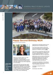
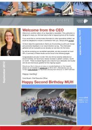
Happy Second Birthday MUH<br />
Welcome from the CEO, Carol Bryant<br />
Welcome to another edition of our GP<br />
e-newsletter. This month we are<br />
celebrating our second birthday. A lot has<br />
happened in these last two years and we<br />
have been grateful to you all for supporting<br />
us in this venture.<br />
The first half of the year has been spent<br />
talking with our stakeholders and asking<br />
them what they need from us to provide an<br />
exceptional service. We recently sent all<br />
our GPs a hospital survey, asking you what<br />
you thought of us and what you needed<br />
from us. We had an overwhelming<br />
response and I would like to thank you for<br />
taking the time to complete the survey.<br />
If you did not get a chance to fill out the<br />
survey and would like to provide feedback,<br />
please don’t hesitate to email me here.<br />
In the event you would like to find out more<br />
about us, I encourage you to attend one of<br />
our GP educational sessions, or visit our<br />
hospital website and view our virtual tours.<br />
Alternatively, you can contact our Business<br />
Development Director who will visit your<br />
practice with a <strong>Macquarie</strong> <strong>University</strong><br />
<strong>Hospital</strong> Specialist so you can find out<br />
more about what we can offer you.<br />
I would also be delighted to chat with you<br />
personally should you have any questions.<br />
I can be reached on 9812 3011.<br />
I do hope you enjoy this month’s<br />
newsletter.<br />
Carol Bryant, CEO, MUH.
GP NEWS<br />
2<br />
Congratulations Professor Rowe<br />
Order of Australia<br />
Professor Dominic Rowe, founder and director of<br />
<strong>Macquarie</strong> Neurology and MND Service at <strong>Macquarie</strong><br />
<strong>University</strong> was named a Member (AM) in the general<br />
division of the Order of Australia for his service to<br />
neurology and for his tireless work on Motor Neuron<br />
Disease.<br />
Motor Neuron Disease (MND) is a rapidly progressive,<br />
terminal neurological disease, with no known cure. An<br />
estimated 1400 people have MND in Australia and<br />
thousands more families and carers live daily with its<br />
effects.<br />
To serve people living with MND, Professor Dominic<br />
Rowe has set up a multi-disciplinary MND service at<br />
<strong>Macquarie</strong> <strong>University</strong>. The MND Service at <strong>Macquarie</strong><br />
<strong>University</strong> accepts referrals from all geographical areas,<br />
including rural and remote areas.<br />
"Studies show that coordinated care doubles the<br />
survival of people living with MND," Professor Rowe<br />
explains. "Our patient-centric model helps us, as<br />
clinicians, to provide this care and anticipate problems<br />
before they arise."<br />
Each person living with MND has unique difficulties with<br />
communication, mobility or speech. In a session at the<br />
clinic, they might see a broad range of allied health<br />
professionals – including speech and language<br />
therapists, respiratory physicians, dietitians, nurses,<br />
occupational therapists, physiotherapists, orthotic<br />
specialists, social workers and neurologists.<br />
Bringing these clinicians to the one place means less<br />
time traveling between medical offices, but Professor<br />
Rowe also sees a significant cost benefit in the service.<br />
For more<br />
information<br />
click here or<br />
to make an<br />
appointment<br />
please call<br />
02 9812 3720<br />
"Seeing all these clinicians normally wouldn't give you<br />
much change from $1000, however, because this is a<br />
clinic funded nearly entirely from donations a visit to<br />
see all ten practitioners costs only $100. After<br />
Medicare, the net cost to the patient is just over $30."<br />
Additionally, when living with a disease like MND,<br />
access to expert care is another huge relief, says<br />
Professor Rowe – especially with a disease that is<br />
relatively uncommon.<br />
"All of the staff in the clinic have experience in MND,<br />
whereas most GPs and community allied health<br />
practitioners will have perhaps one person with MND to<br />
manage," he explains. "The MND Service enables a<br />
concentration of experience to help the patient and their<br />
family."<br />
Based on a US model, the clinic was first implemented<br />
in Australia by Professor Rowe at Sydney's Royal North<br />
Shore <strong>Hospital</strong> in 1998. He recently moved his clinic to<br />
<strong>Macquarie</strong> <strong>University</strong>, where he could combine clinical<br />
practice with research as part of the Australian School<br />
of Advanced Medicine (ASAM).<br />
"Excellence in clinical service provision and research<br />
go hand in hand. Our patients with neurological<br />
diseases must have access to the best therapies and<br />
research programs available," says Professor Rowe.<br />
The clinic is funded through tax-deductible gifts, which<br />
can be donated through the <strong>Macquarie</strong> Foundation and<br />
ASAM. To find out more please click here.
<strong>June</strong> <strong>2012</strong><br />
3<br />
Musculoskeletal training could help millions<br />
Many Australians are suffering<br />
from pain related to treatable<br />
musculoskeletal conditions that are<br />
difficult for primary care doctors to<br />
diagnose without further<br />
postgraduate training.<br />
An RACGP survey estimates six<br />
million people across the country<br />
suffer from musculoskeletal<br />
problems, costing the health<br />
system and economy many<br />
millions of dollars every year. Many<br />
of these people have soft tissue<br />
pain sources that are not always<br />
identified, in their joints, muscles<br />
and ligaments.<br />
“Soft tissue pain is a great mimic,”<br />
says <strong>Macquarie</strong> <strong>University</strong>’s<br />
Associate Professor Michael<br />
Creswick. “These disorders can<br />
mimic many other health problems<br />
and yet are diagnosable and quite<br />
often very treatable if doctors are<br />
appropriately trained. Appropriate<br />
early intervention can minimise<br />
chronic pain and long term<br />
medication such as antiinflammatories<br />
and opioids.”<br />
Sometimes musculoskeletal pain<br />
can be so severe that the patient<br />
requires urgent or semi-urgent<br />
admission to MUH for pain relief<br />
and for rapid assessment.<br />
Availability of world class facilities<br />
including imaging is very helpful of<br />
course. These can be utilised to<br />
direct appropriate conservative<br />
care, including injection<br />
techniques, or surgical care when<br />
necessary.<br />
He and Associate Professor Rod<br />
Ayscough have helped establish<br />
the country’s only Master of<br />
Medicine Degree in<br />
Musculoskeletal Medicine at<br />
<strong>Macquarie</strong> <strong>University</strong>’s Australian<br />
School of Advanced Medicine.<br />
The course specifically trains<br />
qualified GPs to better identify and<br />
treat musculoskeletal disorders.<br />
Such training is growing in the<br />
United States and Europe as more<br />
is understood about<br />
musculoskeletal conditions.<br />
Creswick believes that doctors,<br />
after ruling out serious causes of<br />
pain, often refer patients to<br />
therapists for treatment of soft<br />
tissue pain without a specific<br />
diagnosis. This is because the<br />
source of pain is not always<br />
apparent from the application of a<br />
standard medical approach.<br />
“Our course participants are<br />
trained in recognising pain referral<br />
patterns, performing a detailed<br />
physical examination and applying<br />
advanced physical medicine<br />
treatment skills. These include<br />
gentle manual therapy, the<br />
injection of muscular “trigger<br />
points” if clinically indicated and<br />
specific exercise prescription.<br />
“There has been a worldwide<br />
medical trend towards treating pain<br />
as an entity in itself, and to<br />
diagnose persisting pain as<br />
intractable, due to nerve damage.<br />
However some people who have<br />
been told they have “neuropathic”<br />
pain have been found to have<br />
ongoing unrecognised but<br />
treatable musculoskeletal, soft<br />
tissue disorders, without nerve<br />
damage,” Creswick says.<br />
Ayscough and Creswick train GPs<br />
to look for possible sources of pain<br />
from soft tissues, including<br />
muscles, ligaments, the bony<br />
attachment points of muscles, as<br />
well as associated nerves. “How<br />
these structures interact in relation<br />
to each patient’s posture and<br />
movement is an important part of<br />
diagnosis and treatment.”<br />
“GPs already have an excellent<br />
overview of the whole patient and<br />
knowledge of underlying serious<br />
diseases. We use a medical model<br />
of diagnosis, so we only train<br />
experienced GPs. Often, with<br />
further training at ASAM, known<br />
pain patterns will be evident on<br />
examination, and our graduates<br />
can immediately treat a<br />
musculoskeletal problem.<br />
If the patient responds and stays<br />
pain-free, then they can avoid<br />
expensive and sometimes<br />
unnecessary tests. One of the<br />
great advantages of primary care<br />
is the ability of the doctor to treat<br />
patients and observe progress<br />
over short timeframes, and adjust<br />
management based on those<br />
observations.<br />
If a patient doesn’t respond as<br />
expected, a revised diagnosis may<br />
be apparent, or it may be<br />
appropriate to order radiological<br />
imaging or blood tests to aid<br />
diagnosis,” says Creswick.<br />
“Many patients have muscular and<br />
postural patterns that are leading<br />
to their pain, and that’s what we<br />
train our graduates to identify and<br />
treat.”<br />
A/Prof. Michael Creswick<br />
Musculoskeletal Medicine<br />
Special Interest: Muscular Pain &<br />
Dysfunction Including Cervicogenic<br />
Headache.<br />
To make a referral, please<br />
call 02 9481 9585. For<br />
more information, please<br />
click here
GP NEWS<br />
4<br />
Advances in Breast<br />
Cancer Surgery help<br />
patients cope much<br />
better with treatment<br />
Total skin sparing<br />
mastectomies<br />
As an experienced breast<br />
surgeon, I witness how breast<br />
surgery has evolved dramatically<br />
over the last few years. As the<br />
newer techniques of immediate<br />
breast reconstruction offer superior<br />
cosmetic results, it is now very<br />
uncommon that my patients would<br />
choose delayed breast<br />
reconstruction when deciding to<br />
have a mastectomy. Patients can<br />
choose to either have an<br />
autologous “flap” or tissue<br />
expander / implant for immediate<br />
reconstruction after we completed<br />
the total skin-sparing mastectomy.<br />
The patient wakes up with a<br />
reconstructed breast with minimal<br />
scarring in the recovery ward.<br />
My patients say they are so amazed<br />
that by being able to minimise<br />
scarring and to save all their breast<br />
skin including their nipple, the<br />
psychological impact of a<br />
mastectomy is much reduced. It<br />
allowed them to cope much better<br />
with their overall oncological<br />
treatment.<br />
Many studies have shown that<br />
immediate breast reconstruction in<br />
well-selected patients is safe and<br />
does not affect survival.<br />
As a mastectomy operation<br />
generally causes denervation of the<br />
chest wall, most patients have<br />
relatively minimal pain. Patients<br />
typically are discharged within 2-3<br />
days after their operation. They are<br />
fully ambulant and are doing most<br />
daily activities within 2-3 weeks.<br />
Therapeutic<br />
Mammaplasties<br />
For patients with breast cancers<br />
in tricky areas, and patients with<br />
large breasts, we individualise<br />
our technique to resize and<br />
reshape the breast after removal<br />
of the tumour and lymph nodes<br />
staging.<br />
Particularly for patients with very<br />
large breasts, radiotherapy may<br />
cause severe and permanent<br />
fibrosis. They will be left with a<br />
Dr Deborah Cheung<br />
Breast Surgeon<br />
Clinical Care Centre,<br />
Suite 302<br />
<strong>Macquarie</strong> <strong>University</strong> Clinic<br />
To make a referral please<br />
call 02 9887 8899.<br />
hard and fibrosed breast for the<br />
rest of their lives. The results of<br />
breast reduction surgery after<br />
radiotherapy are unpredictable<br />
and have a much higher<br />
complication rate. Hence, it is<br />
logical to reduce both breasts<br />
before radiotherapy than<br />
afterwards.<br />
This procedure is medicare and<br />
health fund rebatable and is<br />
performed at the same time as<br />
breast cancer surgery. Patients<br />
have a high satisfaction rate.<br />
Most patients tell me how much<br />
better they feel and how little<br />
discomfort they have after the<br />
operation.
Short Scar Breast Reduction<br />
By Dr Steve Merten<br />
In the last 15 years there have been<br />
significant advances in breast reduction<br />
techniques, with major advantages to our<br />
patients. The “traditional” breast<br />
reduction technique, popular since the<br />
1960s, was an effective and safe<br />
operation, however had drawbacks of<br />
frequent healing problems, due to tight<br />
skin closure, in the short term and, being<br />
bottom-heavy with potential for<br />
“bottoming out” in the long term.<br />
Fundamental shifts in the design<br />
concepts of breast reductions have led to<br />
major changes, with the development of<br />
the “short scar” breast reduction, also<br />
termed “vertical” breast reduction or<br />
“lollipop” breast reduction, as the final<br />
scar pattern is around the circular<br />
areolar, then vertically down to the fold<br />
beneath the breast.<br />
This is in contrast to the traditional<br />
“anchor” shaped scar, which has, in<br />
addition, a long scar in the fold beneath<br />
the breast. Short scar breast reduction<br />
differs from the traditional technique in<br />
that the internal blood supply to the<br />
Dr. Rami Sorial (Orthopaedic Surgery),<br />
A/Prof Des. Bokor ( Orthopaedic<br />
Surgery), A/Prof Glen Croxson,(OHNS)<br />
Retiring senior Examiner, A/Prof Ray<br />
Sacks, (OHNS) Senior Examiner, A/Prof.<br />
Graham Gumley (Orthopaedic Surgery)<br />
Senior Examiner.<br />
5<br />
nipple is based high in the breast and the<br />
heavy tissue from the lower breast is<br />
completely removed. The breast is then<br />
reduced and reconfigured in a conical<br />
shape, then sewn so that its new<br />
construction, and not the skin<br />
envelope, maintains the shape of the<br />
breast.<br />
This allows less skin to be removed<br />
and elimination of the scar beneath the<br />
breast. As well as less scars, however,<br />
the even greater benefits are a<br />
shapelier breast and better long-term<br />
maintenance of the result.<br />
RACS PART 2<br />
Surgical fellowship examination<br />
At the recent RACS Part 2<br />
Surgical Fellowship examination in<br />
Brisbane, it was clear that<br />
<strong>Macquarie</strong> <strong>University</strong> had a strong<br />
representation on the Court of<br />
Examiners. The Examiners are<br />
responsible for the development,<br />
setting and evaluation at the end<br />
of training examinations in their<br />
fields. Examiners serve for 8 years<br />
on the Court and take up to 10<br />
days of unpaid time to serve the<br />
profession and community in this<br />
way, upholding the professional<br />
standards of surgery.<br />
The short scar breast reduction is best<br />
suited for moderate sized breast<br />
reductions, however by combining the<br />
internal reconstructive concepts with a<br />
scar beneath the breast, even the largest<br />
breasts can be successfully reduced with<br />
the shape advantages of this technique.<br />
Dr Merten was one of the first surgeons<br />
in Australia to be trained and<br />
experienced in the short scar breast<br />
reduction, during his additional training in<br />
Canada in 2002, and has been<br />
exclusively using these techniques since<br />
that time.<br />
Dr Merten has been training the next<br />
generation of plastic surgeons these<br />
techniques for the last 10 years.<br />
Click here to see before and<br />
after photos<br />
To make a referral to Dr Steve<br />
Merten, please call 02 9812<br />
3890 or click here for more<br />
information.<br />
RACS Annual Scientific Congress<br />
On 6 - 10 May this year, the RACS<br />
Annual Scientific Congress was held in<br />
Kuala Lumpur. A number of MUH<br />
specialists were chairing and presenting<br />
seminars at this activity including;<br />
Professor Marcus Stoodley<br />
Professor John Cartmill<br />
Dr Michael Vallely<br />
Dr Paul Bannon<br />
Dr Michael Wilson<br />
Dr Jonathan Clark<br />
A/Prof Reginald Lord<br />
A/Prof Greg Falk<br />
Dr Matthew Rickard<br />
Dr Carsten Palme<br />
Dr Michael Stephen<br />
Dr John McGuinness
<strong>June</strong> <strong>2012</strong><br />
6<br />
Breast Program<br />
Our Breast Cancer Program is well<br />
underway with multi-disciplinary team<br />
meetings occurring every fortnight. In<br />
the first two meetings in <strong>June</strong>, the<br />
team has discussed 15 new cases,<br />
exploring the best available treatment<br />
options for each patient, as well as<br />
developing a planned approach to<br />
their care. Doctors in attendance<br />
included surgeons Dr Deborah<br />
Cheung, Dr Elisabeth Elder, Dr<br />
Jeremy Hsu, with input from radiation<br />
oncologists Professor John Boyages,<br />
Associate Professor Susan<br />
Pendlebury, Dr Annie Ho and medical<br />
oncologist Dr Pirooz Poursoltan and<br />
plastic surgeon Dr Thomas Lam.<br />
Pathologist, Dr Jessamine Reddy<br />
from Douglass Hanly Moir and a<br />
consultant from <strong>Macquarie</strong> Medical<br />
Imaging are also in attendance,<br />
ensuring all the necessary experts<br />
are available in one room, to offer a<br />
truly coordinated approach to cancer<br />
treatment.<br />
Professor John Boyages, Director of<br />
the <strong>Macquarie</strong> <strong>University</strong> Cancer<br />
Institute (MCI) and Professor of<br />
Breast Oncology at the Australian<br />
School of Advanced Medicine<br />
(ASAM) is delighted with what has<br />
been achieved over the last few<br />
months. “It’s a very exciting time for<br />
us, as the breast cancer program<br />
grows from strength to strength.”<br />
Breast Assessment Clinic<br />
A daily breast assessment clinic is<br />
opening in Suite 302 at the<br />
<strong>Macquarie</strong> <strong>University</strong> Clinic.<br />
Appointments to this service can be<br />
made on 9887 8887.<br />
For all appointments and<br />
enquiries, please<br />
telephone 02 9887 8899<br />
Professor John Boyages, Megan Southwell<br />
Lymphoedema Treatments<br />
at MCI<br />
A suite of lymphoedema treatments<br />
are now available through the<br />
<strong>Macquarie</strong> <strong>University</strong> Cancer Institute<br />
(MCI), from early detection and<br />
intervention methods to new surgical<br />
options, including liposuction.<br />
Last month, high school teacher,<br />
Megan Southwell was the first patient<br />
at MUH to be treated for<br />
lymphoedema with liposuction.<br />
The operation was a resounding<br />
success and was covered by channel<br />
10 news. To view the program click<br />
here.<br />
Clinical Psychologist<br />
joins MCI<br />
Dr Jemma Gilchrist, a leading<br />
Clinical Psychologist has joined the<br />
team at the <strong>Macquarie</strong> <strong>University</strong><br />
Cancer Institute. She is a key<br />
addition to our multi-disciplinary<br />
team of experts, as we recognise<br />
the importance of psycho-social<br />
and supportive care in providing<br />
comprehensive cancer<br />
management.<br />
Dr Gilchrist has particular expertise<br />
in oncology. Common concerns<br />
<strong>Macquarie</strong><br />
<strong>University</strong> Cancer<br />
Institute<br />
Hotline<br />
02 9887 8887<br />
The team of cancer experts involved<br />
in her treatment including:<br />
Dr Thomas Lam<br />
Cosmetic & Plastic Surgery<br />
Dr Quan Ngo<br />
Cosmetic & Plastic Surgery<br />
Dr Helen Mackie<br />
Rehabilitation Medicine<br />
Louise Koelmeyer<br />
Occupational Therapy<br />
Asha Heydon-White<br />
Physiotherapist<br />
For further information about these<br />
options in lymphoedema treatment,<br />
please contact the MCI Cancer<br />
Hotline (during business hours) on<br />
9887 8887.<br />
that she assists with are:<br />
adjustment to diagnosis, treatment,<br />
chronic disease or disability;<br />
managing stress and anxiety;<br />
sadness and depression; sleep<br />
difficulty; body image and sexuality;<br />
communication and decisionmaking;<br />
grief and loss; relationships<br />
and living with uncertainty.<br />
Dr. Gilchrist holds fortnightly clinics<br />
on Wednesday at the Clinical Care<br />
Centre. Patients may be eligible for<br />
a rebate from Medicare if referred<br />
by their GP.
7<br />
Ear Drum Surgery<br />
Clinical Associate Professor<br />
Nirmal Patel<br />
<strong>Macquarie</strong> <strong>University</strong><br />
Definition & Natural History<br />
Tympanic membrane perforations<br />
are holes in the ear drum that most<br />
commonly occur as a consequence<br />
of either ear infections or trauma to<br />
the ear.<br />
Tympanic Membrane Anatomy<br />
The pars flaccida region of the<br />
tympanic membrane is that region<br />
above the short process of the<br />
malleus (Figure 1). Typically this<br />
area is called the attic region and is<br />
clean and smooth with a few blood<br />
vessels noted.<br />
If this region is not smooth and free<br />
of wax/ debris and in particular if a<br />
perforation can be seen in the attic,<br />
then a cholesteatoma may be<br />
suspected. The annulus of the ear<br />
drum delineates the margin. A<br />
marginal perforation is a sign of an<br />
unsafe ear.<br />
Box Out – Marginal and Attic<br />
Perforations (The Unsafe Ear)<br />
When either a marginal or attic<br />
perforation is seen, this is an unsafe<br />
situation. A marginal perforation is<br />
one where the hole reaches the<br />
annulus of the ear drum (Figures<br />
2&3). An attic perforation is a hole<br />
above the short process of the<br />
malleus.<br />
Both of these situations can disturb<br />
the normal flow of epithelium from<br />
the tympanic membrane out of the<br />
external auditory canal and may be a<br />
sign of cholesteatoma (epithelial<br />
inclusion cyst of the temporal bone -<br />
see previous therapy update article),<br />
which often requires surgical<br />
intervention.<br />
Red flags in the Diagnosis of<br />
Tympanic Membrane<br />
Perforations<br />
• Marginal perforations<br />
• Attic perforations<br />
• Chronically discharging or bleeding<br />
ear.<br />
• Perforation with significant clinical<br />
hearing loss<br />
For more information on<br />
Clinical Associate Professor<br />
Nirmal Patel click here or to<br />
make an appointment please<br />
call<br />
Management<br />
Most acute perforations from<br />
infection will spontaneously heal with<br />
culture directed antibiotic treatment<br />
of the ear infection. The majority of<br />
acute traumatic perforations will also<br />
heal spontaneously. If the middle ear<br />
is soiled then antibiotics (such as<br />
amoxicillin) are used for a week.<br />
The patient should maintain strict<br />
water precautions and be regularly<br />
checked until the hole heals,<br />
usually in a few weeks.<br />
Occasionally the acute perforations<br />
will not heal and then they are<br />
defined as chronic.<br />
If a patient is asymptomatic then<br />
most dry safe central perforations<br />
are left alone.<br />
Dry safe chronic perforations are<br />
managed surgically if the patient is<br />
symptomatic, in the form of either<br />
discharge from the ear or hearing<br />
loss.<br />
Ear Drum Surgery requires a<br />
tympanoplasty (repair of the ear<br />
drum and/ or ear bones). Two<br />
newer techniques that we employ<br />
are the onlay technique and<br />
cartilage tympanoplasty. Onlay<br />
Tympanoplasty is a technique we<br />
employ for large holes or anterior<br />
ear drum holes<br />
(Figure 4).<br />
Cartilage tympanoplasty is used<br />
when the middle ear pressure is high<br />
causing retraction of the residual ear<br />
drum. At <strong>Macquarie</strong> <strong>University</strong><br />
<strong>Hospital</strong> we also use the latest<br />
techniques of employing High<br />
Definition (HD) camera endoscopy<br />
to perform minimally invasive ear<br />
surgery to try and reduce surgical<br />
trauma and pain as well as<br />
improve healing times.<br />
Acknowledgements:<br />
Dr John Kelly for two of the<br />
otoscopic pictures.<br />
For more surgical videos of ear drum<br />
hole repair, visit Youtube and search<br />
“Dr Nirmal Patel” or<br />
scan the following QR<br />
code into your<br />
smartphone.<br />
Figure 1 Otoscopic view of a normal<br />
left ear – the oblique line illustrates the<br />
demarcation between pars flaccida<br />
(superior) and pars tensa (below). The<br />
solid white arrow begins on the short<br />
process of the malleus and extends up<br />
to show a normal non-diseased attic<br />
region.<br />
Figure 1 Otoscopic view of a normal left<br />
ear – the oblique line illustrates the<br />
Figure 2 Otoscopic view of a left ear<br />
with a margin (unsafe) perforation –<br />
note the perforation extending to the<br />
annulus of the tympanic membrane and<br />
also inferiorly, the white squamous<br />
epithelium migrating into the middle ear.<br />
Figure 3 Otoscopic view of a left ear<br />
with a marginal unsafe perforation –<br />
if untreated this ear may develop into<br />
cholesteatoma.<br />
Figure 4 Otoscopic View of a Left Ear<br />
with a Central Perforation – the ear is<br />
inflamed and moist.