
You also want an ePaper? Increase the reach of your titles
YUMPU automatically turns print PDFs into web optimized ePapers that Google loves.
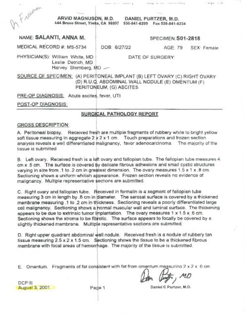
-•ARVID MAGNUSON M.D. DANIEL PURTZER, M.D.444 Bruce Street, Yrel a, CA 96097 530-841-6209 Fax 530-841-6234NAME: SALANTI, ANNA M.MEDICAL RECORD #: MS-5734SPECIMEN:S01-2818DOB: 6/27/22 AGE: 79 SEX' FemalePHYSICIAN(S): William White, MDLeslie Detrich, MDHarvey Sternberg, I\|1DDATE OF SURGERY:SOURCE OF SPECIMEN: (A) PERI ONEAL IMPLANT (B) LEFT OVARY (C) RIGHT OVARY(D) R.U.C . ABDOMINAL WALL NODULE (E) OMENTUM (F)PERITONEUM, (G) ASCITESPRE-OP DIAGNOSIS. Acute ascites!, fever, UTIP05T-OP DIAGNOSIS.GROSS DESCRIPTION:SURGICAL PATHOLOGY REPORTA. Peritoneal biopsy. Received freih are multiple fragments of rubbery white to bright yellowsoft tissue measuring in aggregate 2 < 2 x 1 cm Touch preparations and frozen sectionanalysis reveals a well differentiated nalignancy, favor adenocarcinoma The majority of thetissue is submittedB. Left ovary. Received fresh is a lejft ovary and fallopian tube. The fallopian tube measures 4cm x .5 cm. The surface is covered tiy delicate fibrous adhesions and small cystic structuresvarying in size from .1 to .2 cm in greatest dimension. The ovary measures 1.5x1 x .8 cm.Sectioning shows a uniform whitish amalignancy. Multiple representative spearance. Frozen section reveals no evidence ofections are submitted.C. Right ovary and fallopian tube. R eceived in formalin is a segment of fallopian tubemeasuring 3 cm in length by ,8 cm in diameter. The serosal surface is covered by a thickenedmembrane measuring .1 to .2 cm in tr ickness. Sectioning reveals a poorly differentiated largecell malignancy. Sectioning shows a normal muscular wall and luminal surface. The thickeningappears to be due to extrinsic tumor i nplantation. The ovary measures 1 x 1.5 x .6 cm.Sectioning shows the stroma to be fibfatic The surface appears to focally be covered by aslightly thickened membrane. Multip e representative sections are submitted.D. Right upper quadrant abdominal vail nodule. Received fresh is a nodule of rubbery tantissue measuring 2.5 x 2 x 1.5 cm. ectioning shows the tissue to be a thickened fibrousmembrane with focal areas of hemorrhage The majority of the tissue is submitted.E. Omentum. Fragments of fat consistent with fat from omentum measurino 2 x 2 x 6cmDCPrlllAugust 3, 2001 Page 1 Daniel C Purtzer, M.D.
ARVID MAGNUSdN, M.D.444 Bruce Street, Yre|ca, CA 96097DANIEL PURTZER, M.D.530-841-6209 Fax 530-841 -6234NAME: SALANTI, ANNA M.SPECIMEN.S01-2818MEDICAL RE'CORD #: MS-5734 DOB: 6/27/22 AGE: 79 SEX: FemaleE. Omentum. Fragments of fat conisistent with fat from omentum measuring 2 x 2 x 6 cm.The external surface is covered by a thick hemorrhagic membrane. Sectioned, fixed, andrepresentative sections are submittese described above.E. On the serosal surface of the fat, there are aggregates of tumor cells with characteristicssimilar to those described above. The tumor nodules range from 0.1 to 0.3 cm in greatestdimension.F. On the serosal surface of the fat, there are multiple small tumor nodules with characteristicssimilar to those described above.G. There are multiple clusters and individual large and malignant cells with characteristicssimilar to those described above.DIAGNOSIS:A. Peritoneal implant: Poorly differentiated large cell malignancy.DCP:lliAugust 3, 2001 Pade2 Daniel C Purtzer, M.D.
ARVID MAGNUS 3N, M.D. DANIEL PURTZER, M.D.444 Bruce Street, Yre|(a, CA 96097 530-841-6209 Fax 530-841-6234NAME: SALANTI, ANNA M.MEDICAL RECORD #: MS-5734B. Left ovary:1) Corpus albicans.2) Endosalpingiosis, benign.DOB: 6/27/22SPECIMEN.S01-2818AGE: 79SEX: FemaleC. Right ovary:1) Corpus albicans.2) Endosalpingiosis, benign.3) Serosal surface implant of a poorjly differentiated large cell malignancy.D. Right upper quadrant abdominal v/ail nodule: Poorly differentiated large cell malignancy.E. Omentum: Poorly differentiated \iF. Peritoneum: Poorly differentiatedG. Peritoneal fluid: Positive for largerge cell malignancy,arge cell malignancy.cell malignancy.COMMENT: This large cell malignancy most likely represents malignant mesothelioma. Onecannot completely exclude the possib lity of metastatic adenocarcinoma. However, this tumorexhibits characteristics that are unusual for most adenocarcinomas that metastasize to theabdominal cavity. The most striking 1nding is the multiple appearances of the tumor, thevariation in size and shape of the turrior cells, as well as the finding of giant tumor cells. Thereare additional special studies which can be performed on the tumor which may help furtherdelineate the etiology of this maligna cy. Consultation with an oncologist is recommended.Once an oncologist has been selected, the slides can be forwarded to an adjacent pathology labthat can perform these stains. The si des will be reviewed by Dr. Arvid Magnuson.DCP:lljAugust 3, 2001Pae3Daniel C Purtzer, M.D.