electronic reprint Crystal structure and charge distribution of ... - CRM2
electronic reprint Crystal structure and charge distribution of ... - CRM2
electronic reprint Crystal structure and charge distribution of ... - CRM2

Create successful ePaper yourself
Turn your PDF publications into a flip-book with our unique Google optimized e-Paper software.
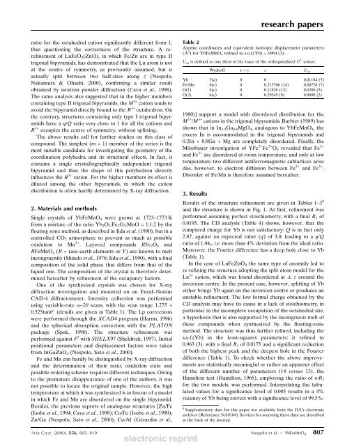
esearch papersratio for the octahedral cation signi®cantly different from 1,thus questioning the correctness <strong>of</strong> the <strong>structure</strong>. A rere®nement<strong>of</strong> LuFeO 3 (ZnO), in which Fe/Zn are in type IItrigonal bipyramids, has demonstrated that the Lu atom is notat the centre <strong>of</strong> symmetry, as previously assumed, but isactually split between two half-sites along z (Nespolo,Nakamura & Ohashi, 2000), con®rming a similar resultobtained by neutron powder diffraction (Cava et al., 1998).The same analysis also suggested that in the higher memberscontaining type II trigonal bipyramids, the M 3+ cation tends toavoid the bipyramid directly bound to the R 3+ octahedron. Onthe contrary, <strong>structure</strong>s containing only type I trigonal bipyramidshave a q/Q ratio very close to 1 for all the cations <strong>and</strong>R 3+ occupies the centre <strong>of</strong> symmetry, without splitting.The above results call for further studies on this class <strong>of</strong>compound. The simplest (m = 1) member <strong>of</strong> the series is themost suitable c<strong>and</strong>idate for investigating the geometry <strong>of</strong> thecoordination polyhedra <strong>and</strong> its structural effects. In fact, itcontains a single crystallographically independent trigonalbipyramid <strong>and</strong> thus the shape <strong>of</strong> this polyhedron directlyin¯uences the R 3+ cation. For the higher members its effect isdiluted among the other bipyramids, in which the cation<strong>distribution</strong> is <strong>of</strong>ten hardly determined by X-ray diffraction.2. Materials <strong>and</strong> methodsSingle crystals <strong>of</strong> YbFeMnO 4 were grown at 1723±1773 Kfrom a mixture <strong>of</strong> the ratio Yb 2 O 3 :Fe 2 O 3 :MnO = 1:1:2 by the¯oating zone method, as described in Iida et al. (1990), but in acontrolled CO 2 atmosphere to prevent as much as possibleoxidation to Mn 3+ . Layered compounds RFe 2 O 4 <strong>and</strong>RFeMnO 4 (R = rare-earth elements or Y) are known to meltincongruently (Shindo et al., 1976; Iida et al., 1990), with a ®nalcomposition <strong>of</strong> the solid phase that differs from that <strong>of</strong> theliquid one. The composition <strong>of</strong> the crystal is therefore determinedhereafter by re®nement <strong>of</strong> the occupancy factors.One <strong>of</strong> the synthesized crystals was chosen for X-raydiffraction investigation <strong>and</strong> mounted on an Enraf±NoniusCAD-4 diffractometer. Intensity collection was performedusing variable-rate !±2 scans, with the scan range 1.275 +0.525tan (details are given in Table 1). The Lp correctionswere performed through the XCAD4 program (Harms, 1996)<strong>and</strong> the spherical absorption correction with the PLATONpackage (Spek, 1990). The <strong>structure</strong> re®nement wasperformed against F 2 with SHELX97 (Sheldrick, 1997). Initialpositional parameters <strong>and</strong> displacement factors were takenfrom InGaZnO 4 (Nespolo, Sato et al., 2000).Fe <strong>and</strong> Mn can hardly be distinguished by X-ray diffraction<strong>and</strong> the determination <strong>of</strong> their ratio, oxidation state <strong>and</strong>possible ordering scheme requires different techniques. Owingto the premature disappearance <strong>of</strong> one <strong>of</strong> the authors, it wasnot possible to locate the original sample. However, the hightemperature at which it was synthesized is in favour <strong>of</strong> a modelin which Fe <strong>and</strong> Mn are disordered on the single bipyramid.Besides, the previous reports <strong>of</strong> analogous <strong>structure</strong>s [Zn/Fe(Isobe et al., 1994; Cava et al., 1998); Co/Fe (Isobe et al., 1990);Zn/Ga (Nespolo, Sato et al., 2000); Cu/Al (GeÂrardin et al.,Table 2Atomic coordinates <strong>and</strong> equivalent isotropic displacement parameters(A Ê 2 ) for YbFeMnO 4 re®ned to s.o.f.(Yb) = 0964 (3).U eq is de®ned as one third <strong>of</strong> the trace <strong>of</strong> the orthogonalized U C tensor.Wyck<strong>of</strong>f x = y z U eqYb 3(a) 0 0 0.01184 (5)Fe/Mn 6(c) 0 0.215706 (14) 0.00728 (7)O(1) 6(c) 0 0.12826 (13) 0.0208 (5)O(2) 6(c) 0 0.29345 (8) 0.0098 (2)1980)] support a model with disordered <strong>distribution</strong> for theM 2+ /M 3+ cations in the trigonal bipyramids. Barbier (1989) hasshown that in In 1.2 Ga 0.8 MgO 4 , analogous to YbFeMnO 4 , theexcess In is accommodated in the trigonal bipyramids <strong>and</strong>0.2In + 0.8Ga + Mg are completely disordered. Finally, theMoÈ ssbauer investigation <strong>of</strong> YFe 2+ Fe 3+ O 4 revealed that Fe 2+<strong>and</strong> Fe 3+ are disordered at room temperature, <strong>and</strong> only at lowtemperature two different antiferromagnetic sublattices arisedue, however, to electron diffusion between Fe 2+ <strong>and</strong> Fe 3+ .Disorder <strong>of</strong> Fe/Mn is therefore assumed hereafter.3. ResultsResults <strong>of</strong> the <strong>structure</strong> re®nement are given in Tables 1±3 1<strong>and</strong> the <strong>structure</strong> is shown in Fig. 1. At ®rst, re®nement wasperformed assuming perfect stoichiometry, with a ®nal R 1 <strong>of</strong>0.0195. The CD analysis (Table 4) shows, however, that thecomputed <strong>charge</strong> for Yb is not satisfactory: Q is in fact only2.87, against an expected value (q) <strong>of</strong> 3.0, leading to a q/Qratio <strong>of</strong> 1.04 5 , i.e. more than 4% deviation from the ideal ratio.Moreover, the Fourier difference has a deep hole close to Yb(Table 1).In the case <strong>of</strong> LuFeZnO 4 the same type <strong>of</strong> anomaly led tore-re®ning the <strong>structure</strong> adopting the split atom model for theLu 3+ cation, which was found disordered at z around theinversion centre. In the present case, however, splitting <strong>of</strong> Ybeither brings Yb again on the inversion centre or produces anunstable re®nement. The low formal <strong>charge</strong> obtained by theCD analysis may have its cause in a lack <strong>of</strong> stoichiometry, inparticular in the incomplete occupation <strong>of</strong> the octahedral site,a hypothesis that is also supported by the incongruent melt <strong>of</strong>these compounds when synthesized by the ¯oating-zonemethod. The <strong>structure</strong> was thus further re®ned, including thes.o.f.(Yb) in the least-squares parameters: it re®ned to0.963 (3), with a ®nal R 1 <strong>of</strong> 0.0175 <strong>and</strong> a signi®cant reduction<strong>of</strong> both the highest peak <strong>and</strong> the deepest hole in the Fourierdifference (Table 1). To check whether the above improvementsare statistically meaningful or rather an apparent effect<strong>of</strong> the different number <strong>of</strong> parameters (14 versus 13), theHamilton test (Hamilton, 1965), employing the ratio <strong>of</strong> wR 2for the two models, was performed. Interpolating the tabulatedvalues for a signi®cance level <strong>of</strong> 0.005 results in a 4%vacancy <strong>of</strong> Yb being correct with a signi®cance level <strong>of</strong> 99.5%.1 Supplementary data for this paper are available from the IUCr <strong>electronic</strong>archives (Reference: NA0104). Services for accessing these data are describedat the back <strong>of</strong> the journal.Acta Cryst. (2000). B56, 805±810 Nespolo et al. YbFeMnO 4 807<strong>electronic</strong> <strong>reprint</strong>