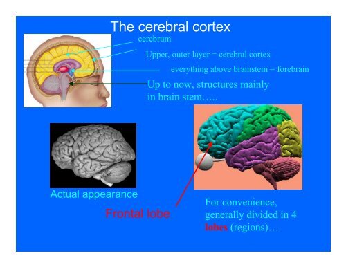
The cerebral cortex
The cerebral cortex
The cerebral cortex
- No tags were found...

Create successful ePaper yourself
Turn your PDF publications into a flip-book with our unique Google optimized e-Paper software.
Inhalt0. Ein Hintergrund, eine Figur, eine Gestalt Seite 21. Anliegen Seite 32. Danke Seite 43. Zur Entstehung der Idee der Geschwister Camps Seite 54. Vom Hintergrund zur Figur zu Figur-Hintergrund Seite 65. Feldtheoretisches Seite 136. Konzept der Geschwister Camps Seite 196.1. Zielgruppe Seite 196.2. Auftrag Seite 206.3. Gruppengröße Seite 226.4. Leitung Seite 226.5. Dauer und Design Seite 236.6. Medien Seite 246.7. Orte und Plätze Seite 276.8. Ablauf eines Geschwister Camps Seite 286.9. Jonas Seite 337. Zusammenfassung Seite 378. Und auch für mich - oder von der Figur in den Hintergrund Seite 399. Literaturverzeichnis Seite 401
• Appear to be 2 regions important forjudgment and decision-makingSocial-personal: requireemotional inputAbstract orimpersonal decisionsJudgment in this realm can remainlargely intact even thoughjudgment in social-emotional realmdisturbed
Motor <strong>cortex</strong>Other functions of frontal lobe• Controls muscles of body• Each part of the body controlled by specific region ofmotor <strong>cortex</strong> (as in lower-left drawing)
Primary motor <strong>cortex</strong> was first “mapped” by Dr.Wilder Penfieldusing technique of electrical stimulationWilder Penfield, M.D., MontrealNeurological Institute
Two basic principles:1) Amount of <strong>cortex</strong> devoted to each part of bodyproportional to amount of information-processingrequired•In this case, number of muscles to becontrolled
2) Connections between brain and body arecontralateralopposite side(Implications for stroke:What if patientparalyzed on right-handside of body?)
• Another function of frontal lobe: One of thebrain’s 2 language centers located here =Broca’s Area (discuss discovery by Broca)ote: Almost always in left hemisphere• Damage can lead to Broca’s Aphasia• Problems with production (e.g., speaking) butgenerally few problems with comprehension[ad lib example]
• Additional functions of frontal lobes:• Short-term memory• Reasoning• Regulation of emotions (e.g., inhibition offear responses)
Parietal lobe• Major function is “body sense” perception• Occurs in region = somatosensory <strong>cortex</strong>first mapped by Penfield using electrical stimulation
• Receives input from receptors in skin, joints, internal organs• responsive to hot, cold, pressure, touch, pain, etc• Same 2 principles apply as for motor <strong>cortex</strong>1) Amount of <strong>cortex</strong> devoted to part of body depends onamount of info-processing required• in this case, number of sense receptors; sensitivity2) Connections to body are contralateral
• Effects of damage• Loss of sensitivity (e.g. to pain)• Loss of ability to “feel” body part
Occipital lobe• Arrives first in primary visual <strong>cortex</strong>• Preliminary processing done (e.g., contours,boundaries, rough shape)• But won’t make much sense until furtheranalysis and processing done• in surrounding areas (visual association areas)• Required to assess size, motion, identity, etc.
PET scan, S performing visualtask:much of the visual association<strong>cortex</strong> is active• Destruction of primary visual <strong>cortex</strong> >>blindness• Over 30 specialized regions in association<strong>cortex</strong>• Damage can lead to various forms of visualagnosias (loss of recognition)• E.g., patient who had great difficultyrecognizing crayon, lock
• Extreme form of agnosia = Neglect Syndrome[Discuss: Eating food on plate;shaving]
Temporal lobePrimary function:processing of auditoryinformation• Similar to situation in occipital lobe• Information first arrives in primary auditory <strong>cortex</strong>• initial processing• Sent on for more complex processingto surrounding auditory associationareas
• Another important speech center (located intemporal lobe) = Wernicke’s Area• Damage leads to Wernicke’s aphasia• problems with both production andcomprehension of speech (and writing)• [speech sample here]Note from Dec ’06 while reading Intro papers- they think speech could be replaced by written lang or signing. Need to verifythen add info on effects on written language and signing. <strong>The</strong> issue is language processing – not speech per se
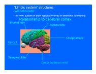
Subcortical forebrain structures• Entire area lying above brainstem is forebrain• By far, largest structure is cerebrum• We have been focusing on most importantpart, outer layer = <strong>cerebral</strong> <strong>cortex</strong>
fMRI showing highest concentration mu-opiod receptors- Zubieta nicotine study• But several important forebrain structures liebeneath <strong>cortex</strong> (< subcortical)• A number of them involved in emotional andmotivational functioning (“limbic system”)
Amygdala• Small structure, plays very importantrole in emotional functioning• Especially fear• Amygdala responds to signs of danger,threat
Amygdala response to fear stimulus3 views of brain, normal S,viewing fearful face
Amygdala-damaged Ss have difficultyrecognizing facial expression of fear“On a scale of 1 to6, how frightenedwould you say thisperson is?”- Normal S: “ 5 or 6”- Amygdala damagedS: “I don’t know,maybe 2?”
• Amgydala also crucial for our ability to quicklylearn to fear dangerous situations (through“classical conditioning”(explain fear conditioningprocedure); explain whatgraph is showing fornormal STest results for S.P.(ormal response to electric shock, but no fearreaction to what should have become a “ConditionedStimulus” (blue square associated with shock))
Other important subcortical structuresHippocampus• Often considered part of “limbic system”• Although, main role in memory• Recently discovered: plays important role inregulating stress response• Can be damaged by protracted stress
Hypothalamus• Regulation of “drives” (e.g., hunger, sex)Lateral Nucleus (Appetetive Centerhunger,thirst)VentroMedial Nucleus (Satiation ofhunger; also involved in sexual arousal)
How is the brain “mapped”?• 1) Study effects of damage to specificregions or structures• E.g. Phineas Gage• E.g., Broca’s discovery of Broca’s Area• 2) Electrical stimulation of brain (ESB)• E.g., Hess’s original work• E.g., Olds & Milner’s discovery of “pleasurecenters”• E.g., Wilder Penfield’s mapping ofsomatosensory & motor <strong>cortex</strong>
3) Brain scanning (imaging)• 2 typesA) Structural scans (CT and MRI)• Give hi resolution 3D images of bodystructures, including brain• of some limited use to psychologists (e.g.studies of schizophrenics)
far more useful• B) Functional scans (functional imaging)• PET scans and fMRI scans (functionalmagnetic resonance imaging)• Indicate which regions of brain most activeunder particular circumstances• Colors represent different levels of activityPETscan
• Imaging studies allow researchers to answerquestions previously unanswerable; e.g.,fMRI scan from Singer et al(2004) expt on empathy forpain