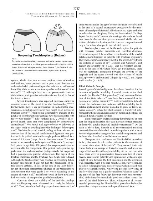
NUMBERD2756dTHE J OURNAL OF B ONE &JOINT S URGERY JBJS. ORGd dVOLUME 90-A 12 ECEMBER 2008PATELLAR I NSTABILITYflexion 54,59,60 , instead of at the lower flexion angles (30° to 45°)that have been recommended by other authors 53,57 , has beenadvocated to avoid overtightening of the graft <strong>and</strong> to ensurethat the patella has engaged the trochlea. LeGr<strong>and</strong> et al. recommendedapplying tension at 45° to 60° of flexion <strong>and</strong> alsochecking that there is symmetric medial <strong>and</strong> lateral translationof the patella at 20° of flexion 64 . Farr <strong>and</strong> Schepsis described an‘‘anatomometric’’ placement of the graft: tensioning the graftwith the knee in 30° of flexion so that it becomes more lax withfurther flexion <strong>and</strong> tighter in terminal extension 57 .A dynamic reconstruction of the medial patellofemoralligament has been proposed as an alternative that is better than astatic reconstruction. Ostermeier et al. performed a dynamicreconstruction by transferring the distal end of the semitendinosusbehind the proximal aspect of the medial collateral ligamentto the medial margin of the patella 53 . <strong>The</strong> authors foundthat a static reconstruction medialized the patella significantlymore than the dynamic reconstruction did (p < 0.01). Thus, adynamic reconstruction could theoretically protect againstovertensioning of the graft. Deie et al. 56 found that dynamicreconstruction provided a significant improvement (p 0.05).Fracture of the patella after fixation of the graft through abone tunnel has been described 54,58 . In a study of twenty-fourknees treated with reconstruction of the medial patellofemoralligament, Mikashima et al. reported two patellar fractures,both of which occurred through bone tunnels in the patella 58 .<strong>The</strong> authors recommended suturing the graft to the patellarperiosteum in all patients except those with a thin periosteum.However, we are not aware of any biomechanical studies comparingtunnel with suture-anchor fixation.Reconstruction of the medial patellofemoral ligament hashad good results in terms of preventing future subluxations ordislocations 54,58,59 . However, not all patients with recurrentpatellar instability may benefit from this reconstruction. Nomura<strong>and</strong> Inoue evaluated twelve knees in twelve patients at anaverage of 4.2 years (range, 3.1 to 5.6 years) after reconstructionof the medial patellofemoral ligament 59 . Using the Insallscale, they found only fair results in patients with preexistingchondromalacia patella. Thus, they recommended reconstructionof the medial patellofemoral ligament for patients withoutadvanced changes in the patellar cartilage.Biomechanically, reconstruction of the medial patellofemoralligament provides more stability than a medial tibialtubercle transfer does. Ostermeier et al. evaluated patellar kinematicsin cadaver knees after either a medial transfer of thetibial tubercle or a reconstruction of the medial patellofemoralligament with a semitendinosus autograft 67 . <strong>Patellar</strong> movement<strong>and</strong> strain in the medial patellofemoral ligament were measuredwith <strong>and</strong> without a 100-N lateral subluxation forceunder both testing conditions. While loading of the nativemedial patellofemoral ligament was greatest in full extension,the reconstruction of the medial patellofemoral ligament reducedthe ligament load <strong>and</strong> lateral patellar displacementcompared with those parameters after the medial transfer ofthe tibial tubercle, regardless of the knee flexion angle. On thebasis of their results, the authors concluded that reconstructionof the medial patellofemoral ligament was better thanmedial transfer of the tibial tuberosity for stabilizing patellarmovement under a laterally directed force. However, reconstructionof the medial patellofemoral ligament does notaddress potential osseous problems <strong>and</strong> can also result in overloadof the medial patellofemoral cartilage 60,61 .TrochleoplastyTrochleoplasty has been used with equivocal results, as reportedin the European literature. Concerns about possibleserious <strong>and</strong> irreversible articular <strong>and</strong> subchondral injury to thetrochlea have limited its use in the United States.Indications for a sulcus-deepening trochleoplasty includeabnormal patellar tracking with a J-sign, usually manifested by atibial tubercle-trochlear groove distance of greater than 10 to20 mm 23 , <strong>and</strong>/or a dome-shaped trochlea noted on a perfectlateral radiograph with overlap of the posterior condyles in apatient with recurrent patellar instability 68 . In a trochleoplasty,cancellous bone is exposed in the trochlea by elevating a stripof cortical bone around the edge of the trochlea. <strong>The</strong> newtrochlear sulcus is created proximal <strong>and</strong> 3° to 6° lateral to theprevious sulcus by removing cancellous bone. <strong>The</strong> trochlearbone shell is then impacted into the new sulcus <strong>and</strong> fixed withtwo small staples (Fig. 4). <strong>The</strong> bone can also be secured withresorbable sutures 69,70 .Verdonk et al. reported equivocal results at eighteenmonths (range, eight to thirty-four months) after trochleoplastyin thirteen knees in twelve patients 71 . <strong>The</strong>ir indicationfor the operation was patellar pain with or without recurrentpatellar instability. According to the Larsen-Lauridsen scoring