Bioluminescence therefore is a specialtype of luminescence that performsvarious functions in flora and fauna,including the luring of prey and alsoof partners. Further functions arewarning, threat and communicationsignals. Nature also uses bioluminescencefor deterrence and camouflage.Familiar phenomena includemarine luminescence, caused byplankton, such as dinoflagellate noctiluca,that react to current changesby emitting light. Fireflies, krills andfish use bioluminescence for variousfunctions. Bioluminescent bacteriaare also known for this phenomenon.<strong>The</strong> Vibrio Fischeri bacterium reproduceson dead saltwater fish, forexample, and its growth can be easilyobserved on a dead salted herring. Inbioluminescence, energy is frequentlyreleased in the form of light resultingfrom the oxidation of luciferin withthe help of the luciferase enzyme.<strong>The</strong> aequorea victoria jellyfish andthe renilla reniformis coral producelight slightly differently, namely byusing photoproteins. Aequorea victoriauses aequorine, a Ca 2+ dependentprimary photoprotein. Unlike luciferin,it is not chemically transferredin the course of the reaction, butreturns to its original condition afterthe emission of light, enabling it tobe reused again and again. <strong>The</strong>green luminescence of these jellyfishis generated by the combination ofGFP (Green Fluorescent Protein) andaequorine.GFP variants modified by geneticengineering play a major role in currentresearch in the natural sciences.As early as 1994, it was demonstratedthat GFP can be coupled withother proteins without loss of the fluorescentfeatures of GFP or the functionsof the GFP-coupled proteins.Further types of luminescence includeelectro and carthodoluminescence.In electroluminescence, excitationis achieved through an electricalcurrent, such as in the case of lightemittingdiodes. In carthodoluminescence,excitation is achieved throughelectron bombardment (example: TVscreen).specialFluoresScience from Carl Zeiss<strong>Fluorescence</strong> microscopy – the key technology formany modern methods in the life sciences enabling lifefunctions to be made visible. Increasingly differentiatedfluorescence applications have enabled science totrack down molecular interactions within cells imageby image.Carl Zeiss has pioneered the development of fluorescencemicroscopy right from the start. Today, newoptical systems visualize weakest fluorescence signals,separate overlapping fluorescence spectra anddocument dynamic life processes at highest recordingspeed. Complex applications are made accessible toa wide variety of users.<strong>The</strong> growing demands made on optical systems canbe easily implemented with LSM 5 LIVE and AxioImager. Our focus on key techniques for the researchof life has now been given a name: FluoresSciencefrom Carl Zeiss.Fig. 2:Cerebellum (rat), superoxidedismutase (blue),Glial Fibrillary AcidicProtein (yellow)Institute for MedicalNeurobiology,Magdeburg, Germany.In 1911, Max Haitinger(1868-1946), who isconsidered the founder ofmodern fluorescencemicroscopy and thefluorochroming technique,coined the term“fluorochrome”.In 1935, AlexanderJablonski (1898-1980)presented his diagram toexplain fluorescence:a fluorochrome featuresdifferent energy states,the so-called singlets(S0, S1, S2...).Beginning in 1950,Albert Hewett Coons(1912-1978) and MelvinKaplan developed theimmunofluorescencetechnique that led tosensational results in cellresearch, an area ofnatural sciences.Max HaitingerAlexander JablonskiAlbert Hewett Coons1868–1946 1898 –19801912 –1978Innovation 14, Carl Zeiss, 20047
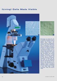
Fig. 3:Endothelial cells, nucleus(blue: DAPI), F-actin(green: BODIPY FL),Mitochondria(red: mitotracker red).<strong>Fluorescence</strong> is the basis of manymodern methods in the life sciences.Fluorescent proteins in particularhave become the key method in thesearch for the secrets of life in theearly 1990s. Today, an increasingnumber of new, more differentiatedfluorescence applications such asFRET, FRAP or FlAsH allow tracking ofmolecular interrelations inside thecell.In the beginning, fluorescence microscopywas performed as transmitted-lightfluorescence microscopy.<strong>The</strong> far more efficient epi-fluorescencemicroscopy then replaced thetransmitted-light technique almostcompletely.<strong>The</strong> modern reflected-light techniquehas one major optical benefit:the objective also acts as a condenser,thus ensuring optimum alignmentof the light beam.detailsNominated for GermanFuture Prize<strong>The</strong> development teamaround Dr. Ulrich Simon,Dr. Bernhard Zimmermann andRalf Wolleschensky from theMicroscopy Group was nominatedfor the 2004 German Future Prizefor the market-ready developmentof the LSM 510 META laserscanning microscope.<strong>The</strong> German Future Prize, awardedby the German Federal President,is an annual award honoringoutstanding innovations in technology,engineering and naturalsciences within Germany. It is notpossible to apply for the prize.<strong>The</strong> right to make recommendationsfor the German Future Prizelies with leading German establishmentsin science and industry.<strong>The</strong> “LSM 510 META” project wasnominated by the Federal Associationof German Industry (BDI).www.zeiss.dewww.deutscher-zukunftspreis.dewww2.uni-jena.de/biologie/zoologie<strong>Fluorescence</strong>-optical instruments from Ca1904August Köhler published his study reportsabout the ultraviolet microscope.1913Beginning in 1913, H. Lehmann and Stanislausvon Prowazek promoted development of thefirst apparatus allowing visualization and alsomeasurement of fluorescence. <strong>The</strong> first opticalinstrument – still called a luminescencemicroscope – was introduced by Carl Zeiss asearly as 1913.1935<strong>The</strong> Ellinger-Hirt luminescence microscopewas mainly used for intravital microscopy.1982<strong>The</strong> development of the first laser scanningmicroscope by Carl Zeiss laid the foundationsfor today's ultramodern threedimensionalreconstruction of cell structuresin fluorescence microscopy.8Innovation 14, Carl Zeiss, 2004