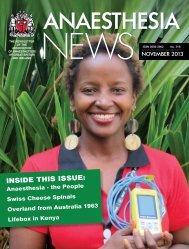
Download - aagbi
Download - aagbi
Download - aagbi

You also want an ePaper? Increase the reach of your titles
YUMPU automatically turns print PDFs into web optimized ePapers that Google loves.
ABSTRACTSOF THEGATANNUALSCIENTIFICMEETING03–05 APRIL 2013OXFORD, UKwww.gatasm.org
Abstracts of theGAT Annual Scientific Meeting03–05 April 2013Oxford, UKThis abstract book has been produced using author-supplied copy. Editing has been restricted to some correctionsof spelling and style where appropriate. No responsibility is assumed for any claims, instructions, methods or drugdosages included in the abstracts: it is recommended that these are verified independently. The contents containedherein are correct at the time of printing and may be subject to change.All submitting authors have declared that appropriate ethical approval has been obtained and that written informedconsent has been obtained from research subjects, and written consent for publication from patients for case reports.These abstracts will appear online on the GAT website:www.gatasm.org/content/oral-poster-prizesLIST OF POSTERSCase Presentation PostersNo. Title Author(s)1 Ultrasound guidance helps avoid wrong operation and improves patient safety. V Athanassoglou, C Morris2 A cause of uvular necrosis never before seen.3Peripartum cardiomyopathy: A likely cause of profound hypotension and myocardial ischaemia following spinal anaesthesia for an electivecaesarean section.V Athanassoglou, A Waddell,M Blahut-ZugajJ Blackshaw, H Hall4 Uterine dehiscence following B Lynch suture. O Boyle, D Milne5 A case study and guidance for the management of a patient with epidermolysis bullosum. A Bradley, R Croft6 A case report of necrotising, haemorrhagic pneumonia caused by panton-valentine leukocidin positive staphylococcus aureus. C Bullen, N Ahmed7 Hyperlipidaemic pancreatitis of pregnancy. M Chazapis8 A case of negative pressure pulmonary oedema: understanding pathophysiology a key to understanding management. C Conroy9 Management of an unusual adult case of acute stridor in a small district general hospital. T Crilley, J Snelling, J Everatt10 Severe adult respiratory distress syndrome and extracoporeal membrane oxygenation.R De Las Casas, L Forbes,A Chakladar, S Marstin11 Visualisation of an epidural septum that prevented the bilateral spread of contrast in the lumbar epidural space. V Dhokia, S Smith, S Ramani12 Ventricular tachycardia complicating the anaesthetic management of cauda equina syndrome. C Dickson, T Sams, I Kannan13 One-lung ventilation in a patient with sickle cell disease: A case report. K Francis, J Collins, D PigottLIST OF FREE PAPERSDräger Oral Presentation PrizeTitlePresentingAuthor(s)14 Retrograde extradural catheter “traction technique” for awake difficult tracheal intubation. S Ghaffar, D Ball15 A case report of concurrent thyrotoxic crisis and diabetic ketoacidosis. V Humphrey, B Slater16 Perioperative presentation of traumatic carotid artery dissection. B Ivory17 Infection due to tuberculosis bovis in ulcerative colitis patient treated with infliximab. V Mandava, C Webb, B Das18 Anaesthetic management in a parturient with uncorrected Tetralogy of Fallot. J Parker, C Grange19 Midazolam-induced hyperactivity treated with flumazenil.20 Life threatening intraoperative hypoxemia in a patient with thalassemia major.S Potru, A Joseph,R RajendramK Grange, S Kannan,S Sivasubramaniam21 GHB causes GBH when withdrawn. N Pritchard, R Hewson22 Seizures post-partum due to an acquired long QT Syndrome. R RajendramThe effect of anaesthetist led sedation training on the quality of sedation practices in Derriford hospital cardiology departmentDr B Ivory23 Not all smokers who are short of breath have COPD. R Rajendram, R ParkerA new method of securing epidural catheters using histoacryl® skin adhesiveDr S Leach24 Treating Cushing’s: A renaissance for etomidate infusions in ICU.R Rajendram, S Schirru,J GriffithsThe ‘go-between’ study: the Traffic Lights tool as a means of effective communication between anaesthetic staffDr S MacDougall-Davis25 Spinal anaesthesia for elective caesarean section in a woman with multiple sclerosis.C Richardson, R Jones,J GreenwoodImproving ITU discharge documentationDr C Parikh26 Overdose of ethylene glycol with an unexpected neurological recovery.W Rutherford, D Quemby,T GuestReturning to work in the West Midlands Deanery: a survey of recent trainee experience and introduction of a return to work programmeManagement of post operative surgical epidural analgesiaCase Presentation Oral PrizeA broken heart in day case – do anaesthetists know enough?A role for intravenous lipid emulsion in cardiac arrest secondary to life-threatening carbamazepine overdosePost operative cerebral salt wasting syndrome following spinal tumour resectionAnaesthesia History Prize winner presentationThe historical role anaesthesia has played in discovering the nature of consciousnessRSM Essay Prize winner presentation (supported by the RSM Section of Anaesthesia)Leading change in anaesthesia in a multi-disciplinary settingDr E PlunkettDr H YusuffDr B SpoonerDr A SudDr R WestDr M JohnDr C Johnston27 Just a little tongue biopsy... C Seeley, I Driver, I Hatcher28 Awake fibreoptic intubation in a patient with isolated laryngeal fracture. S Shotter, G Sommerville29Phaechromocytoma presenting under anaesthesia for day surgery: The role of magnesium in the patient’s subsequent management and acomplication possibly related to its use.E Sleap, M Jenkins, A Wade,J Wells30 Regional obstetric anaesthesia in non-surgical scoliosis patients. S Stobbs, S Cross, R Burns31 Major haemorrhage from scalp laceration. A Sykes, P Dowson32 Abdominal hypertension masking propofol infusion syndrome.E Traer, R Rajendram,J Mandeville, J Griffiths,J Westbrook33 Normoglycaemic diabetic ketoacidosis in pregnancy. S Vasdev, J Salim, T Jovaisa34 Chylous ascites: An unusual cause of a pleural effusion. J Wakeford, P Farquhar-Smith35 Back pain. A cautionary tale.C Williams, T West, S Bell,L DeLloyd, S Harries, R Baraz36 TOE scan or not TOE scan (on GICU). Ask and it shall be given to you; seek and you will find. C Williams, S Ahmed37 Starting a remifentanil infusion at the end of surgery to facilitate smooth emergence from anaesthesia in a potentially difficult airway. M Wilson, C Burnand2 Abstracts of GAT ASM 2013 Abstracts of GAT ASM 2013 3
No. Title Author(s)100 Daily sedation holds in the intensive care unit: an audit of local practice following introduction of a sedation protocol.C Goh, G Barker, S Alderson,S McKechnie101 High fidelity simulation training for the primary FRCA OSCE. R Harvey, C Gillan, S Edgar135 Prolonged clear fluid fasting times on the gastroenterology suite: the unintended consequence of the recommended fasting time.136 Regional anaesthesia for facial plastic surgery.K Richardson, J Smith,A PearsonA Riskalla, P Modayil,A Joseph, R Rajendram,A D'Souza102 Lung care during general anaesthesia: A survey of anaesthetic practice in the UK. D Horner, A Lumb137 Northern deanery primary FRCA trainee survey. T Sams, C Dickson103 Text message reminder to prompt administration of pain relief after children’s day surgery – a proof of concept initiative.L Hulatt, O Hussain, V Allen,D Mason138 Data storage and backup: An audit of trainees’ habits. A Simpson104 Anaesthesia and the environment: an audit of anaesthetic waste disposal in the operating theatre. C Ivermee, T Katawala105 The projected cost savings with the use of recycling bins on an ICU in the UK. B Ivory106 Setting up a regional trainee committee and teaching programme in the Oxford Deanery.107 Implementation of Thromboelastography® Platelet Mapping to reduce cancellations in surgical patients on antiplatelet therapy.108 Safety cannulas - one cannula to rule them all?109 ‘A pain in the neck’: establishing a safe system for changing tracheostomy tubes in hospitalised patients.110 Shared mental models, team expertise and patient safety in anaesthesia.C Janes, M Rowland,J Shorthouse, O DyarR Kasivisvanathan, K Mitchell,S MallettR Khirwadkar, A Bhalla,C Dragomir, C ChevanesseM Kigozi, S Esprit, T Moorthy,C Bradley, L Clarke, R Yorke,J Dunne, C Holland, S UddinN Lau, S Malhotra,M Burtscher, N Sevdalis111 Total knee arthroplasty: local infiltration of anaesthetic versus other methods of analgesia. S Liddle, V Annam112 Audit of anaesthetic volatile use at a large tertiary referral centre in Northern England.S Lobaz, M Hamilton,A Sweenie113 Have lessons been learnt? An audit of difficult airway planning and experience in anaesthetists. J Longbottom, S Varshney114 Aseptic precautions in paediatric caudal anaesthesia. A Maddock, P Jefferson, D Ball115 End-of-life care in four adult intensive care units in the Oxford University Hospitals Trust.N Makris, T Kauhanen,W Seligman116 An audit of money saved with the purchase of a new ultrasound machine. J Mathers, D Patel, A Hayward117 Audit of preoperative fasting times for elective caesarean section: Are we compliant with new guidelines? V Nalawade, R Thompson118 Prospective caesarean section GA audit: Are we compliant with revised RCOA standards? V Nalawade, U Misra, Z Arfeen119 Anticonvulsant use in traumatic brain injury (TBI).120 Elective asleep fibreoptic intubation for teaching. A retrospective analysis of 136 cases.121SMART-COP score for patients admitted with community acquired pneumonia (CAP) to an ICU in a district general hospital: A smarter way ofidentifying patients with severe CAP?G Nickols, D Janssen, AManaraC Oscier, T Pepall, PSidebottomM Pachucki, F Kelly, A Padkin122 Improving access to critical incidents management guidelines in district general hospital theatres. M Pachucki, P Hersch139 The impact of electronic prescribing in a district general hospital intensive care unit on medication error rates – a completed audit cycle. J Strachan140 Informed consent for general anaesthesia – an audit of risks discussed with patients. L Talbot, G Paddle141 Setting up an ‘ introduction to tracheostomy management course’ for health care professionals in the west of Scotland. K Tober, K Owen142 Improving morbidity and mortality in the emergency laparotomy patient. A Vaughton, S Baker143 Audit of timing of epidural top-ups prior to delivery; a completed audit cycle. J Wakeford, M Stevens144 An audit of hydrocortisone use in the treatment of septic shock. D Whitmore145 Anaesthesia For organ donation in brainstemdead patients: A UK national survey. M Wilson, J Cupitt146 Re-audit of Management of fracture neck of femur. Are we improving?Medical Students Prize Poster CompetitionNo. Title Author(s)147 Provision for the higher risk surgical patient, the Royal Cornwall Hospital perspective. D Bunce148 Are their faces really smiling? - Paediatric pain assessment.C Yeow, P Poh, E Pillai,J DedhiaH Donaldson, H Laycock,C Bantel149 Privacy and dignity in the recovery room after surgery audit at the Bradford Royal Infirmary (BRI). T Foulcher, S Griffith150 Epidural anaesthesia and analgesia in hepatic resection. T Jarbawi151 Audit on the emergency induction checklist.M Jones, D Maloney,RM Knights152 In-situ patient simulation as a tool for prehospital training. R McCarthy, S Mitra153 A cry that can’t be heard.I Savage, Z Sheng,B Macpherson, C Connell,G Kennedy, C Marshall,S Mckerron, I Ross154 The relationship between stigmatization and self-esteem in chronic pain patients. M Wall, D Hegarty123 An integrative review of physical rehabilitation on ICU.124 Assessment of how well the safer surgery WHO theatre checklist is completed.125 Survey of anaesthetists’ preferred method for postoperative oxygen supplementation following simple general anaesthesia.126 Intraoperative CPR in the prone position, would you perform it?127 Prevention of cough at tracheal extubation: A meta-analysis of six techniques.R Parker, J Halliday,R Rajendram, J GriffithsJ Parmar, N Hickman,T BourneJ Parmar, W Russell,O WilliamsS Phillips, F Lamb,M MackenzieR Rajendram, A Joseph,SK Ramachandran128A randomized, crossover, observational simulation-based survey comparing use of a needle-guide with the freehand technique for in plane USguided regional anaesthesia.R Rajendram, J McGrath129 The provision of an on call service for regional anaesthesia.130 Optimising fasting of children prior to elective cardiac surgery.131 Readmissions to ICU after small bowel transplantation.R Rajendram, J McGrath,S Stamatakis, A JosephR Rajendram, B Clevenger,E HaxbyR Rajendram, I Edmond,T Thomas, J Millo132 Improving use of the WHO Surgical Safety Checklist. R Rajendram, A Joseph133 Readmission to ICU after kidney and pancreas transplantation. R Rajendram, G Hadjipavlou134 Agreement on ECG rhythm strip interpretation by anaesthetists.R Rajendram, S Kale, S Patel,V Nangalia6 Abstracts of GAT ASM 2013 Abstracts of GAT ASM 2013 7
FREE PAPER ABSTRACTSDräger Oral Presentation PrizeReturning to work in the West Midlands Deanery: a survey ofrecent trainee experience and introduction of a return to workprogrammeE. V. E. Plunkett 1 , C. L. Baxendale 2 , N. Osborn 2 , J. Budd 1 , K. Cullis 3and A. Malins 4Worcestershire Royal Hospital, Worcester, UK, Heartlands Hospital,Birmingham, UK, Queen Elizabeth Hospitals, Birmingham, UK andPostgraduate School of Anaesthesia, Critical Care and EmergencyMedicine, West Midlands Deanery, UKemmaplunkett@doctors.org.ukThere have been several documents published recently on the subjectof Returning to Work: an update of the Royal College of Anaesthetists(RCoA) Return to Work Guidance [1] and a new guidance documentfrom the Academy of Medical Royal Colleges (AoMRC) [2]. Thesegive recommendations regarding what a re-introduction to theworkplace should involve. The Wessex Anaesthetists have developeda Return to Work Programme [3] which has been recognised as “anexample of good practice” by the RCoA. There has also been arecent increase in Return to Work (RTW) courses for anaesthetistsreturning to work after a break. Our aim was to establish what theexperience of our local trainees has been, with a view to improvingthis whilst also accounting for recently published guidance.MethodsWe designed an online survey which was sent out in May 2012 to alltrainees in the West Midlands who had had a recent return to workfollowing a period of maternity leave.ResultsThe survey was sent to 25 trainees and there was a 72% response rate(18/25). Four surveys were incomplete. The return to work dateranged from December 2008 to February 2012. 78% (14/18) ofrespondents returned less than full time and the average length of timeoff was 10.5 months (range of 7-13 months). Most (72%) traineeswere ST5+. 39% (7/18) stated they did not receive any adviceregarding their RTW. Only three trainees used Keeping in Touch(KIT) days and no trainees did specific RTW courses. No respondentsdid extra assessments as part of their return to work and there was anaverage (mean) of 2.7 weeks before starting on calls (range 0-7 weeks,median and mode = 2). Trainees returning full time found theirconfidence returned rapidly (1-3 weeks) but this was more variable forLTFT trainees. 10/14 (71%) reported one or more problems onreturning to work.DiscussionOn the basis of these findings, an action plan of ways that trainees’RTW could be improved has been agreed. This includes increasingawareness of RTW guidance, KIT days and RTW courses. Mostimportantly an RTW programme has been introduced for anaesthetictrainees. This is similar to the Wessex programme but is extended toinclude “Pre-leave planning” in addition to a “Record of Reintroductionto Duties”. Our associated paperwork includes guidancefor trainees and their supervisors and all the relevant items from theAoMRC checklist, formatted in a way to make it user friendly for ourspeciality and region. A generic deanery “Return to Training” policyis also in preparation based on the anaesthetic programme.References1. Royal College of Anaesthetists. Returning to work after a periodof absence. May 20122. Academy of Medical Royal Colleges. Return to PracticeGuidance. April 20123. King W, Haigh F, Aarvold A, Hopkins D, Smith I. Returning towork the Wessex Way. Anaesthesia News June 2012; 299: 18-19Management of post-operative surgical epidural analgesiaH. Yusuff 1 , S. Chadwick 1 and I. Clegg 11-North Manchester General Hospital,Manchester UKEmail- haty_67@yahoo.co.ukIn 2002, a patient had an epidural abscess and a number ofcontributory factors in the postoperative management werehighlighted in an audit. A trust wide protocol was produced andimplemented across the three hospitals in the trust. We conducted anaudit to evaluate the management of surgical patients receivingepidural analgesia over a 5 year period to find out if practice haschanged. We also analysed the microbiology results of all the epiduralcatheter tips.MethodsWe retrospectively analysed 1932 patients who received postoperative epidural analgesia between 2007 and 2011. We excludedobstetric patients. Data was extracted from clinical notes by the acutepain nurses and the laboratory results data base. The data wasanalysed using Microsoft excel and stats directResultsFive hundred and hundred and eighty two patients had a positivemicrobiology catheter result of which most were normal commensals.Eighty four patients (14%) grew pathogenic organisms (fig 1). Fiftypercent of Catheters kept in for longer than 5 days belonged topatients in the7 th and 8 th decade. Twenty one percent of patients withpathogenic catheters had diabetes and a similar percentage had anactive infection in a separate anatomical location, the most commonsite being respiratory. Hepatobiliary (27%) and Colorectal (23%)surgery were the most prevalent specialties in the pathogenic group.There was no case of epidural abscess.Table 1: Organisms cultured from epidural cathetersOrganism NumberStaph. Aureus 22including MRSAEnterococcus 12Pseudomonas. A 3Escherichia. Coli 8Coagulase 157negative staphDiscussionFrom the results the elderly were more likely to have the catheters leftin situ for longer, a risk factor which can be modified. It is especiallynecessary to modify this risk factor as the elderly have a highermortality when they develop epidural abscess 2 . It is likely that patientswith cancer having received adjuvant chemo-radiotherapy are atincreased risk as demonstrated by a high prevalence of positivecatheters in patients who have had Hepatobiliary and Colorectaloperations. We have changed the trust wide protocol to includesending epidural tips for culture and early removal of cathetersespecially in high risk patients.References1. T. M. Cook, D. Counsell and J. A. W. Wildsmith on behalfof The Royal College of Anaesthetists Third NationalAudit Project. Major complications of central neuraxialblock British Journal of Anaesthesia 2009; 102 (2): 179–902. Sendi P, Bregenzer T, Zimmerli W. Spinal epiduralabscess in clinical practice. Quarterly Journal of Medicine.Jan 2008;101(1):1-12.Case Presentation Oral PrizeA broken heart in day case – do anaesthetists know enough?BB Spooner 1 , AJ Gait 1 , R Huggett 1Russells Hall Hospital 1 , Dudley, West Midlands, UKbspooner@doctors.org.ukWe report the unusual case of a 37 year old female patient withDowns syndrome who suffered cardiac arrest immediately followinginduction of general anaesthesia. She was later found to have a stressinduced cardiomyopathy (also known as broken heart syndrome ortakotsubo cardiomyopathy). Stress induced cardiomyopathy has beenreported in the perioperative period [1], but not in a patient with alearning difficulty.Case reportThe patient was scheduled to have dental extractions under generalanaesthesia as a day case procedure. She did not have anycardiovascular complications of Downs syndrome. She received nopremedication. Before induction of anaesthesia her heart rate was 120beats min -1 and her blood pressure 160/110 mmHg. She wasuncooperative and had an inhalational induction. Following inductionof anaesthesia she became profoundly bradycardic and then sufferedan asystolic cardiac arrest. Cardiopulmonary resuscitation wascommenced with 1mg of adrenaline which led to a return ofspontaneous circulation within two minutes. Electrocardiogramshowed ST elevation in leads I and aVL without reciprocal changes.Echocardiogram showed apical akinesia of her left ventricle. Her peaktroponin was elevated at 5.0 µg/l ( 40 mg/L,as in our case, are associated with coma, respiratory depression,arrhythmias and seizures. ILE is an effective treatment, acting as a‘lipid sink’, for cardiovascular collapse associated with localanaesthetic toxicity, and has also been reported as beneficial incardiovascular collapse caused by several lipophilic drugs e.g.verapamil, quetiapine and propranolol [2, 3]. However, in theliterature to date, there are no documented cases of ILE resulting inreturn of spontaneous circulation in cardiac arrest, secondary tocarbamazepine overdose. This case also highlights the need to initiateintravenous sodium bicarbonate and multi-dose activated charcoalwhilst carbamazepine levels are established, regardless of the patient’shistory.References1. Hojer J, Malmlund H and Berg A. Clinical Features in 28Consecutive Cases of Laboratory Confirmed Massive Poisoningwith Carbamazepine Alone. Clincial Toxicology 1993; 31: 449-458.2. Weinberg G. Lipid Emulsion Infusion: resuscitation for localanesthetic and other drug overdose Anesthesiology 2012; 117:180-7.3. Cave G, Harvey M and Graudins A. Review article: Intravenouslipid emulsion as antidote: A summary of published humanexperience. Emergency Medicine Australasia 2011; 23: 123-141.FREE PAPER ABSTRACTS10 Abstracts of GAT ASM 2013 Abstracts of GAT ASM 2013 11
FREE PAPER ABSTRACTSCase Presentation Oral PrizePost operative cerebral salt wasting syndrome following spinaltumour resectionR West 1 , R Pilling 2 and P. Gunning 21. Royal Free London Hospital, London, UK, 2. Royal NationalOrthopaedic Hospital, Stanmore UKraharahmanwest@googlemail.comCerebral salt wasting syndrome (CSWS) is a disorder of natriuresisresulting in hyponatraemia and hypovolaemia. It frequently occursfollowing intracranial pathologies, such as subarachnoid haemorrhageor cerebral space occupying lesions. We report an extremely rareclinical presentation of CSWS in the postoperative period followingspinal tumour resection. Informed consent was completed.Case report: A 56 year old gentleman presented with back pain foundto be caused by cord compression from a metastatic deposit to his 5 ththoracic (T5) vertebra. He underwent an elective partial posterolateralvertebrectomy of T5 and fusion of T2 to T8. The tumour wascompletely resected, and right T5 nerve root was sacrificed. His pastmedical history included a left pneumonectomy the previous year forprimary adenocarcinoma of the lung. Medication history comprised ofanalgesia and dexamethasone (4mg) reducing dose for 3 weeks priorto the surgery. On day 1 post-op he presented with polyuria of500ml/hr, hypotension and tachycardia. Blood results showeddecreased sodium (131 mmol/L) and osmolality (276mOsmol/kg).Urinalysis showed increased urinary sodium (208 mmol/L) andosmolality (555 mOsmol/kg). Twenty four hour urine output was 5600ml. A diagnosis of CSWS was made due to the high volume ofconcentrated urine and high urinary Na together with normal serumosmolality. Normal thyroid function, liver function, preoperative renalfunction and marginally suppressed cortisol level aided us inexcluding other causes of hyponatraemia. The high urinary sodiumcontent also excluded diabetic insipidus (DI) and Syndrome ofinappropriate release of antidiuretic hormone (SIADH). We treated thehypovolaemia with 0.9% saline to replace hourly volume of urineoutput. We also commenced fludrocortisone 100mcg TDS 12 hourspost presentation. On day 3 we started slow release sodium tablets of40 mmol 4 times a day. Over the next 5 days serum sodium returnedto normal and high renal sodium loss ceased.Discussion: To our knowledge, this is the first case report associatingCSWS with surgical resection of spinal tumour. The differentialdiagnosis between CSWS and SIADH can be confusing as both maypresent with similar laboratory and clinical findings. The fundamentaldifference is the extra cellular fluid volume status. The key feature ofCSWS is extra cellular volume depletion and natriuresis. SIADH ischaracterized by dilutional hyponatremia secondary to increased totalbody water and high levels of antidiuretic hormone. Thepathophysiology of renal sodium loss in CSWS is unclear. Postulatedmechanisms includes natriuretic peptide induced natriuresis anddisruption of sympathetic neural input to the kidney. [1]Fludrocortisone is thought to work via direct action on the renal tubuleto reduce sodium excretion. Our case demonstrates that CSWS couldoccur without intracranial pathology following spinal tumourresection. Postoperative hyponatraemia may be a common finding inthe postoperative period. However it may rarely be due to a CSWS – acondition that can be clinically challenging. Making a distinctionbetween CSWS and SIADH is of crucial importance given thedivergent nature of therapy.References1. Yee AH, Burns JD, Wijdicks E F.M. Cerebral Salt Wasting:Pathophysiology, Diagnosis, and Treatment. Neurosurgery clinics ofNorth America 21 2010; 339-352Anaesthesia History Prize winner presentationThe historical role anaesthesia has played in discovering thenature of consciousnessDr M JohnThis essay is available on the GAT website:www.gatasm.org/content/oral-poster-prizesRSM Essay Prize winner presentation(supported by the RSM Section of Anaesthesia)Leading change in anaesthesia in a multi-disciplinary settingDr C JohnstonThis essay is available on the GAT website:www.gatasm.org/content/oral-poster-prizes1 2Ultrasound guidance helps avoid wrong operation and improvespatient safetyV. Athanassoglou 1 , C. Morris 1Wycombe General Hospital, High Wycombe, UKathanass@mac.comWe present a case where ultrasonography changed the operation thepatient had consented for and was critical in the patient’s management.Case reportA 61-year-old woman had been placed on the emergency list for a leftbelow knee amputation. She appeared ill. She declined a spinalanaesthetic. We planned to perform a general anaesthetic, popliteal andsaphenous nerve blocks for postoperative analgesia. On positioning forthe nerve block, a preliminary ultrasound scan revealed gas in the softtissues over the popliteal fossa. On further scanning it became obviousthe disease process had spread proximally. The extent of the disease wasnot obvious from external limb examination. Only the ultrasounddisplayed the real extent. A below knee amputation was the wrongoperation to perform as the disease had already spread distally.However, on review of the consent form the patient had not beenconsented for an above knee amputation. Therefore, on full consultationthe surgeon, anaesthetist, and theatre team we decided to not proceedwith the operation and wake up the patient. Once the patient recoveredfrom the anaesthetic they were consented for an above knee amputationand the procedure was performed later that evening. The anaesthetic andoperation were uneventful and the patient was safely discharged home14 days later.DiscussionThe use of ultrasound guided popliteal and saphenous nerve blocksprovide good anaesthesia and analgesia for below knee, ankle and footsurgery. 1 Ultrasound is not a common mode of investigation for theextent of gas gangrene, despite it being a specific and sensitive modalityto detect subcutaneous and intramuscular air. 2 It has aided the earlydiagnosis of patients with scrotal swelling of unknown origin, 3 lowerlimb subcutaneous emphysema of abdominal origin, 4 and necrotizingfasciitis. 5 This is the first reported case of ultrasound aiding thediagnosis of lower limb gas gangrene. While evidence accumulates forincreased efficacy and speed of regional anaesthesia under ultrasoundguidance, an increased safety profile has yet to be confirmed. Theclassical technique for popliteal and saphenous nerve blocks is relativelystraightforward, and ultrasound may appear to add little to an alreadyeffective and low risk procedure. However, the use of ultrasound in ourcase allowed us to alter management and avoid the wrong operation. Inthis instance the use of ultrasound undoubtedly improved patient carebut the ethical implications of incidental ultrasound findings is still aconcern and hindrance for scanning all patients.References1. Salinas FV. Ultrasound and review of evidence for lowerextremity peripheral nerve blocks. Regional Anesthesia PainMedicine 2010;35: S16-25.2. Butcher CH, Dooley RW, Levitov AB. Detection of subcutaneousand intramuscular air with sonography: a sensitive and specificmodality. Journal Ultrasound Medicine 2011;30: 791-795.3. Morrison D, Blaivas M, Lyon M. Emergency diagnosis ofFournier’s gangrene with bedside ultrasound. American JournalEmergency Medicine 2005;23: 544-547.4. Jager GJ, Rijssen HV, Lamers JJH. Sucutaneous emphysema ofthe lowere extremeity of abdominal origin. GastrointestinalRadiology 1990;15: 253-258.5. Anreasen TJ, Green SD, Childers BJ. Massive infectious softtissue injury: diagnosis and management of necrotizing fasciitisand purpura fulminans. Plastic & Reconstructive Surgery2001;107: 1025-1034.A cause of uvular necrosis never seen beforeV. Athanassoglou 1 , A. Waddell 2 , M. Blahut-Zugaj 2Wycombe General Hospital, High Wycombe, UK, Great WesternHospital, Swindon, UKathanass@mac.comSore throat is common after operations. Several factors contribute topostoperative sore throat. The incidence varies with the method ofairway management [1]. However, persistent or worsening painassociated with the feeling of something in the throat is likely toindicate localised trauma to either the uvula or other oropharyngealstructures. The i-gel supraglottic airway, an alternative supraglotticdevice with a small, noninflatable, anatomically shaped cuff, has beenfound to result in a lower incidence of sore throat [2]. Uvular necrosisis thought to result from ischaemic necrosis due to mechanicalinterruption of the blood supply.Case reportWe report a case where the use of an i-gel caused uvular trauma. A 43year old presented for a hip arthroscopy. On review of her previousanaesthetic charts she had had i-gel supraglottic devices before. Weused a size 4 i-gel device, which was appropriate according to thepatients weight and size. Throughout the procedure the patient wasbreathing spontaneously on the i-gel and airway pressures did notexceed 2cmH 2O. The lubricated airway was inserted easily on the firstattempt without difficulties. The procedure lasted 90 minutes. She wasdischarged home later that day. Seven days after her operation herhusband contacted our anaesthetic department and showed us a pictureof his wife’s throat showing marked redness and soft palate swelling.The symptoms started on the second postoperative day but they weretolerable. We referred her to an Ear, Nose and Throat (ENT) clinic,where a diagnosis of uvular necrosis was made. She was kept underreview in the ENT clinic but never needed any specific treatment.DiscussionUvular necrosis has been reported after tracheal intubation as far backas 1978 [3]. It has occurred after LMA use [1], long-term intubation[4], and blind suctioning [5]. Review of the literature did not producea case where uvular necrosis has been produced due to an i-gel. Thereare 14 reported cases from 1984 where routine perioral interventionshave led to uvular necrosis [6]. LMA cuffs are permeable to nitrousoxide and carbon dioxide, which could potentially lead to increasedcuff volume and pressure during long procedures, resulting inpostoperative sore throat due to uvular and pharyngeal oedema.However, the i-gel due to its small noninflatable cuff should obviatethat risk and therefore localized airway complications should be lesswith a well-placed i-gel. Whatever the mechanism of trauma leadingto uvular necrosis, potentially severe airway compromise may occur.Therefore, it is vitally important whatever airway device we use toplace it carefully minimizing oropharyngeal trauma.References1. McHardy FE, Chung F. Postoperative sore throat: cause,prevention and treatment. Anaesthesia 1999;54: 444–53.2. Keijzer C, Buitelaar DR, Efthymiou KM, et al. Acomparison of postoperative sore throat and neckcomplaints after the use of the i-gel® and the LaPremiere® disposable laryngeal mask: A double-blinded,randomized, controlled trial. Anesthesia Analgesia2009;109: 1092-1095.3. Ravindran R, Priddy S. Uvular edema, a rare complicationof endotracheal intubation. Anesthesiology 1978;48: 374.4. Harris MA, Kumar M. A rare complication of endotrachealintubation. Lancet 1997;350:1820-1821.5. Bogetz MS, Tupper BJ, Vigil AC. Too much of a goodthing: uvular trauma caused by overzealous suctioning.Anesthesia Analgesia 1991;72: 125-6.6. S. R. Emmett, S. D. Lloyd, M. N. Johnston, Uvular traumafrom a laryngeal mask, British Journal of Anaesthesia,2012;109: 468.POSTER ABSTRACTS12 Abstracts of GAT ASM 2013Abstracts of GAT ASM 2013 13
POSTER ABSTRACTS3 4 5 6Peripartum cardiomyopathy: a likely cause of profoundhypotension and myocardial ischaemia following spinalanaesthesia for an elective caesarean section.W. J. Blackshaw, H. HallWarrington and Halton NHS Foundation Trust, Warrington, UKjonblackshaw@doctors.org.ukPeripartum cardiomyopathy (PPCM) is an idiopathic cardiomyopathythat presents with heart failure secondary to left ventricular systolicdysfunction toward the end of pregnancy or in the months afterdelivery, in the absence of any other cause of heart failure [1].Symptoms suggestive of cardiac disease are often experienced duringnormal pregnancy; this, combined with the late and often subtle onsetof symptoms, makes diagnosis of PPCM difficult in the antenatalperiod. An abnormal cardiovascular response to neuraxial anaesthesiamay be the first indication that cardiac function is impaired.Case ReportA 34 year-old woman, G2P1, attended hospital for an electivecaesarean section on week 39 of a normal pregnancy. She had a pastmedical history of ankylosing spondylitis. Induction of spinalanaesthesia was achieved with 2.2 mls of 0.5% hyperbaricbupivacaine and 300 mcg of diamorphine at L3/L4 level. A period ofprofound hypotension ensued which was refractory to fluid, ephedrineand metaraminol boluses. During this period, the patient sufferedchest pain with ECG evidence of global myocardial ischaemia. Earlyarterial and central venous catheter insertion with inotropic supportwere achieved before delivery of a healthy baby by caesarean section.The mother required three days of inotropic support but wasdischarged from hospital after one week. Serial echocardiogramsshowed initial moderate left ventricular dysfunction with recovery ofnormal function after two months.DiscussionThis report discusses the anaesthetic management of the unexpectedcardiovascular instability resulting from spinal anaesthesia on a likelybackground of reduced cardiac contractility. It highlights the need forprompt multidisciplinary decision making to achieve the mostfavourable outcome and examines whether steps could be taken tobetter identify women with this problem.References1. Sliwa K, Hilfiker-Kleiner D, Petrie MC et al. Current state ofknowledge on aetiology, diagnosis, management, and therapy ofperipartum cardiomyopathy: a position statement from the HeartFailure Association of the European Society of Cardiology WorkingGroup on peripartum cardiomyopathy. Eur J Heart Fail Aug 2010;12(8): 767-78.Uterine dehiscence following B Lynch sutureO Boyle, D MilnePinderfields Hospital, Wakefield, UKOwenboyle@doctors.org.ukIntroduction: A B-Lynch suture is a commonly used method [1] fortreating an atonic uterus preventing the need for hysterectomyfollowing caesarean section. We describe a case where complicationsensued 10 years post procedure in a subsequent pregnancy withdevastating effect.MethodsCase report: In 2002 a 22 year old women who was at 29 weeksgestation attended the emergency department as an unrestrainedpassenger involved in a road traffic collision. Significant abdominalinjuries were sustained that led to haemodynamic compromise andfoetal death. Following a massive transfusion an emergency caesareansection was performed. Due to uterine atony a B-Lynch suture wasplaced which successfully provided haemostasis and preserved theuterus. Subsequently a splenectomy and liver laceration repair wereundertaken. In 2012 the same patient presented to hospitalcomplaining of right sided abdominal pain at a gestation of 31 weeks.She had injected heroin earlier that morning which masked theseverity of her pain leading to a presumptive diagnosis of appendicitissecondary to an elevated white cell count. Foetal heart rate wasdetected and the patients observations were normal. Afterapproximately five hours the patient rapidly deteriorateddemonstrating signs of hypovolaemic shock. Due to the patientshistory of intravenous drug use venous access was very difficult. Thepatient was transferred to recovery on labour ward so central venousaccess and resuscitation could take place. Prior to transfer the patientbecame profoundly hypotensive and bradycardic. The majorhaemorrhage protocol was initiated. A consultant radiologist notedextensive free fluid in the abdomen. The patient was immediatelytransferred to theatre for an emergency laparotomy. On explorationshe was found to have complete dehiscence of the upper and posterioraspects of the uterus with the foetus demised and free in the abdomen.The uterus was repaired and haemostasis was achieved and the patientwas discharged from hospital a few days later.DiscussionThroughout the pregnancy there was no evidence of uterine pathologyand until this point the pregnancy had been uneventful. This casehighlights the potential risk of B-Lynch sutures obtunding the bloodsupply to areas of the uterus potentially weakening the uterine wallendangering the life of mothers in subsequent pregnancies. The use ofheroin prior to attending hospital may have masked her pain earlier inthe progression of her pathology. Earlier detection may have resultedin a better outcome given foetal heart sounds were detected onadmission. This case also highlights the difficulties associated withresuscitating intravenous drug users and difficulty obtaining venousaccess.References1. Grotecut CA, Larsen FW, Jones MR, Livingston E. Erosion of aB-Lynch suture through the uterine wall: a case report. Journal ofReproductive Medicine 2004;49: 849 -52.A Case Study And Guidance For The Management Of A PatientWith Epidermolysis BullosumA. Bradley 1 , R Croft 11 Royal Manchester Children’s Hospital, Manchester, UKEmail of corresponding author: anthony.bradley@nhs.netEpidermolysis Bullosa encompasses a group of inherited disordersthat are characterised by the generation of blisters at the site ofmechanical trauma. Genetic changes cause defective connective tissueformation and present shortly after birth. This dermatologicalcondition provides challenges during the perioperative period.Unfortunately, as of yet, there is no medical cure.Case ReportA 3 year-old child presented with extensive scald injuries from bathwater. She was known to have a background history of EpidermolysisBullosa and was receiving specialist care in another tertiary centre.During her admission, she required multiple surgical interventions,which presented many difficulties for the anaesthetists concerned.Guidance for her care was sought from her specialist centre.DiscussionEpidermolysis Bullosa affects all squamous cell epithelium includingthe oral mucosa, potentially posing the biggest issue to theanaesthetist. Repeated trauma, either iatrogenic (e.g. airwayinstrumentation) or spontaneous (e.g. brushing teeth or mastication),causes blister formation and then subsequent scarring. This can resultin a difficult airway, seen in up to a third of all these patientsundergoing procedures or surgery. Other issues include fixing ofdressings, as well as monitoring and handling of the patient. Securingintravenous (I.V.) devices may be difficult due to shearing forcesfound between adhesive dressings and the epidermis below, riskingfurther bullae formation. Cardiac monitoring is of concern, as thetransducers are traditionally adhesive to ensure adequate contact. Thisresults in the same issues as for I.V. or surgical dressings. It has, forthis reason been noted that minimal standards of the Association ofAnaesthetists of Great Britain and Ireland have at times beendisregarded. Dealing with an anaesthetised patient requires manualhandling. This must be undertaken with the greatest degree of care toavoid shearing forces on the skin. Handling and consoling by wellmeaningrecovery staff has been associated with the development ofnew blisters therefore effective handover is essential. With ourexperience of regularly anaesthetising this child for her frequentsurgical procedures, combined with other centres’ recommendations,we have managed to find ways of overcoming all the above issues.We have therefore been able to provide safe and effective anaesthesiafor patients with this condition.SummaryAt first glance Epidermolysis Bullosa looks very challenging to theAnaesthetist. But other than a potentially difficult airway, all otherissues can be readily overcome with careful attention to detail,planning and communication with all team members. This means thata safe and AAGBI-compliant anaesthetic should be available to allpatients with Epidermolysis Bullosa.ReferencesWA Ames, BJ Mayou and K Williams; Guidance for anaestheticmanagement of patients with Epidermolysis Bullosa – British Journalof Anaesthesia 1999 82 (5): 746-51Lohom G, Lyons B; Anaesthesia for children with EpidermolysisBullosa: a review of 20 years’ experience – European Journal ofAnaesthesiology. Nov 2001; 18(11): 754-54Elliot Krane MD; Lucile Packard children’s hospital Stanforduniversity, paediatric anaesthesia and pain management – Guidelinesfor anesthetic management of EBA case report of necrotising, haemorrhagic pneumonia caused bypanton-valentine leukocidin positive staphylococcus aureusC Bullen¹, N Ahmed²Croydon University Hospital, London, UKCarolineb@doctors.org.ukPanton-Valentine Leukocidin (PVL) is a cytotoxin associated withincreased virulence of certain strains of staphylococcus aureus. Itoften leads to skin and soft tissue infections, but can also cause asevere, necrotising pneumonia the mortality rate of which is up to75% [1]. Here we present a case of rapidly progressive respiratoryfailure, haemoptysis and sepsis in a patient with influenza B and acommunity acquired PVL-positive MRSA pneumonia.Case Report:A previously healthy 46 year old lady presented with a 3 day historyof flu-like symptoms, and new onset haemoptysis the day ofadmission. On examination she demonstrated signs of septic shockand respiratory failure with oxygen saturations of 63% on air.Investigations revealed neutropenia and leucopoenia with elevatedCRP, and a progressively falling platelet count. Chest radiographshowed bilateral dense infiltrates, with diffuse alveolar haemorrhagereported on CT. She deteriorated rapidly in accident and emergencywhere she was intubated, with heavily blood stained secretionspouring from the endotracheal tube. She was transferred to intensivecare where she was treated aggressively with antibiotics, steroids,intravenous immunoglobulin and recombinant factor VIIa(Novoseven). She continued to deteriorate with increasing inotropicand vasopressor requirements, and worsening respiratory failure –Po2 of
POSTER ABSTRACTS7 8 9 10Hyperlipidaemic Pancreatitis of PregnancyM. ChazapisUniversity College Hospital, London, UKm.chazapis@yahoo.comA case report of a rare complication of pregnancy, requiring veryclose multidisciplinary care between obstetricians, general surgeons,anaesthetists and intensivists.MethodsA 36 year old primigravida, with an uncomplicated pregnancypresented at 34+4 weeks gestation with acute epigastric pain. She wasadmitted, and quickly deteriorated, with increasing pain resistant tomorphine, maternal tachycardia and falling oxygen saturations. Lipidprofiling revealed a grossly lipaemic sample, with a cholesterol of37.8 mmol/L, and triglycerides >57 mmol/L. An abdominalultrasound was unremarkable.ResultsAfter discussion with the medical team, general surgeons andobstetricians, following a deteriorating clinical picture of the patient, adecision was made to proceed to emergency Caesarean section, usinga midline incision, for direct inspection of possible pathology. Thepancreas looked inflamed, and there was gross ascites. The baby wasdelivered safely, and the mother taken intubated to the intensive careunit. A baseline insulin infusion was started, and haemofiltration toreduce the triglyceride levels was considered, but the lipid profilenormalised over 24 hours following delivery. The patient left hospitalten days later.DiscussionPancreatitis in pregnancy is an unusual but serious disease, associatedwith a maternal mortality of over 30% 1 . Most cases are secondary togallstones. In pancreatitis associated with hyperlipidaemia, the effectof delivery on the decline of plasma triglyceride levels can beimmediate and dramatic.References1. Wilkinson EJ. Acute pancreatitis in pregnancy: A review of 98cases and a report of 8 new cases. Obstetrical and GynecologicalSurvey 1973; 28: 281-303.A case of negative pressure pulmonary oedema: understandingpathophysiology a key to understanding management.Dr Clare ConroyEaling Hospital, Southall, UKcjconroy@doctors.org.ukNegative pressure pulmonary oedema (NPPO) is a well recognisedcomplication of laryngospasm and other forms of upper airwayobstruction [1,2,3]. An understanding of the pathophysiology involvedhas implications for its management.Case ReportA 35 year old ASA 1 male presented to the surgeons with a one dayhistory of severe abdominal pain. A CT demonstrated a grosslydistended stomach measuring 26 cms. He underwent an emergencylaparotomy for acute bowel obstruction. A standard rapid sequencewas performed, he was intubated with a size 8mm endotracheal tubeand a nasogastric tube was inserted. The intraoperative course wasuneventful and he was easily ventilated and oxygenated. Onextubation, the patient became very restless and bit down hard on hisendotracheal tube before self extubating. He continued to be veryrestless and agitated, his oxygen saturations began to drop and hedeveloped signs of laryngospasm. Initial management involvedpositive pressure mask ventilation with 100% oxygen and two bolusesof 50mg propofol. The oxygen saturations initially improved, howeverhe kept developing intermittent laryngospasm with periods ofhypoxia. About 20 minutes after initial extubation the patient wasatraumatically re-intubated and sedated. On auscultation bilateralcrackles were present and suctioning from the endotracheal tuberevealed pink frothy sputum. A diagnosis of possible NPPO was madeand the patient was transferred to ICU. Supportive management whichincluded CPAP was given and he was successfully extubated within12 hours and non invasively administered continuous positive pressure(CPAP) ventilation was continued. A toxicology screen was positivefor cocaine, cannabinoids and benzodiazepines. His oxygenationimproved over the next 16 hours and he was discharged from the ICUafter 72 hours.DiscussionThis case report shows a probable diagnosis of NPPO following acutelaryngospasm post extubation. NPPO has been widely reported inyoung, athletic and healthy men who suffer upper airway obstructionand reports have shown an increase in alveolar permeability in crackcocaine users [1]. NPPE is characterised by an acute onset with arapid recovery with 12-24 hours [1]. Management is normallysupportive utilising ventilatory support whether it is intubation orCPAP and controversially the use of loop diuretics [2]. The mainmechanisms thought to be involved in the pathophysiology of NPPEare mechanical. The generation of a large negative intrathoracicpressure, of the order of 100 – 150 cm H2O [2], with a concomitantmassive increase in transmural pressure leads to stress failure at thealveolar capillary membrane [1, 3] as well as an increase in cardiacafterload and not only large fluid shifts. Because of this the use ofdiuretics in euvolaemic patients is probably unwarranted.References1. Schwartz D, Maroo A, Malhotra A, Kesselman H. NegativePressure Pulmonary Haemorrhage. Chest 1999; 115: 1194-1197.2. Krodel D, Bittner A, Abdulnour R, Brown R, Eikermann M.Case Scenario: Acute Postoperative Negative PressurePulmonary Edema. Anaesthesiology 2010; 113: 200-7.3. Broccard A, Liaudet L, Aubert JD, Schnyder P, Schaller MD.Anesthesia and Analgesia 2001; 92(1): 273-54. West JB. Cellular Responses to Mechanical Stress. InvitedReview: Pulmonary capillary stress failure. Journal of AppliedPhysiology 2000; 89: 2483 - 248Management of an unusual adult case of acute stridor in a smalldistrict general hospitalT. Crilley 1 , J. Snelling 2 and J. Everatt 1Horton General Hospital, Banbury, UK 1 and John Radcliffe Hospital,Oxford, UK 2trina.crilley@gmail.comStridor is a sign of turbulent airflow through a partially obstructedupper airway. Frequently seen in children, acute stridor is rarer inadults.Case ReportA 49 year old male with chronic alcoholism presented to our districtgeneral hospital with a two day history of persistent vomiting. Whilstawaiting review he developed acute inspiratory stridor following anepisode of vomiting and complained of something lodged in histhroat. There was no preceding history of fever, cough or sore throat.The anaesthetic team were fast bleeped and on initial examination thepatient was pale and sweating. He was sat upright, breathing throughan open mouth, and using accessory muscles. He was tachycardic (142bpm), tachypnoeac (32 bpm), hypertensive (BP 190/90) and apyrexial.Oxygen saturations were 100% on high flow O 2, dropping below 89%on air and there was an audible inspiratory stridor. Investigationsrevealed a raised white cell count and CRP plus renal impairment. Anarterial blood gas revealed hypercapnia with a hypochloraemicalkalosis secondary to vomiting (on 10 L.min -1 O 2; pH 7.48, PO 2 66,PCO 2 7.0, BE 14.5, HCO 3 - , 14.5). Chest x-ray was unremarkable withno evidence of a foreign body. Initial management includedhumidified high flow O 2, nebulised adrenaline and i.v. hydrocortisone.ENT at the tertiary centre 27 miles away were contacted andimmediately proceeded to the Horton. Nasendoscopy performed byENT revealed a swollen epiglottis. The patient was transferred totheatre for intubation, where the surgical team were scrubbed inpreparation for surgical tracheostomy in case of failed intubation.Induction of anaesthesia with sevoflurane was performed by theConsultant Anaesthetist. At laryngoscopy a Cormack and Lehanegrade 3 view was obtained and an enlarged epiglottis noted.Intubation, with a size 7 cuffed endotracheal tube (ETT; bougieguided), was successful on first attempt. On day 3 post intubation aleak developed around the ETT cuff and endoscopy revealed a normalepiglottis. The patient was then successfully extubated. Allmicrobiology cultures were negative.DiscussionAcute stridor as a sign of upper airways obstruction in adults is bothrare and a medical emergency. Causes include recent head and necksurgery, infection, anaphylaxis, tumours, airway trauma, prolongedintubation and inhaled foreign bodies/blood/vomitus. 1 In this case thecause is unclear but is likely due to physical and/or chemical trauma tothe epiglottis from violent vomiting. Management of patients withacute stridor is based mainly on experience in paediatrics 2 and earlysenior involvement is imperative. It includes humidified high flowoxygen, nebulised adrenaline (5 mg) and i.v. dexamethasone (8 mg).Heliox ® may play a role as may endoscopy to determine aeitology.Definitive management involves securing the airway. Gaseousinduction with sevoflurane is preferred, as it avoids muscle relaxantuse, preserving spontaneous ventilation as mask ventilation may notbe possible. Provision for surgical tracheostomy is essential. Sincepatient transfer is not usually appropriate this case highlights theimportance of a multi-disciplinary team approach and timelycommunication with ENT in district hospitals lacking these services.References1. Robinson N, Hall G, Fawcett V. How to Survive in Anaesthesia,Fourth Edition. Wiley and Sons, UK, 2011.2. Maloney E, Meakin G. Acute stridor in children. ContinuingEducation in Anaesthesia, Critical Care & Pain, 2007; 7:183-86.Severe Acute Respiratory Distress Syndrome and ExtracorporealMembrane OxygenationDe Las Casas R, Chakladar A, Forbes L, Marstin SSt Richard’s Hospital, Chichester, UKruthdlc@doctors.org.ukCommunity acquired pneumonia (CAP) accounts for approximately6% of admissions to adult Intensive Care Units (ICU) in the UK [1].There is considerable experience in the management of these patientswithin such ICUs. In cases of refractory hypoxic respiratory failureretrieval for Extracorporeal Membrane Oxygenation (ECMO) is anincreasingly available option. We report a case of severe AcuteRespiratory Distress Syndrome (ARDS) secondary to CAP, who wasretrieved for ECMO on day eight of his admission.Case ReportA 74 year old previously fit gentleman presented to A&E with a oneweek history of increasing shortness of breath. Within 12 hours ofarrival, his clinical condition necessitated admission to the ICU forrespiratory support. He was treated for a severe CAP but deteriorateddespite treatment with antibiotics and continuous positive airwaypressure. On day two he was intubated and ventilated due toworsening gas exchange and exhaustion. Following a period ofrelative stability, he deteriorated on day six of admission, and lungprotective ventilation became difficult. On day eight peak inspiratorypressure was over 40cmH 20 but achieving only tidal volumes of300mls, and despite positive end expiratory pressure of 15cmH 20 andincreasing of Fi02 to 0.9, hypoxia continued to worsen. Deepsedation, paralysis, permissive hypercarbia, prostacylin infusion, andadjustment of ventilation parameters proved ineffective, and ourpatient began to desaturate significantly during basic nursing care.His Murray score was 3.5 and he was referred to, and accepted by theregional ECMO team. Initiation of the ECMO, and transfer to theregional centre occurred within eight hours. Unfortunately, fourweeks after retrieval, he developed multi-organ failure and treatmentwas withdrawn.DiscussionThe conventional management of ARDS with mechanical ventilationis increasingly recognised to potentially exacerbate underlying lunginjury via high airway pressures and oxygen concentrations. WhilstNICE guidance in 2011 was unable to draw firm conclusionsregarding the efficacy of ECMO [2], the Intensive Care Societyreview in 2012 found, in light of the CESAR trial, that ECMOoutcomes and safety are improving, and predicts it will become astandard critical care therapy in non-specialist units [3]. Criteria forreferral for ECMO, and explanation of the Murray Score, are clearlylaid out on the websites of ECMO centres. We found the ECMO teamwelcomed discussion, encouraged early referral, and were keen todemonstrate and explain the initiation of ECMO to our ICU team. Inour experience ECMO was an accessible escalation when we reachedthe ceiling of critical care therapy that we could offer.References1. Woodhead M, Welch CA, Harrison DA, Bellingan G, Ayres JG.Community-acquired pneumonia on the intensive care unit:secondary analysis of 17,869 cases in the ICNARC Case MixProgramme Database. Critical Care 2006; 10(Suppl 2):S12. NICE (2011). Extracorporeal membrane oxygenation for severeacute respiratory failure in adults [IPG391]. London: NICE3. Hung M, Vuylsteke A, Valchanov K. Extracorporeal membraneoxygenation: coming to an ICU near you. Journal of theIntensive Care Society 2012; 13: 2-9.POSTER ABSTRACTS16 Abstracts of GAT ASM 2013 Abstracts of GAT ASM 2013 17
POSTER ABSTRACTS11 12 13 14Visualisation of an epidural septum that prevented the bilateralspread of contrast in the lumbar epidural spaceV. Dhokia 1 , S. Smith 1 and S. Ramani 1Northampton General Hospital, Northampton, UKvishal.dhokia@doctors.org.ukAn epidural septum, or the plica mediana dorsalis (PMD), is a midlineband of connective tissue within the dorsal epidural space. Despitethe appearance of the structure in different studies [1, 2], itsprevalence, exact nature and clinical significance remainscontroversial. It is quoted as one cause of a ‘unilateral epidural’;however, anaesthetic has been shown to cross the midline even in itspresence [3] and cases of unilateral epidurals have most frequentlybeen attributed to other causes such as anterior or transforaminalplacement of an epidural catheter [4].Case ReportA 35 year old female presented to the chronic pain clinic with chronicback pain with bilateral nerve root symptoms. She had previouslybeen treated with a lumbar epidural steroid injection with good effectwhich was to be repeated. The patient was positioned prone and withfluoroscopic guidance the L4-L5 epidural space was identified by aloss of resistance to saline technique using a right para-medianapproach. An epidurogram was performed using contrast to confirmsatisfactory needle position. Contrast spread was confined to the rightside and a midline septum was delineated (Figure 1). A left paramedianapproach was then used to access the left side; similarly thecontrast was unable to spread to the contralateral side.Figure 1.DiscussionThis case provided a rare demonstration of an anatomical barrier,consistent with an epidural septum, or PMD, that prevented thebilateral spread of contrast in the lumbar epidural space. For epiduralsteroid injections we would advocate the routine use of contrast toidentify such anatomy.References1. Blomberg RG, Olsson SS. The lumbar epidural space in patientsexamined with epiduroscopy. Anesthesia and Analgesia 1989;68: 157-160.ame A, Name B.2. Savolaine ER, Pandya JB, Greenblatt SH, Conover SR.Anatomy of the human lumbar epidural space: new insightsusing CT-epidurography. Anesthesiology 1988; 68: 217-220.3. Stevens DS, Balkany AD. Appearance of plica mediana dorsalisduring epidurography. Pain Physician 2006; 9(2): 268-270.4. Asato F, Goto F. Radiographic findings of unilateral epiduralblock. Anesthesia and Analgesia 1996; 83: 519-22.Ventricular tachycardia complicating the anaestheticmanagement of cauda equina syndrome.C.F Dickson 1 , T.E Sams 2 and I. Kannan 3Royal Victoria Infirmary, Newcastle upon Tyne, UK 1 , FreemanHospital, Newcastle upon Tyne, UK 2 , and Royal Victoria Infirmary,Newcastle upon Tyne, UK 3 .drcfdickson@hotmail.comWe describe the anaesthetic management of right ventricular outflowtract ventricular tachycardia (RVOT-VT) complicating the emergencyout of hours neurosurgical management of cauda equina syndrome.Case ReportA 22-year-old woman presented with a two day history of worseninglumbar back pain, bilateral sciatic leg pain, difficulty initiatingmicturition, altered bowel habit, and altered saddle sensation. Shedescribed a history of RVOT-VT and had undergone two previousablation procedures. On examination she was tachycardic at 135 beatsper minute (BPM), blood pressure was 135/86 mmHg, there waslumbar spine tenderness, lower limb weakness, absent lower limbreflexes, bilateral sciatic leg pain, and reduced perianal sensation andanal tone. Magnetic resonance imaging demonstrated central L4/5 discherniation causing cauda equina compression. A decision foremergency surgical decompression of incomplete cauda equinasyndrome was made. Following anaesthetic review a 12 lead ECGwas performed which demonstrated ventricular tachycardia (VT) (116BPM) and left bundle branch block (LBBB). Cardiologist advice wassought and a plan to control the VT, restore sinus rhythm, andproceeded with surgery was established. Invasive arterial monitoringand central venous access were obtained. Loading and maintenancedoses of amiodarone and magnesium sulphate were administeredwhich slowed the VT. Defibrillation pads were applied and inductionachieved with fentanyl, propofol and rocuronium, maintained withisoflurane then desflurane. In the prone position a lumbar L4/5discectomy was performed. Reversion to sinus rhythm occurredduring the procedure and she remained normotensive throughout.Postoperatively she was extubated and transferred to HDU and thencoronary care. She made a full neurological recovery, was commencedon bisoprolol, and underwent further radio frequency ablation therapy.DiscussionRVOT-VT is a variant of idiopathic VT which is a rare form ofmonomorphic VT found in structurally normal hearts. It is morecommon in females and is usually seen in the third to fifth decade oflife. In the majority of cases it follows a benign course with a very lowrisk of sudden cardiac death [1]. Symptoms include palpitations,syncope, dizziness and chest pain. It is associated with a characteristicECG morphology of VT with LBBB and inferior axis. Acutetermination of RVOT-VT may be achieved with adenosine, carotidsinus massage, verapamil, and lignocaine. Betablockers, calciumchannel blockers, and amiodarone are effective first line therapies.Radiofrequency ablation carries a high curative success rate. Caudaequine syndrome is rare, but potentially devastating if symptomspersist. Evidence supports urgent surgical decompression particularlyin incomplete cases [2]. This case describes the management of tworare conditions out of hours. The priority was safe and timely lumbardecompression. A multispecialty approach was essential. We couldfind no other case report of RVOT-VT complicating anaesthesia forcauda equina syndrome.References1. Babhwar N, Scheinman MM. Idiopathic ventricular tachycardia:Diagnosis and management. Current Problems in Cardiology2007; 32(1): 7-43.2. Lavy C, James A, Wilson-MacDonald J, Fairbank J. Caudaequina syndrome. British Medical Journal 2009; 338: 881-4.One-lung ventilation in a patient with sickle cell diseaseK. Francis 1 , J. Collins 2 and D. Pigott 31 Buckinghamshire Healthcare NHS Trust, High Wycombe, UK2 Royal Berkshire NHS Foundation Trust, Reading, UK3 Oxford University Hospitals NHS Trust, Oxford, UKKat_francis@hotmail.comWe describe the safe management of a 19 year-old female Afro-Caribbean patient with sickle cell disease requiring one-lungventilation for video-assisted thorascopic thymectomy. Although thistechnique is associated with a risk of hypoxaemia and therefore sicklecrisis, our technique resulted in minimal disruption to haemoglobinoxygen saturations, and no immediate rise in the sickle haemoglobinfraction. We have found no previous reports of the management ofsickle cell disease patients presenting for surgery requiring one-lungventilation.Case ReportRecently diagnosed with myasthenia gravis, the patient’s medicalhistory included a preoperative intensive care admission forpneumonia warranting non-invasive ventilation. During that episodeher haemoglobin concentration ([Hb]) was 7.0g/dL and the sicklehaemoglobin fraction (HbS) was 91.3%. This was managed withexchange transfusions such that preoperative [Hb] was 11.1g/dL andHbS 18.1%. On the day of surgery, a thoracic epidural was sited, andfollowing pre-oxygenation, anaesthesia was induced with fentanyl,propofol and low-dose vecuronium. A 37Ch double lumen tube (DLT)was sited and satisfactory isolation of the lungs achieved. Anaesthesiawas maintained with isoflurane in oxygen and air (FiO2 0.5) andremifentanil. Intra-operatively, peripheral haemoglobin oxygensaturation remained 100% during two-lung ventilation, dropping to96-99% during one lung ventilation (the right lung having beencollapsed). Following uneventful surgery the DLT was exchanged fora single lumen tracheal tube prior to transfer to the cardiothoraciccritical care unit. Full neuromuscular recovery had been demonstratedprior to extubation. The epidural provided good analgesia and she wasdischarged to the ward the following morning. Immediate postoperativeblood results showed [Hb] of 10.3g/dL and HbS 18.9%. Thepatient was discharged home on the second postoperative day. Bloodtests approximately two weeks postoperatively demonstrated [Hb]11.2g/dL, and a rising HbS at 31.9%.DiscussionSickle cell disease (SCD) is an autosomal recessive hereditaryhaemoglobinopathy. Sickle haemoglobin polymerises under specificphysiological conditions, including hypoxia, hypothermia,dehydration, infection and acidosis. The mainstay of anaesthetisingpatients with SCD focuses on the maintenance of normal oxygenation,temperature, and circulating volume. The potential for surgery toexacerbate SCD has been recognised since the first major review of itsanaesthetic implications in 1955 1 though such operations as majorvascular surgery 2 and cardiopulmonary bypass 3 are described inpatients with SCD without complication. There exists somecontroversy as to whether preoperative transfusion of SCD patientsreduces the perioperative complication rate. The risk of transfusionrelatedcomplications must be balanced against the potential reductionin SCD-related events. Further research is required into bestanaesthetic management of SCD patients undergoing one-lungventilation.References1. Shapiro ND, Poe MF. Sickle cell disease: an anesthesiologicalproblem. Anesthesiology 1955; 16: 771-802. Vipond AJ, Caldicott LD. Major vascular surgery in a patient withsickle cell disease. Anaesthesia 1998; 53: 1204-6.3. Marchant WA, Wright S. Aortic cross-clamping in sickle celldisease. Anaesthesia 2001; 56: 286-7.Retrograde extradural catheter “traction technique” for awakedifficult tracheal intubationS Ghaffar 1 , DR Ball 2Dumfries and Galloway Royal Infirmary, Dumfries, UK.sadiaghaffar@nhs.netRetrograde tracheal intubation is an alternative form of airwaymanagement [1]. We report a modified retrograde technique [2] forawake tracheal intubation for an adult with a difficult airway, wherevisual techniques were predicted to be problematic.Case ReportA previously well male patient fell from a roof, sustaining multipleinjuries, including unstable thoracic and lumbar vertebral fractures,fractures of midface, pelvis, fibula, ribs and elbow. After stabilisation,he presented for fixation of his compound elbow fracture. There wasbleeding into the oral cavity and neck movement was limited, but nocervical fracture was reported. We performed an awake retrogradeapproach using remifentanil and clonidine sedation with topical localanaesthesia. Head and neck position was maintained in a neutralposition. Standard monitoring was used. There were five basic steps.First, needle cricothyroidomy was done by one of us (SG) using a 16GTouhy needle angulated in a cephalad direction. Second, an extraduralcatheter was passed through the needle and the catheter was extractedfrom the mouth by the operator (SG). Third, the catheter was tied tothe side hole of a tracheal tube (6.5 mm Fastrach, Laryngeal MaskCo, Mahe, Seychelles). Fourth, the tube was passed into the mouth,guided by pulling the catheter at the neck. When tube advancementstopped, it was pushed to complete passage through the larynx. Thiswas confirmed by capnography. Fifth, once the catheter had beendirected from the neck, it was secured to the chest with a transparentdressing to prevent accidental removal and to keep it clean. Thesuccessful intubation sequence was well tolerated. Surgery andemergence were uneventful. Awake tracheal extubation was chosenand tube, with catheter, pulled out of the patient’s mouth. He wastransferred to another hospital for pelvic fracture management eightdays later, and subsequently discharged to home several weeks later.DiscussionWe selected this technique for two reasons. First, airway soiling withblood and secretions limited a visual technique and second, neckmovement was limited. Retrograde techniques may have clinicaladvantages over other approaches for difficult airways, includingfibreoptics. They can be used when any airway view is obscured byblood or secretions, do not require extension of the neck and areusually easy to perform. The two main limitations are unfavourableneck anatomy or high-grade trismus. The “traction technique” weused involved pulling the tube rather than sliding it over a guide.Orthodox retrograde methods use some form of guide (wire orcatheter) upon which the tracheal tube is threaded to direct the tubethrough the airway. Tension on the guide, however, may cause holdup of the tube at periglottic structures: a “cheese wire” complication[1]. The “traction technique” reduces this risk [2]. Also, the cathetermay be left in place after tracheal extubation and is available for arepeat intubation if needed. This option is not possible when a guideis used. In general, retrograde approaches are uncommonly used, andthe method we report has not received much clinical attention. Webelieve that retrograde approaches in general, and this method inparticular, offer distinct clinical advantages and deserve widerapplication.References1.Dhara SS. Retrograde tracheal intubation. Anaesthesia 2001; 56:1094-1104.2. Abu-Madi MN, Trop D. Pulling versus guiding: a modification ofretrograde guided intubation. Canadian Journal of Anaesthesia 1989;36: 336-9.POSTER ABSTRACTS18 Abstracts of GAT ASM 2013 Abstracts of GAT ASM 2013 19
POSTER ABSTRACTS15 16 17 18A case report of concurrent thyrotoxic crisis and diabeticketoacidosisV. Humphrey 1 , B.Slater 11 Victoria Hospital, Kirkcaldy, UKEmail correspondence to: vanessa.humphrey@nhs.netWe present a case of thyrotoxic crisis and concurrent diabeticketoacidosis (DKA) in a 15 year old girl who was admitted to ourintensive care unit via the hospital accident and emergencydepartment. Although cases of simultaneous presentations havepreviously been reported [1], it is clinically unusual and this caseresulted in both diagnostic and management challenges. Previous casereports demonstrate that delays in identification and prompt treatmentof the two conditions can lead to serious complications and mortality[2]. We therefore believe that presentation in this forum will raiseawareness with important lessons for clinical practice.A 15 year old girl with a history of Type I diabetes mellitus and oncarbimazole treatment for Grave’s disease presented to accident andemergency with deteriorating Glasgow Coma Scale (GCS), pyrexia of>38 ◦ C, agitation, extreme sinus tachycardia of 200-220 beats perminute and hypertension of 160/100 mmHg. There was no overt signof cardiac failure on presentation. She had significantly raised bloodglucose of 34mmol/l, ketonuria and a metabolic acidosis (arterial pHof 7.04). Despite appropriate initial insulin and fluid therapy for DKA,the patient remained agitated and confused with GCS 10-11 (E 1-2, V 4,M 5). There were no focal neurological signs and no history of druguse. Urine toxicology screens returned negative. Due to agitation shewas intubated and transferred for computerised tomography (CT) scanof her head, which was reported as normal. Suspicion was raised of athyrotoxic crisis and laboratory tests confirmed a thyroid stimulatinghormone (TSH) level of
POSTER ABSTRACTS19 20 21 22Midazolam-induced hyperactivity treated with flumazenilS. Potru 1 , A. Joseph 2 and R. Rajendram 3Jackson Memorial Hospital 1 , University of Miami, Florida, USA,Princess Alexandra Hospital 2 , Harlow, UK, and John RadcliffeHospital 3 , Oxford, UKCorrespondence: rajkumarrajendram@doctors.org.ukBenzodiazepines are characteristically anxiolytic, amnestic, andsedative. However paradoxical reactions may cause restlessness,agitation, or aggression. This case report describes successfulmanagement a postoperative paradoxical reaction to midazolam withthe benzodiazepine-receptor antagonist flumazenil.Case Report68-year-old Caucasian male presented for elective robot-assistedlaparoscopic prostatectomy. He had osteoarthritis, hypertension,hypercholesterolemia, obesity, coronary artery disease but had had acoronary artery bypass graft and obstructive sleep apnoea but did notrequire CPAP. His regular medications included aspirin, atorvastatin,finasteride, losartan, metoprolol, mometasone furoate, nitroglycerin,terazosin, and meloxicam. He denied previous adverse drug reactionsand previous complications with general anaesthesia.He was premedicated with midazolam (2 mg i.v.) and notably wasrelaxed and drowsy thereafter. The patient was induced withoutincident using fentanyl 200 mcg, propofol 250 mg and vecuronium 10mg. Anaesthesia was maintained with isoflurane, remifentanil infusionand intermittent boluses of morphine (total 10 mg). Dexamethasoneand diphenhydramine were administered for prophylaxis againstpostoperative nausea and vomiting. The procedure was difficult andwas aborted after two hours. Residual neuromuscular blockade wasreversed, isoflurane was stopped and the patient was extubateduneventfully. However, prior to transfer to the post-anaesthesia careunit (PACU), the patient became agitated and aggressive, and insistedon urinating in the restroom despite having a urinary catheter. Hepulled at the oxygen facemask and required manual restraint toprevent self harm from falling out of bed. After administration of 1mg of midazolam the agitation increased. He struggled against therestraints and continued to try to get out of bed, despite reassurancethat he had a catheter in his bladder. His vital signs remained stablethroughout this period. Ten minutes later another 1 mg of midazolamwas given and he appeared to settle, so transfer to recovery wasinitiated. En route he became more restless and struggled against hisphysical restraints; so, another 1 mg of midazolam was given. Onarrival in the PACU, the patient’s vital signs were all stable (BP147/83, HR 75, SaO2 96%, RR 14, T 36.4) and there was no focalneurological deficit but physical restraint was still required. Despite afurther 1 mg of midazolam the patient continued to struggle.Ultimately, flumazenil 0.5 mg was given in divided doses to reversethe effects of midazolam. Within three minutes the patient calmed,stopped struggling, answered all questions appropriately and asked forremoval of the restraints. He subsequently denied having anyrecollection of the preceding events.DiscussionThe aetiology and mechanism of benzodiazepine-induced paradoxicalhyperactivity have yet to be elucidated, although a central cholinergicmechanism has been suggested. This report and others [1] demonstrateeven small amounts of flumazenil can be effective in treating this.Despite the risks associated with the use of flumazemil werecommend its use for both diagnostic and therapeutic purposes.Life threatening intraoperative hypoxemia in a patient withthalassemia major.K. Grange 1 , S. Kannan 1 and S. Sivasubramaniam 11 City Hospital, Birmingham, U.K.s.kannan@nhs.net.Patients with thalassemia major presenting for surgery usually do nothave serious systemic dysfunction which significantly impact ongeneral anaesthesia. However, they may be susceptible tocomplications in certain situations. We report a patient who sufferedlife threatening intraoperative hypoxemia. There have been no similarreports in the literature.ReportA 34 year old Asian woman, weighing 55 kg, presented for drainageof an abscess in her left great toe. Medical history included transfusiondependent thalassemia major, hepatic haemosiderosis,hypothyroidism, hypogonadism and osteoporosis. There was noevidence of pulmonary or cardiac haemosiderosis. She rated herexercise tolerance as good. Hb concentration was 10 g % and liverfunction tests were normal. After preoxygenation, anaesthesia wasinduced with 150 mg propofol and 50 microgram of fentanyl. A size3 laryngeal mask airway was inserted and anaesthesia was maintainedusing oxygen, air and sevoflurane, under controlled ventilation.Following skin incision, the airway pressures increased and theETCO 2 trace on the monitor disappeared. Attempts at manualventilation were difficult due to high airway resistance. Additionaldose of 50mg propofol, 50 microgram fentanyl and higherconcentration of sevoflurane were administered without success. Dueto falling SpO 2, trachea was intubated using suxamethonium. Manualventilation with 100% oxygen continued to be difficult requiring peakairway pressures of 35 mmHg and SpO2 fell to 50%. Systolic bloodpressure read 50 mmHg at this point. 500 ml intravenous Hartmann’ssolution fluid bolus was commenced and 500mcg IV metaraminol wasgiven first. Suspecting anaphylaxis, 50 micrograms of IV adrenalinewas given along with intratracheal salbutamol. SpO 2 improved to 76%on FiO 2 of 1.0. Arterial blood gas showed a PaO 2 6.5 kPa and PaCO 28 kPa. Auscultation now revealed bilateral air entry with scatteredwheeze with no clinical evidence of pneumothorax. Blood pressurenormalised and did not require any further dose of vasopressors.Airway pressures continued to remain high at 35 mmHg with a tidalvolume of 350ml. Chest X ray did not show any pneumothorax andCT Pulmonary Angiogram was negative. Thirty minutes later, SpO 2improved to 95%. The patient was kept sedated and ventilated inrecovery due to lack of availability of critical care bed. Patientcontinued to improve gradually and four hours later, blood gas andairway pressure normalised. The patient was then extubated.DiscussionIt is likely that the first episode of high airway pressures was related toinadequate analgesia. However, in the absence of pulmonaryembolism, the prolonged duration of hypoxemia with failure torecover was unexpected. Patients with thalassemia can havesubclinical pulmonary hypertension [1]. The initial response to painwould have led to spasm of the airway. However, pulmonary vascularresponse to hypoxia and sustained efforts to increase the mean airwaypressure would have led to the prolonged episode of hypoxemia. Inpatients with thalassemia, it is important to remember potential forsubclinical pulmonary hypertension and efforts must be taken tominimise further increases in pulmonary vascular pressures.GHB causes GBH when withdrawnN. Pritchard 1 , R. Hewson 1Royal London Hospital, London, UKpritchardnaomi@hotmail.comWe report a case of gamma hydroxybutyrate (GHB) withdrawal; adiagnosis carrying high morbidity and mortality, and easy to missgiven the nature of its presentation.Case ReportA 42 year old brittle asthmatic presented agitated, tachycardic,wheezing and tachypnoeic; having recently taken GHB. Initiallytreated for exacerbation of asthma, he retained CO 2 requiringintubation and ventilation. He was sedation tolerant and two attemptsat extubation failed due to aggression and sympatheticoverstimulation. Research revealed his symptoms were consistent withGHB withdrawal, a new diagnosis to the treating team. He wasresedated, treated with benzodiazepines and methadone. He had atracheostomy and was discharged 20 days later.DiscussionGHB, (endogenous gamma aminobutyric acid (GABA) precursor anddirect GABA B receptor agonist) is used clinically but more commonlyabused recreationally [1]. Withdrawal may be caused by downregulationof GABA receptors [2], occurs after 7 days use, peaks at 24hours post cessation, lasting up to 14 days [3]. Severe agitation,seizures, rhabdomyolysis and hallucinations accompany autonomicinstability, tachycardia, hypertension, tremor and temperaturedysregulation [3]. Benzodiazepines are recommended for psychoticagitation with barbiturate and propofol sedation to decrease muscularactivity [3]. GABA B agonist Baclofen may be useful. β-blockade andantipsychotic medications may be harmful. Diagnosis is distracted byco-morbid life-style factors, credible alternative diagnoses andvariable symptomatology. Quicker recognition may have promptedearlier tracheostomy, avoided failed extubations and nursing anaggressive, under-sedated patient. With increasing availability ofGHB products, tolerance and dependence may increase.References1. Wax AU. Just a click away: recreational drug Websites onthe Internet. Pediatrics. 2002: 109(6)2. Benzer TI, Cameron S, Russi CS. Gamma-Hydroxybutyrate Toxicity.http://emedicine.medscape.com/article/820531-overview#a0104 (accessed 26.8.2012)3. Dyer JE, Roth B, Hyma BA. Gamma-hydroxybutyratewithdrawal syndrome. Annals of Emergency Medicine.2001: 37(2):147-53.Seizures post-partum due to an acquired long QT SyndromeR. RajendramJohn Radcliffe Hospital, Oxford, UKCorrespondence: rajkumarrajendram@doctors.org.ukSeizures caused by global cerebral ischaemia due to reduced cardiacoutput may be clinically indistinguishable from those caused byepilepsy. In this case arrhythmias induced by an acquired long QTsyndrome presented as seizures.Case ReportA 43 year old woman underwent elective Caesarean section forplacenta praevia 36 weeks into her second pregnancy and wasdischarged home five days later. She was readmitted with sepsis dueto pyelonephritis ten days post-partum. The patient was managed in aroom on the labour ward so she could be with her baby. She wastreated with intravenous cefuroxime. She continued to deterioratedeveloping diarrhoea, vomiting and became confused and agitated.This was treated with cyclizine and haloperidol. Two days lateradmission urine and blood cultures grew Pseudomonas. Cefuroximewas replaced with intravenous ciprofloxacin and the patient improved.Three days later the patient had a tonic-clonic seizure whilst in bed.This was witnessed by a midwife who reported that the seizureterminated spontaneously within one minute and that the bloodglucose was 6.0mmol/L. The patient was reviewed by a neurologistwho requested MRI of the brain to exclude cerebral venous sinusthrombosis. However, whilst awaiting the MRI the patient had asecond seizure which was followed by cardiorespiratory arrest.Defibrillation of VF restored sinus rhythm and spontaneouscirculation (ROSC) within three minutes of cardiac arrest. On ROSCthe Glasgow Coma Score was 13 (E4, V4, M5). Physical examinationwas otherwise unremarkable. Blood taken during CPR revealed:potassium 2.7mmol/L and magnesium 0.5mmol/L. An ECG revealedmarked prolongation of the corrected QT interval (QTc 550ms). Asthe ECG recorded on admission to hospital was normal (Figure 5;QTc 380ms) the long QT syndrome (LQTS) was thought to be due tohaloperidol, ciprofloxacin, hypokalaemia and hypomagnesaemia.Ciprofloxacin was stopped and replaced with tazocin. Correction ofmagnesium and potassium over the next 24 hours reduced the QTc to450ms. The patient was then discharged to the coronary care unit withno significant neurological or cognitive deficit. Two days after theciprofloxacin was changed to tazocin the QTc returned to the prearrestduration of 380ms, confirming that the LQTS was acquiredrather than an unmasking of a concealed congenital LQTS. There wereno further convulsions or arrhythmias and the patient was dischargedhome.DiscussionRisk factors for developing the acquired LQTS are common incritically ill patients. Prevention, detection and treatment of QTinterval prolongation must be considered for all patients.Measurement and documentation of the QTc interval is therefore animportant aspect of cardiac monitoring in ICU. Predisposing drugsshould be avoided if possible and discontinued if the QT intervalprolongs. An up-to-date list of that drugs that prolong the QT intervalis available at www.qtdrugs.org.[1]Furthermore all patients who have arrhythmias, pre-syncope, syncope,or seizures must have a 12 lead ECG recorded. If the QTc interval isprolonged treatment with magnesium and correction of electrolyteabnormalities is required to prevent TdP.POSTER ABSTRACTSReferences1. Thurston TA, Williams CGA. Reversal of a paradoxical reaction tomidazolam with flumazenil. Anesthesia and Analgesia 1996;83:192.Reference1. Aessopos A, Farmakis D. Pulmonary hypertension in β thalassemia.Annals of the New York Academy of Sciences. 2005; 1054: 342- 9.References1. Arizona Center for Education and Research on Therapeutics. QTdrug lists. QTdrugs www.QTdrugs.org. Accessed 08/08/12.22 Abstracts of GAT ASM 2013 Abstracts of GAT ASM 2013 23
POSTER ABSTRACTS23 24 25 26Not all smokers who are short of breath have COPDR. Rajendram 1 and R. Parker 2John Radcliffe Hospital 1 , OxfordAintree University Hospital 2 , Liverpool, UKCorrespondence: rajkumarrajendram@doctors.org.ukType II respiratory failure is ventilatory failure causing alveolarhypoventilation. Whilst COPD is a common cause, any part of therespiratory pathway from the cerebral cortex to the respiratorymuscles can be responsible. Muscle weakness pre-existing or acquired(e.g. ICU myopathy) should be considered in all patients.Case ReportA 52-year-old woman with a 38 pack-year history of cigarettesmoking presented in extremis. One month earlier a similarpresentation had required invasive ventilation for type II respiratoryfailure. CTPA revealed bibasal collapse and consolidation. Sheimproved with salbutamol and ipratropium, steroids and antibiotics.Weaning ventilatory support was difficult so a tracheostomy wasformed. Two weeks later she was transferred to a respiratory ward.The tracheostomy was decannulated and she was discharged homewith a diagnosis of COPD. After discharge her breathlessness,weakness and general malaise progressed. One week after discharge,she was found on the floor, cyanosed and unable to stand. Onreadmission O 2, salbutamol and ipratropium, steroids and antibioticswere administered. Arterial blood gases revealed pH 7.07, pCO 211kPa, pO 2 33kPa and BE 4mmol/l. Hypercapnoea resulted in coma(GCS < 8) so the tracheostomy was recannulated and mandatoryventilation was initiated. Head CT found only cerebral oedema andafter readmission to ICU the patient’s GCS gradually improved to E4,VT, M6. Respiratory effort was limited and ventilatory support wasstill required. The patient was transferred to a respiratory ward whereshe weaned onto nocturnal non-invasive ventilation (NIV) and thetracheostomy was decannulated. Spirometry and sniff nasalinspiratory pressure confirmed severe inspiratory muscle weakness.Despite the smoking history, her flow volume loop excluded air flowobstruction. The patient reported progressive weakness over severalyears and had a waddling ‘myopathic’ gait. A neurologist notedweakness of all muscle groups (4 + /5), absent deep tendon reflexes andflexor plantar responses but could not demonstrate fatigability orfasciculation. Cranial nerves were normal and sensation was intact.The diagnosis of acid maltase deficiency (AMD) was confirmed byanalysis of peripheral blood lymphocytes, muscle biopsy and enzymeassay. The patient was discharged home seven weeks afterreadmission. Six months later she was tolerating the nocturnal NIVwell with controlled daytime arterial blood gases (pCO 2 6.5kPa; BE5.1mmol/l) but unfortunately had to stop working as a domesticassistant due to physical incapacity.DiscussionAdult onset AMD is autosomal recessive with a frequency ofapproximately 1:40000.[1] Glycogen accumulates in cells becauseacid maltase (α1,4 glucosidase) is deficient. This affects skeletalmuscle and patients may present with respiratory failure or peripheralmuscle weakness. Prognosis is highly variable and presentation isoften insidious depending on the enzyme activity. As no cure isavailable, supportive therapies and screening of family members arethe cornerstones of management. However, enzyme replacement isbeing developed and the results of initial studies are promising.[1]References1. Winkel LP, Van den Hout JM, Kamphoven JH, et al. Enzymereplacement therapy in late-onset Pompe’s disease: a three-yearfollow-up. Annals of Neurology 2004; 55: 495–502.Cushing’s syndrome: returning to etomidate infusions in ICUR. Rajendram, S. Schirru and J. GriffithsChurchill Hospital, Oxford, UKCorrespondence: rajkumarrajendram@doctors.org.ukEtomidate is a carboxylated imidazole that is a potent inhibitor of theadrenal enzyme 11 β hydroxylase. Etomidate was widely used forsedation of critically ill patients until it was found to increasemortality in these patients due to associated hypoadrenalism.[1] Wedescribe the use of etomidate to treat hypercortisolism.Case ReportA 57 year old woman with Cushing’s syndrome secondary to adrenalcarcinoma with unresectable liver metastasis underwent bilateraladrenalectomy and splenectomy. Prior to adrenalectomy the cortisolhad been controlled with an oral block and replace regime ofmetyrapone and hydrocortisone. This was stopped on the day ofsurgery but reinstated post-operatively after confirmation of ongoingcortisol secretion from the liver metastases. Two weeks after theinitial surgery she was admitted to ICU with intrabdominal sepsis. Atlaparotomy a large pelvic collection was drained. Three drains wereleft in situ, the abdomen was packed and left open and i.v. coamoxyclavand metronidazole were started. Noradrenaline wasrequired to maintain end organ perfusion so hydrocortisone 50 mg qdsi.v. was given to ensure that levels of cortisol were adequate.However, 24 hours after admission to ICU the cortisol was very high(2291 mcg/dL). Two days after laparostomy the packs were removedand the abdomen was closed. Metyrapone was then given via anasogastric tube but absorption was not reliable and so an etomidateinfusion was started (2.5 mg/hr i.v.) and seven hours after starting theinfusion the cortisol was 1489 mcg/dL. Within 24 hours the patientwas weaned off noradrenaline and then extubated. Metyrapone andi.v. boluses of hydrocortisone were initially continued alongside theetomidate infusion but enteral absorption remained erratic and cortisolremained >500 mcg/dL so these were stopped. Unfortunately two daysafter extubation she developed sepsis resulting in type 1 respiratoryfailure from acute lung injury. She was reintubated and noradrenalinewas restarted. Cultures of fluid from the pelvic collection grewPseudomonas and a Candida so co-amoxyclav was changed topiperacillin-tazobactam and fluconazole was started. Thenoradrenaline was weaned as she improved. During this time thehypercortisolism was difficult to treat. Various regimens were triedbut the best control was achieved with an i.v. block and replaceregimen. A fixed rate infusion of etomidate (1 mg/hr) and a variablehydrocortisone (1-2 mg/hr) infusion were titrated to maintain cortisollevels of 400-500 mcg/dL. Three days after re-intubation she wasextubated. When oral intake was established the infusions werereplaced with metyrapone and oral hydrocortisone.DiscussionManagement of unwell patients with Cushing’s syndrome in ICU orHDU facilitates rapid control of cortisol with etomidate if the enteraltherapy is unreliable.[1] We recommend the use of an i.v. block andreplace regimen using a fixed rate etomidate infusion and variablehydrocortisone infusion. In the present case this controlled cortisolwithin the target range and was the easiest to monitor and titrate of thevarious regimens that were tried. In the 21 st century experience withthe use of etomidate infusions is limited. The complete block ofectopic cortisol production by a low dose etomidate infusion in thiscase highlights the profound adrenal suppression that occurs ifetomidate is used for induction of anaesthesia (0.1-0.2 mg/kg i.v.bolus) or sedation (5-20 mcg/kg/minute i.v.).Reference1. Preda VA, Sen J, Karavitaki N, Grossman AB. Etomidate in themanagement of hypercortisolaemia in Cushing's syndrome: a review.European Journal of Endocrinology. 2012;167:137-43.Spinal anaesthesia for elective caesarean section in a woman withmultiple sclerosis.CL Richardson, R Jones, J GreenwoodWorcestershire Royal Hospitalrhidianjones@hotmail.comAlthough obstetric patients with multiple sclerosis (MS) have anincreased risk of requiring instrumental/operative delivery, there islittle and conflicting evidence in the literature as to the optimal choiceof anaesthetic technique. The effects of local anaesthetics ondemyelinated fibres and disease progression are not known.Considering the beneficial effects of neuroaxial anaesthesia onmother/neonate, consistent evidence as to whether this may modulatedisease is lacking.The caseA 33-year-old Caucasian female (G1 P0) with active MS underwent aplanned caesarean section pregnancy for breech presentation at 38weeks. Spinal anaesthetic using 0.5% Marcain was performed with noneurological sequelae or change from baseline symptomatologyacutely or over a 3-month telephone follow-up period.DiscussionTo our knowledge, our case report represents the second reported casein the literature of successful use of spinal anaesthesia for electivecaesarean section in a patient with co- existing MS. Nevertheless,most obstetric anaesthetists would preferentially use a regionaltechnique in such a scenario 1 . Our case demonstrates stability of MSsymptoms in the context of active disease over a substantial follow upperiod. There is a need for further reporting of outcomes in suchpatients in the form of case series in order to inform practice.References1. Drake E, Drake M, Bird J, Russell R. Obstetric regional blocksfor women with multiple sclerosis: a survey of UK experience.International Journal Obstetric Anesthesia. 2006 Apr;15(2):115-23Overdose of ethylene glycol with an unexpected neurologicalrecovery.W. Rutherford, D. Quemby and T. GuestTorbay Hospital, Torquay, UK.william.rutherford@nhs.netCase ReportEthylene glycol (EG) is a readily available poison with highly toxicmetabolites which requires rapid treatment. We report a case of EGpoisoning with delayed neurological recovery and prolongedunconsciousness.A 57-year-old male presented to the Emergency Department with adecreased level of consciousness requiring emergency intubation.There was no history to aid diagnosis. He was transferred to the ICUfor ventilation and multiple organ support. Blood results revealed asevere metabolic acidosis (pH 6.89) with an anion gap of 34. TheNational Poisons Information Service advised initiation of Fomepizolefor likely EG overdose. Continuous veno-venous haemodiafiltration(CVVHDF) was also required to correct the worsening acidosis and tohelp clear the EG. Blood samples taken on his admission laterrevealed an EG level of 1138mg/L. We estimate this serum level isapproximately 12 hours after ingestion.Despite correcting his biochemistry and returning the EG levels toalmost zero, by day three the patient`s neurological state had notimproved and he remained unconscious after sedation breaks. Thetime course of his recovery was such that withdrawal of support wasconsidered.On day five he showed significant improvement, opening his eyes butexhibiting no response to painful stimuli. An electroencephalogram(EEG) showed findings compatible with a non-specific encephaliticprocess. By day six he appeared to be localising to his tracheal tubewith the left arm. He remained dependent upon CVVHDF for anurickidney injury until day 16, by which time he could move both armsand legs to command. A tracheostomy inserted to aid ventilatorweaning was removed on day 18 and he was discharged to the generalward on day 21. He has since been reviewed in the ICU follow upoutpatient clinic. He has no focal neurological deficit and has made afull renal recovery.The clinical presentation of EG poisoning is well described in theliterature as consisting of three stages. Neurological manifestations areapparent within 0.5–12 hours of ingestion, followed bycardiopulmonary dysfunction and finally renal dysfunction. [1, 2]There are multiple reports of delayed cranial and peripheralneuropathies occurring several days after initial recovery [2], and thisis often considered the fourth stage of the clinical presentation. It isexpected that after a period of intoxication lasting 24-48 hours, grossneurological recovery should occur if the toxic effects do not provefatal.To our knowledge, there are no reports of prolonged depression ofconsciousness followed by complete recovery. Our case providesevidence of a good clinical outcome and justifies the decision tocontinue full support.References1. Eder AF et al. Ethylene glycol poisoning: Toxicokinetic andanalytical factors affecting laboratory diagnosis. ClinicalChemistry 1998;44:168–1772. Rahman SS et al. Autonomic dysfunction as a delayed sequelaeof acute ethylene glycol ingestion: a case report and review ofthe literature. Journal of Medical Toxicology 2012;8(2):124-9.POSTER ABSTRACTS24 Abstracts of GAT ASM 2013 Abstracts of GAT ASM 2013 25
POSTER ABSTRACTS27 28Just a little tongue biopsy...C. Seeley 1 , I. Driver 1 , I. Hatcher 1Ipswich Hospital, Ipswich, UK 1cseeley@doctors.org.ukCase ReportA 55 year old man presented on the elective maxillo-facial list for anEUA and biopsy of base of tongue. Symptoms at referral includeddysphasia, a foreign body sensation in his throat, and a recent onset ofhaemoptysis. An ENT SpR had reported an ulcer on the base of thetongue following microendoscopy. An MRI neck was ordered and thepatient was booked for urgent biopsy.The patient had undergone a pre-operative assessment followingreferral, where examination was reported as unremarkable.The MRI result had not been followed up by either the maxillo-facialor ENT teams.On the day of surgery the patient had deteriorated with worseningdysphasia and shortness of breath. MRI images viewed by theanaesthetist showed almost complete airway obstruction by a 35mmsupra-glottic mass.The case was postponed, steroids started and an urgent ConsultantENT review requested. Following a biopsy of the mass, taken underlocal anaesthetic, he underwent an awake fibreoptic intubation forlaser debulking of the tumour. A fibreoptic video availabledemonstrates the extent of the airway compromise.This case presentation provides learning points regardingcommunication between surgical and anaesthetic teams, theimportance of thorough pre-operative assessment with follow-up ofimaging requests, and the potentially fatal consequences when thisfails.Awake fibreoptic intubation in a patient with isolated laryngealfractureS. Shotter and G. SommervilleTunbridge Wells Hospital at Pembury, Tunbridge Wells, UKsmshotter@doctors.org.ukA 61 year old, ASA 1, female nurse presented to Accident &Emergency following a bicycle accident. She had ridden her bicycleinto the rear of a stationary lorry with the tail gate down, sustaining animpact to the front of her neck. She presented with a swollen neck,dysphonia and hoarseness. She was maintaining oxygen saturations of100% on 15 litres of oxygen through a non-rebreathe mask.MethodsThe patient became frankly stridulous on any palpation of the neck.The neck was swollen and no anterior anatomy was palpable. She wassplinting her airway so lying flat for a CT scan was not an option. Thepatient was transferred to theatre, and the case discussed withanaesthetic and ENT consultants. Flexible nasoendoscopy wasperformed and showed laryngeal oedema and some supraglottic blood.The patient was remarkably calm despite her pathology, and thedecision was made to attempt awake fibreoptic intubation (AFOI). Anurse and ENT surgeon were scrubbed for tracheostomy. The pharynxwas anaesthetised with 2% lignocaine using a ‘spray as you go’technique. A size six cuffed nasal endotracheal tube (ETT) wassuccessfully passed through the vocal cords. 100micrograms offentanyl and 200mg of propofol were then administered.ResultsA CT scan showed an isolated fracture of the thyroid cartilage withextensive surgical emphysema. There was extensive oedema, with nospace around the endotracheal tube at the level of the lesion. The ENTteam felt surgical intervention was not necessary, and so the patientwas managed conservatively on intensive care. She remained sedatedfor 5 days and received high dose dexamethasone and antibioticsbefore a return to theatre for rigid bronchoscopy. This showed theairway oedema had resolved, and demonstrated a healing lesion wherethe fractured cartilage had pierced her trachea. She was reintubatedorally prior to a controlled wean on intensive care. She wassuccessfully extubated with no voice change, and discharged to theENT ward for further monitoring prior to discharge home.DiscussionLaryngeal fracture is a rare injury, with incidence reported to bearound 1 in 30,000 [1]. Anaesthetic approach is not widely discussedin published literature. The concern with AFOI is that it requires a cooperativepatient, which can be difficult in such distressing situations.Furthermore the need to apply local anaesthetic to her vocal cordscould have caused laryngospasm of her sensitised glottis. Theminimum ETT size is also limited by what can slide over the scope. Inthis patient she understood what was happening and was co-operative.An alternative approach would have been to insert a tracheostomyunder local anaesthetic. This would again have necessitated the patientbeing able to lie flat, and would have been technically challenging dueto extensive surgical emphysema. The risks of giving a generalanaesthetic without first securing an airway would be great, as the lossof airway tone could have caused ventilatory compromise with thepossibility of being unable to obtain airway access. AFOI is a usefultool for airway management in laryngeal fracture provided carefulpatient selection is implemented, and an alternative means of securingthe airway surgically is also prepared.References1. Pyeong Kim J., Jae Cho S. Young Son H., Je Park J., Hoon WooS.. Analysis of clinical feature and management of laryngealfracture: recent 22 case review. A 27-year experience. YonseiMedical Journal. 2012;53(5):992-8729 30Phaeochromocytoma presenting under anaesthesia for daysurgery: the role of magnesium in the patient’s subsequentmanagement and a complication possibly related to its use.E. Sleap, M. Jenkins, A. Wade and J. Wells.Basingstoke and North Hampshire Hospital, Hampshire HospitalsFoundation Trust, Basingstoke, UKesmesleap@googlemail.comCase studyA previously fit and well 55 year old shepherd attended the DayTreatment Centre for inguinal hernia repair. After induction ofanaesthesia he developed labile blood pressure up to 180 mmHgsystolic, tachycardia and vasoconstriction. Surgery was abandonedand some stabilisation was achieved with esmolol and GTN, allowingtransfer to ITU. Initial ITU management of the hypertension andtachycardia was with phentolamine and esmolol infusions.Complications of the adrenergic crisis included pulmonary oedema,cardiac ischaemia, renal failure and digital infarcts.Phaeochromocytoma was confirmed by high plasma metadrenalineand normetadrenaline levels and CT scan showing a left adrenal mass.Introduction of an infusion of magnesium sulphate, targeting plasmaconcentrations of 2–4 mmol/litre helped achieve enough stability foropen adrenalectomy 15 days after ITU admission. Magnesiumboluses and infusion were used intraoperatively and the procedure wascompleted without immediate complication. Two days later, fallinghaemoglobin concentration prompted a return to theatre. Clotting andplatelet count were normal. Two litres of blood clot was removedfrom the abdomen but no bleeding point was identified. The patientwas discharged home two months after admission.DiscussionPhaeochromocytomas are rare, catecholamine-secreting tumours,usually arising the adrenal medulla. They present a challenge foranaesthetists, both in phaeochromocytoma surgery, and occasionallywhen an undiagnosed patient suffers adrenergic crisis. In both settingsthe aim is to gain control of hypertension, usually initially with αadrenoceptor blockade (phentolamine in this case), before proceedingto surgery for tumour resection. A β blocker is often needed tocounteract tachycardia. Magnesium can be a useful addition to thesetherapies; magnesium not only blocks catecholamine receptors, butalso has the helpful actions of inhibiting release of catecholaminesfrom the adrenal medulla and peripheral nerves, vasodilatation andantiarrhythmic effect [1]. As this case illustrates, it has a role both inanaesthesia for phaeochromocytoma surgery [2], and in adrenergiccrises [3]. However, there is evidence that pharmacologicalmagnesium levels inhibit platelet function in vitro and vivo [4]. Theclinical significance of this remains uncertain although bleeding timewas prolonged in a study of pregnant women requiring magnesiuminfusions [5]. In the case described, we wonder whether magnesiumrelatedplatelet dysfunction could have contributed to the postoperative bleeding.References1. Herroeder S, Schönherr M, De Hert S, Hollmann M. Magnesium– essentials for anesthesiologists. Anesthesiology 2011; 114:971-93.2. James M. Use of magnesium sulphate in the anaestheticmanagement of phaeochromocytoma: a review of 17anaesthetics. British Journal of Anaesthesia 1989; 62: 616-23.3. James M, Cronje L. Phaeochromocytoma crisis: the use ofmagnesium sulfate. Anesthesia and Analgesia 2004; 99: 680-6.4. Gries A, Bode C, Gross S, Peter K, Bohrer H, Martin E. Theeffect of intravenously administered magnesium on plateletfunction in patients after cardiac surgery. Anesthesia andanalgesia 1999; 88: 1213-9.5. Fuentes A, Rojas A, Porter K, Saviello G, O’Brien W. The effectof magnesium sulphate on bleeding time in pregnancy. AmericanJournal of Obstetrics and Gynecology 1995; 173: 1246-9.Regional obstetric anaesthesia in non-surgical scoliosis patientsSL Stobbs 1 , SJ Cross 1 and R Burns 1Simpson Centre for Reproductive Health, Royal Infirmary ofEdinburgh 1, Edinburgh, UKsarahstobbs@doctors.org.ukScoliosis is a common cause for referral to our High Risk AnaestheticAntenatal Clinic, which serves a unit with >7000 deliveries per year.Referrals can be made by Midwives or Obstetricians and are aheterogeneous group of patients including surgically treated to mildasymptomatic cases. Women with indwelling metalwork arecounselled regarding the alternatives to regional anaesthesia andanalgesia and those with untreated scoliosis are informed of theincreased difficulty in performing regional techniques and theincreased incidence of failure [1]. Our aim was to assess the outcomeof neuroaxial blocks in untreated scoliosis patients.MethodsWith approval from the hospital Quality Improvement Facilitator wesearched the clinic database for scoliosis referrals from 2009 to 2012.The maternity TRAK online electronic note system allowed us toidentify those women who had had regional techniques performed andour department audit data provided follow-up details for assessing theadequacy of anaesthesia and analgesia.ResultsOut of the 39 patients with scoliosis who had regional techniquesperformed, 28 had complete follow up and of these three hadinadequate blocks. One spinal did not reach an adequate level prior toemergency caesarean section and was converted to generalanaesthetic. Two epidurals were inadequate: one patient requiredpudendal block for forceps delivery; the second required a resite of afailed epidural which was also unsatisfactory and required entonox fora forceps delivery.Table 1. Details of findingsNumber of patientsSeen in clinic 2009-2012 85Regional anaesthesia performed 39Follow-up completed 28Lost to follow-up 11Epidural 15Spinal 13Adequate block for delivery 25Inadequate block 3DiscussionIn this service evaluation we have found a 2/15 rate of inadequateblock from epidural anaesthesia for labour and delivery and 1/13 rateof conversion to GA due to inadequate spinal anaesthesia foremergency caesarean section in patients with completed follow-up.This rate is higher than that quoted in the OAA advice leaflets whichis 1/8 and 1/100 respectively for straightforward cases, in particularthe rate of spinal failure in our study is much higher [2]. However, thisstudy was limited by low patient numbers and a considerable numberbeing lost to follow-up. In our clinic we see a number of patients withscoliosis who are untreated and asymptomatic although these resultsmay indicate they are at a significantly increased risk of regionaltechnique failing and therefore it is appropriate for them to becounselled in this way.References1 Smith PS, Wilson RC, Robinson APC et al. Regional blockadefor delivery in women with scoliosis or previous spinal surgery.International Journal of Obstetric Anesthesia 2003;12: 17-22.2 http://www.oaa-anaes.ac.ukhttp://abstracts.oaaanaes.ac.uk//assets/_managed/editor/File/Info%20for%20Mothers/PR_leaflets/2009_ed3_oct_oaa_pain_labour.pdfPOSTER ABSTRACTS26 Abstracts of GAT ASM 2013 Abstracts of GAT ASM 2013 27
POSTER ABSTRACTS31 32 33 34Major Haemorrhage from Scalp LacerationA. Sykes and P. DowsonCumberland Infirmary, Carlisle, UKanna0404@hotmail.comThere are significant time pressures to get patients with a major headinjury from the Accident and Emergency (A&E) resuscitation room into the CT scanner. A concurrent scalp injury, despite being noticed onthe primary survey, can be overlooked as potential for causing lifethreateninghaemorrhage.Case ReportA 19 year old man fell through a glass door, with a history of heavyalcohol consumption. The ambulance journey from the scene to A&Etook one hour and the paramedics were unable to obtain vascularaccess. The paramedics reported that the patient had a head woundbut no other obvious injuries. On arrival the head bandages weresaturated with blood. The patient was tachycardic, hypotensive andresponded only to pain. The arterial blood gas haemoglobin (Hb) was12g/dl. Initial fluid resuscitation included 1000ml crystalloid, 500mlcolloid and two units of O-negative packed red cells (PRC). Hisblood pressure normalised but he remained tachycardic. A CT wasindicated because of a low GCS. He had a rapid sequence inductionand was sedated with propofol. Blood was noted to be soakingthrough the head bandages when in-line manual C-spine stabilisationwas applied for intubation. An arterial line was inserted prior totransfer to CT and a repeat Hb was still 12g/dL. In the CT scanner thepatient became increasingly hypotensive. He received a furthercolloid and cross-matched PRC. The CT showed a massiveextracranial haematoma with no intracranial pathology. He returned tothe A&E resuscitation bay, had multiple bleeding scalp arteriesclamped and received 2 further units of PRC. With the patientstabilised and the bleeding under control he was transferred to theatrefor definitive management. In total the patient required 8 units ofPRC, 4 units of fresh frozen plasma, 2 pools of platelets andintravenous Tranexamic acid. He was kept intubated and ventilatedin the intensive care unit overnight, with uneventful extubation anddischarge to the ward later that day.DiscussionThere can be major haemorrhage from scalp lacerations in adults andtheir ability to cause shock should always be recognised. It may beargued that the patient should not have been transferred to the CTscanner, despite a period of blood pressure stability, without thesource of his initial cardiovascular instability being addressed.Abdominal hypertension masking propofol infusion syndrome.E. Traer, R. Rajendram, J. Mandeville, J. Griffiths and J. WestbrookJohn Radcliffe Hospital, Oxford, UKCommunications: emilytraer@doctors.org.ukWe describe a patient in whom the propofol infusion syndrome (PRIS)was masked by the coincident development of an abdominalcompartment syndrome.Case ReportA 22 year old man was admitted to the neurointensive care unit(NICU) after a RTA which resulted in petrous bone, facial andcervical vertebral fractures, pneumocephalus and diffuse axonal injurybut no other injuries. The patient was intubated, mechanicallyventilated and sedated with propofol and fentanyl. The ICP roserequiring treatment that included sedation (propofol >4mg/kg/hrinfusion and boluses), fluid resuscitation, and noradrenaline tomaintain a CPP of 60mmHg.Five days after NICU admission the ICP was controlled (25cmH 2O) and thenoradrenaline required to maintain the CPP increased up to0.3mcg/kg/min. On day six abdominal compartment syndromedeveloped with a sustained rise in intra-abdominal pressure(>20mmHg) resulting in hypotension and anuric renal failure. RisingICP impaired CPP despite addition of an adrenaline infusion. Urgentabdominal CT scan revealed colonic distension due to a ‘kinked’transverse colon. Whilst awaiting colonoscopy, the patient developedprofound hypotension secondary to a VT that was refractory toamiodarone. To avert impending cardiac arrest a midline laparotomywas performed on the NICU and the distended transverse colon wasdecompressed with an 18 gauge needle. Although this immediatelyrestored sinus rhythm, severe right ventricular impairment was seenon transthoracic echo (TTE) and the hypotension remained refractoryto noradrenaline, adrenaline, milrinone and vasopressin.Reviewing the events leading up to the cardiovascular collapserevealed that on day 2, lactic acidosis and myoglobinuria haddeveloped but were thought to be due to traumatic rhabdomyolysis(CK 5000iu/dL). On day 4, Brugada-like changes were present on theECG and right ventricular dysfunction had been seen on TTE. On day6, potassium was 6.4mmol/L and lactate was 4.0mmol/L before theonset of VT. Blood was also then noted to be lipaemic; PRIS was thusdiagnosed and propofol was changed to midazolam and morphine.The patient remained sedated, on haemofiltration, with a laparostomyfor seven days. During this time, despite thiopentone, atracurium andhypertonic saline the ICP remained high (>25mmHg) for over 36hours and CPP was consistently below 50mmHg. However, CT headdemonstrated preservation of grey-white differentiation and serialTTE subsequently demonstrated almost complete resolution of theright ventricular dysfunction. The laparostomy was successfullyclosed, the renal function gradually improved and the inotropicsupport and vasopressors weaned. On sedation hold the patientrecovered rapidly and was extubated. Two weeks later he wasdischarged home with no significant neurological or cognitive deficitbesides facial nerve palsy related to the initial petrous fracture.DiscussionIn this case both PRIS and abdominal compartment syndrome causedcardiovascular collapse. It is likely that high dose propofol andnoradrenaline contributed to the development of abdominalcompartment syndrome. Decompressive laparotomy was life-savingbut the concomitant PRIS was initially overlooked. Recognition ofthe key features of PRIS[1]: elevated lactate, hepatic dysfunction,renal impairment, raised CK and cardiac failure was critical to thesubsequent management of this patient.ReferenceSmith H, Sinson G, Panayiotis V. Vasopressors and Propofol InfusionSyndrome in Severe Head Trauma. Neurocritical Care 2009;10:166-172.Normoglycaemic diabetic ketoacidosis in pregnancyS. Vasdev 1 , J. Salim 2 and T. Jovaisa 3Queens Hospital Barking Havering and Redbridge Trust, Romford,1, 2, 3UKEmail of corresponding author - svasdev@doctors.org.ukDiabetic ketoacidosis (DKA) is an important complication ofpregnancy that can compromise both mother and fetus. Although theclinical presentation is similar as in non-pregnant women, it can occurwith normal blood sugars which can lead to a delay in diagnosis andinitiation of definitive treatment. We describe two unusual cases ofnormoglycaemic DKA in pregnancy, which were managed in thecritical care department of a district general hospital.Case ReportThe first case was a 26 year old non-diabetic primigravida of 31weeks gestation, who was initially admitted under the medical teamwith suspected pyelonephritis. The urine dipstick demonstrated 2+leucocytes alone. Subsequently, the patient developed a lowerrespiratory tract infection fours days following admission. Her arterialblood gas (ABG) demonstrated a severe compensated metabolicacidosis with normal lactate levels, a raised anion gap, a serumglucose of 4.5 mmol/L and normal renal function. She was admittedto the intensive care unit and within 24 hours her acidosis worseneddespite intravenous antibiotics and aggressive fluid therapy. At thisstage urinalysis was performed which demonstrated significantketonuria, however she remained euyglycaemic. The second case wasa 32 year old primigravida of 23 weeks gestation with establishedgestational diabetes who was admitted to Labour ward HDU withsuspected meningoencephalitis. An ABG was performed whichdemonstrated a moderate metabolic acidosis, normal lactate levels, araised anion gap, a serum glucose of 6.6 mmol/L, and normal renalfunction. Urinalysis revealed 2+ ketones. In each case there was nohistory of significant starvation or vomiting and both patientsappeared clinically euvolaemic. Both patients were commenced oninsulin/dextrose infusions and blood glucose levels were maintainedbetween 4 and 10 mmol/L, with a rapid improvement in the metabolicacidosis seen. Once fully resolved the insulin/dextrose infusions werestopped, and both patients were stepped down to ward-based care.During their critical care stay they were assessed withcardiotocography (CTG) with no evidence of fetal compromise. Theremainder of their hospital stays were uneventful and were bothdischarged home with ante-natal follow up.DiscussionNormoglycaemic DKA is a rare complication of pregnancy. A uniqueset of metabolic changes that occur in women during the late stages ofpregnancy can predispose to the development of DKA [1]. There is adecrease in insulin sensitivity due to the production of insulinantagonistic hormones such as cortisol, and insulin requirementsincrease throughout pregnancy due to the high glucose requirementsof the placenta and fetus. It can present with normal blood glucoselevels which can lead to a delay in diagnosis. The onset of DKA canbe more rapid in the pregnant patient with a more severe acidosis seen[2]. Early initiation of insulin therapy should not be withheld despitenormal blood sugars and patients should be managed in a critical carearea with multidisciplinary input.References1. Chico M, Levine SN, Lewis DF. Normoglycaemic diabeticketoacidosis in pregnancy. Journal of Perinatology 2008;28: 310-312.2. Kamalakannan D, Baskar V, Barton DM, Abdu TAM.Diabetic ketoacidosis in pregnancy. Postgraduate MedicalJournal 2003; 79: 454-457.Chylous ascites: an unusual cause of a pleural effusionJ.Wakeford and P. Farquhar-SmithRoyal Marsden Hospital, London, UKjkwakeford@gmail.comWe present a case of chylothorax post abdominal surgery thatpresented as a pleural effusion. Chylous ascites is well documentedpost abdominal surgery, but is most commonly associated withinterventions such as resection of abdominal aortic aneurysm andretroperitoneal lymph node dissection [1]. Although chylous acitieshas been documented after laparoscopic nephrectomy [2], there are nopublished cases as it presenting as a pleural effusion.Case reportA 56 year old male presented to the critical care unit post-operativelyfollowing a left adrenalectomy and nephrectomy for a nonfunctioningadrenocortical carcinoma. There had been a smalldiaphragmatic injury at the time of surgery. Over the course of thefirst night CPAP was initiated because of increasing oxygen demand.A CXR was done which showed a significant effusion as well as apneumothorax. The pleural fluid and was found to have a proteincontent of 33g/dl and an amylase of 13020 U/l (serum amylase 128U/l). The high amylase content was highly suggestive of anabdominal source. CT was performed and showed some dependentascites in the abdomen with a small residual pneumothorax and lungconsolidation. There was minimal pleural fluid which was consistentwith effective fluid drainage. Discussion with the surgeons revealedthat they had a suspected chyle leak around the region of the superiormesenteric artery during the operation. The diagnosis of chylousascities was strengthened by the drainage fluid turning cloudy aftereating ice cream. The patient was managed conservatively with alow-fat diet and octreotide infusion.DiscussionThere are a myriad of causes of pleural effusion. Chyle is wellregonised as a cause of pleural effusion but is usually secondary toleakage from the thoracic duct or one of the main lymphatics thatdrain to it. Our patient however produced copious amounts of chylefrom an abdominal site remote to the thoracic duct. Thediaphragmatic injury facilitated drainage of the chylous ascites intothe pleural cavity thus resulting in a pleural effusion with thepneumothorax. This thesis was supported by the greatly elevatedamylase in the chest drain fluid. A low fat diet can reduce the flow ofchyle into the lymphatics. If chylous ascites persists despite dietarymanagement, total parenteral nutrition has been advocated [3].Paracentesis can result in immediate symptom relief butreaccumulation of fluid usually follows, and patients may requirerepeated paracentesis [3]. Octreotide has been described in casereports as useful to decrease lymphatic flow [1]. Management goalsfocus on maintaining nutrition and decreasing the formation of chyle.[3] This case also highlights the importance of effectivecommunication between surgeons and anaesthetic/CCU staff, sincesurgical factors can have a direct bearing on the aetiology of postsurgical problems.References1. http://emedicine.medscape.com/article/185777-overview#a12. Aerts J, Matas A, Sutherland D, Kandaswamy R. Chylousascites requiring surgical intervention after donornephrectomy: case series and single center experience.American Journal of Transplanation. Jan 2010;10(1):1243. Aalami OO, Allen DB, Organ CH Jr. Chylous ascites: acollective review. Surgery. Nov 2000;128(5):761-78.POSTER ABSTRACTS28 Abstracts of GAT ASM 2013 Abstracts of GAT ASM 2013 29
POSTER ABSTRACTS35 36Back pain. A cautionary tale…..C. Williams, T. West, S. Bell, S. Harries, L. De Lloyd and R. BarazUniversity Hospital of Wales, Cardiff, UKwilliamscn@cardiff.ac.ukWe present a case of epidural abscess following straightforwardepidural placement for labour analgesia in a healthy primigravida.Case ReportA 37 year old lady underwent induction of labour at 36+2 weeks for amonochorionic di-amniotic twin pregnancy. She had been commencedon aspirin 75mg daily at 12 weeks gestation as per NICE guidelines.Once in established labour she requested an epidural, which wasinserted uneventfully under strict aseptic conditions at L2/3 andprovided effective analgesia for labour and instrumental delivery. Thecatheter was in situ for 15 hours and the block regressed onceanalgesia was discontinued. She was asymptomatic on postnatalreview. Forty-eight hours after delivery the patient developed backpain and redness at the epidural site. She was apyrexial with noneurological findings and had a normal leucocyte count. A diagnosisof superficial site infection was made and oral flucloxacillin wascommenced. The following day her symptoms and signs wereimproving and the patient was discharged home on antibiotics withappropriate advice. She re-presented two days later with severeradicular back pain shooting into her neck and down her legs, pusdischarging from the epidural site, neutrophilia, and raised CRP.There was no neurological deficit. After neurologist review anddiscussion with a radiologist it was agreed that MRI was warrantedbut could be delayed to the following morning. Antibiotic therapy wasamended to intravenous flucloxacillin and vancomycin. MRI thefollowing morning revealed an extradural mass at L2/3 reported as'consistent with an infected haematoma'. Neurosurgical reviewrecommended conservative treatment with close neurologicalmonitoring, serial inflammatory markers and repeated MRI scans tomonitor progress. Cefotaxime was added as advised by microbiology.Staphylococcus aureus was identified as the causative organism.Follow-up MRI four weeks post-delivery showed complete resolutionof the extradural mass.DiscussionEpidural abscess is a rare complication of CNB with an incidence of2.1 per 100 000 and is strongly associated with significant morbidityand mortality [1, 2]. Interestingly this patient was on aspirin whichmay have contributed to development of an epidural haematoma,although MRI cannot differentiate between blood or pus, so thisremains uncertain. Presentation of epidural abscess rarely follows theclassical triad of fever, back pain and neurological deficit. Shootingradicular pain appears to be typical whilst neurological deficit is a latesign associated with poor prognosis [3, 4]. Consequently we believethat MRI should be performed without delay in any patient in whomepidural abscess is suspected. In certain carefully selected patientsconservative management can achieve a good outcome.References1. Grewel S, Hocking G, Wildsmith JAW. Epidural abscess.British Journal of Anaesthesia 2006;96:292-302.2. National audit of major complications of central neuraxial blockin the United Kingdom. Report and findings of the third nationalaudit project of the Royal College of Anaesthetists. January20093. Danner RL, Harmann BJ. Update on spinal epidural abscess; 35cases and review of the literature. Review of Infectious Diseases1987;9(2):265-744. Darouiche RO. Spinal epidural abscess. New England Journal ofMedicine 2006;355(19):2012-20TOE scan or not TOE scan (on GICU)Ask and it shall be given to you; seek and you will find…..C. Williams 1 and S. Ahmed 21 University Hospital of Wales, Cardiff, UK. 2 Royal Albert EdwardInfirmary, Wigan, UKwilliamscn@cardiff.ac.ukThis case highlights the role of echocardiography in the generalintensive care setting and discusses the differential diagnosis ofintracardiac masses.Case reportIn this case report we describe a 54-year-old previously healthypatient who presented in extremis with abdominal pain. Acomputerised tomography scan was performed demonstrating featuresconsistent with mesenteric ischaemia. It also revealed a filling defectin the left ventricle, presumed to be a thrombus. The patientunderwent emergency bowel resection and was subsequently admittedto the intensive care unit (ICU) as a level three patient.Transoesophageal echocardiography (TOE) was performed to furtherinvestigate the left ventricular mass. It showed an intracardiac mass,with appearances consistent with a cardiac tumour. The consensus ofopinion between all the specialities involved, was that the features ofthis mass were consistent with sarcoma, a malignant tumour of theheart, with a very poor prognosis. Despite maximal therapy, thepatient deteriorated. Given the improbability of achieving control ofher intra-cardiac pathology and her acute abdominal deterioration thesituation was deemed unsalvageable and on-going treatment waswithdrawn.DiscussionThis case emphasises the fundamental role of echocardiography in theevaluation of patients with signs and symptoms of cardiac failure,unexplained cardiac murmurs or systemic emboli. It is now consideredan indispensable tool for diagnosis and haemodynamic monitoring incritically ill patients and leads to therapeutic changes in approximately25% of patients [1]. Indications for performing echocardiography(either transthoracic or TOE) in the ICU setting have expanded and itis considered a requirement for critical care physicians to acquirecompetency as reflected by several recently published guidelines [2,3]. With careful attention to mass location and morphology along withappropriate application of clinical information, echocardiography canbe used to distinguish between the three main intracardiac masslesions: tumour, thrombus and vegetation. Tumours of the heart arerare with an incidence of 0.0017% [4]. Intra-cardiac sarcomas arehighly malignant, rapidly infiltrating all layers of the heart andmetastasise widely with a high mortality rate [5]. They have oftengrown to a considerable size before detection. Presentation may bewith systemic emboli and a high index of suspicion is required toachieve timely diagnosis of these patients.References1. Orme RM, Oram MP, McKinstry CE. Impact ofechocardiography on patient management in the intensive careunit: an audit of district general hospital practice. British Journalof Anaesthesia 2009;102(3):340-3442. Expert Round Table on Ultrasound in ICU. International ExpertStatement on Standards for Critical Care Ultrasonography.Intensive Care Medicine 2011;37(7):1077-10833. Neri L, Storti E, Lichtenstein D. Towards an ultrasoundcurriculum for critical care medicine. Critical Care Medicine2007;35(5):290-3044. Luper WE, Klima T, Klima M. Angiosarcoma of the heart.Bulletin of the Texas Heart Institute 1977;4(1): 87-91.5. Silverman NA. Primary cardiac tumours. Annals of Surgery1980;191:127-13837 38Starting a remifentanil infusion at the end of surgery to facilitatesmooth emergence from anaesthesia in a potentially difficultairwayM. Wilson 1 , C. Burnand 2Salford Royal Hospital, Manchester, UK 1 Salford Royal Hospital,Manchester, UK 2dr_mingwilson@yahoo.co.ukThe anaesthetic as well as the surgical management of a case ofprofound epistaxis in a patient with Hereditary HaemorrhagicTelangiectasia (HHT) will be discussed, specifically exploring at theuse of remifentanil. Remifentanil has been shown to facilitate smoothemergence from anaesthesia. This is particularly useful in situationswhere coughing or bucking on an endotracheal tube on emergencefrom general anaesthesia (GA) would be detrimental. In the electivesetting, remifentanil would be commenced at induction of anaesthesiaor immediately following induction. In this case it was started at theend of the surgical procedure in order to facilitate a smooth emergenceand prevention of bleeding. Although this technique has not beenrecognised yet, it maybe developed for use in emergency cases such asthe one presented.MethodsWe present a case report that introduces a novice method of deliveryof general anaesthesia that is specific for ‘difficult’ or ‘challenging’airways. A 52-year-old man with known HHT presented with ahistory of continuous substantial epistaxis. There was significantblood loss and he required resuscitation with intravenous fluids andblood products. He underwent an uneventful GA for attemptedcauterisation of the bleeding points and ligation of the sphenopalatineartery. This was challenging surgically due to the extensive bleedingand the procedure was abandoned. Posterior packing with ballooninsertion was a temporary means of stemming the bleeding. Due to thehigh risk of further re-bleeding, the surgeon specifically requested thatit was crucial that the patient must not be allowed to cough or strain.At the end of the case, a remifentanil infusion was started at an effectsite concentration of 4ng/ml and continued for a further ten minutes.During this time, the patient’s airway was suctioned under directvision. An orogastric tube was inserted to empty the stomach contentsand was then removed. The patient was placed in the head-up positionand was not disturbed. Remifentanil effect site concentration wasreduced to 2ng/ml, the sevoflurane was discontinued and the fresh gasflow increased.This case has been compiled with the written consent of the patient.ResultsThe patient emerged from anaesthesia with a ‘clear-head’ and wascomfortable. There was no coughing, retching, vomiting, sneezing orstraining. The patient was given small boluses of morphine for nasaldiscomfort. He remained haemodynamically stable throughout therecovery period and there was no further bleeding of the nasal cavity.DiscussionThe pharmacokinetic and pharmacodynamic properties of remifentanilmake it an ideal agent to preserve the surgical site on emergence fromanaesthesia. To our knowledge there are no published reports ofstarting remifentanil at the end of a surgical procedure to facilitatesmooth emergence from anaesthesia. Commencing the use ofremifentanil at the end of an emergency case should be considered toassist and promote smooth emergence from anaesthesia.References1. Choi EM, Park WK, Choi SH, et al. Smooth emergence in menundergoing nasal surgery: the effect site concentration ofremifentanil for preventing cough after sevoflurane-balancedanaesthesia. Acta Anaesthesiologica Scandinavica 2012; 56:498-503.ICU patient pain assessments are often limited, with differentfactors affecting medical and nursing reviewsS. Alderson 1 , F. Frame 1 , C. Goh 1 , and S. McKechnie 1John Radcliffe Hospital, Oxford, UKs.m.alderson@gmail.comPain is common but under-recognised in critically ill patients. Dailypain assessment shortens the duration of ventilation and ICU length ofstay, and should therefore be undertaken routinely.[1] We reviewedmedical and nursing pain assessment in ICU.MethodsThe presence and content of a medical and nursing pain assessments,patient length of stay, and barriers to communication wereprospectively collected for one month. Significance was determinedusing chi-squared test.ResultsData for 58 ICU inpatients over 155 patient care episodes (PCE,defined as 24-hour periods of care) was collected. In 64 (41%) ofPCEs, patients did not receive a single medical pain assessment.Patients were less likely to receive a medical pain review the longerthey remained on ICU (n=5
POSTER ABSTRACTS39 40An audit of anaesthetists’ awareness of the contents of the difficultairway trolleyT. Barge and P. DugglebyStoke Mandeville Hospital, Aylesbury, UKEmail: tom.barge@doctors.org.ukThe Difficult Airway Society (DAS) has published guidelines for therecommended management of an unanticipated difficult intubation[1]. As part of theses guidelines, DAS recommend certain pieces ofairway equipment be available on a dedicated difficult airway trolley(DAT) in any location where an anaesthetic is given [2]. Anaesthetistsshould be aware of the equipment on this trolley and be familiar withhow to use it [2].MethodsWe audited anaesthetists’ awareness of the contents of our hospitalsthree DATs. By means of a standardised written questionnaire, 15pieces of airway equipment were surveyed, some of which wereavailable on our hospitals DATs and some of which were not.Responses were “yes”, “no” or “not sure” for each piece of equipment.There was variability in the contents of the three DATs and so ifequipment was available on any one DAT, we allowed a “yes” as acorrect response. Initial and re audit results were compared using acontingency table and the Chi-squared test.ResultsTwelve junior (CT1-2), 16 middle grade (registrars, staff grades andassociate specialists) and 22 consultants were surveyed in the initialaudit (Table 1). Our standard was 100% awareness of availableequipment. After initial audit, we advertised the contents of ourhospitals DATs by means of an oral presentation at the localanaesthetic governance meeting, and recommended consideration begiven to standardising the contents of the DATs. One month later were-audited 10 juniors, 12 middle grades and 15 consultants using thesame questionnaire (Table 1). Awareness of the contents of the DATincreased significantly following intervention (P
POSTER ABSTRACTS43 44Effectiveness of procedure-related pain management duringburns dressing changes – is there a need for distractiontechniques?Y. Mustafa, C. Small and M. BowdenQueen Elizabeth Hospital, Birmingham, UKyassar.mustafa@nhs.netThe changing of dressings in burns patients can be excruciatinglypainful, and has been considered as the most painful from amongst allnon-surgical procedures. 1 The reasons are multi-factorial; in additionto being intrinsically very painful, major burns dressings can take overan hour to change and, furthermore, repeatedly changing them canpromote anxiety, thus making subsequent changes even moredistressing. This audit was undertaken to investigate whether pain andanxiety were adequately controlled during dressing changes, in burnspatients who were not receiving analgosedation by an anaesthetist.The standard employed was that no patient should experience morethan mild pain or anxiety at any point during the procedure.MethodsThis prospective audit was undertaken at a burns centre in a largeteaching hospital over a four week period. All burns patients who didnot require analgosedation provided by an anaesthetist wereinvestigated, including those who did not receive supplementalanalgesia. The study assessed the levels of both pain and anxietyexperienced by these burns patients before, during and after theirdressings were changed. An 11-point pain intensity numerical ratingscale was employed, with a score of greater than four representingmoderate or severe pain/anxiety. Moreover, the body surface area ofthe burn, age of the burn and number of dressing changes were alsorecorded.ResultsForty two burns patients were assessed over the four week period. Ofthese, seven were excluded from the study as they had receivedintravenous ketamine by a consultant anaesthetist. Of the remaining35, eight patients (22.9%) reported an average pain score of greaterthan four, with a median pain score of 2.5 (1-4 [0-9]). Thirteenpatients (37.1%) reported a worst pain score of greater than fourexperienced at some point during the procedure, with a median worstpain score of 3 (2-6 [0-10]). Seven patients (20.0%) reported ananxiety score of greater than four during the procedure, with a mediananxiety level of 1 (0-3 [0-9]).DiscussionOur results indicate that pain and anxiety are inadequately controlledin a significant proportion of dressing changes in burns patients. Wesuggest a review of existing multimodal analgesic regimes, includingoptimising dose and timing of analgesia. We also suggest the use ofalternative pain relieving strategies, such as virtual reality (VR) baseddistraction therapy (e.g. SnowWorld), which intends to divertattention from the painful stimulus, aiming instead to focus on apleasant virtual world. 2 This group plans to trial a novel VR baseddistraction system and evaluate its efficacy in pain management.References1. Diamond AW. Analgesia for burns dressing. Journal of theRoyal Society of Medicine 1982; 75(Suppl 1): 33–35.2. Hoffman HG, Chambers GT, Meyer WJ et al. Virtual reality asan adjunctive non-pharmacologic analgesic for acute burn painduring medical procedures. Annals of Behavioral Medicine April2011; 41(2): 183-191.Risk of readmission to intensive care unitsA. D Patel 1 , B. D Young 11 John Radcliffe Hospital, Oxford, UKCorresponding author: dp606@ic.ac.ukReadmission to the intensive care unit (ICU) is generally believed tobe associated with the same risk factors as death after ICU discharge.The aim of this study was to identify if the risk factors for post-ICUdeath are associated with readmission to ICU.MethodsA total of 100 patients readmitted to two ICUs between September2011 and September 2012 were case matched 1:2 with non-readmittedpatients on age, gender, reason for admission and APACHEII score.Risk factors obtained from a systematic review 1 of post-ICU deathincluding discharge time from ICU, length of ICU stay, mechanicalventilation, primary disease process, biochemical markers, and GCSwere compared between groups.Results:The mortality of readmissions was 10%. No risk factors for post-ICUdeath were shown to be associated with ICU readmission.Discussion:Risk factors identified as predicting post-ICU mortality were notassociated with readmission. The assumption that techniques to reducereadmission will also reduce post-ICU mortality may not be valid.References:1. Rosenberg AL, Watts C. Patients readmitted to ICUs: a systematicreview of risk factors and outcomes. Chest 2000; 118(2): 492-502.45 46Admissions and outcomes of vascular surgery patients admitted toPeter’s Intensive Care Unit before and after service expansionM. Sweeney 1 and M. Parris 11 Ashford and St Peter’s NHS Foundation Trustmark.sweeney@asph.nhs.ukIn August 2011 the vascular surgical service at St Peter’s Hospital wasexpanded and subsequently the vascular surgery admissions to ICUhave increased in line with this. This poster examines the increasedactivity and the outcomes of vascular surgical patients admitted to StPeter’s Intensive Care Unit before and after this service expansionMethodsComparison was made of patients admitted in the 12 monthspreceding service expansion in August 2011 to those admitted in thesubsequent 12 months. The electronic ward based system WardWatcher was used to search for all patients admitted to ICU directlyfrom theatre under the care of a vascular consultant surgeon andelectronic discharge summaries were used to determine the mortalityand morbidity of patients during ITU admission. Chi squared tests wasused to determine mortality significance.ResultsAdmissions to ITU have increased from 37 to 117 annually sinceservice expansion of which the largest increase is seen in infrainguinalsurgery. (Table 1) Overall mortality is significantly improvedpost-expansion (p
POSTER ABSTRACTS47 48Bispectral Index Monitoring in Patients Undergoing ElectiveSurgical Resection of Non-Small Cell Lung Cancer; AnObservational StudyM.A. Aslam 1 , J. Yeung 1 , M. Wilson 1 , B. Naidu 1 , T. Melody 1 , F. Gao 11 Birmingham Heartlands Hospital, Birmingham, UKmudassar.aslam@doctors.org.ukBispectral index (BIS) monitoring is a non-invasive method ofquantifying depth of general anaesthesia (GA). Secondary analysis ofrandomised studies has shown that prolonged “cumulative low BIS”,implying excessive depth of anaesthesia, is associated with excessmortality. [1] However, this relationship is less pronounced whencancer patients are excluded. [2] This may indicate that cancer patientsare more sensitive to excessive anaesthesia, or that the presence ofcumulative low BIS (BIS
POSTER ABSTRACTS51 52Improving consent in anaesthesia: a simple but effective wayK.Bosworth 1 , M.Davies 1 and S.Risebero 1Worcestershire Royal Hospital, Worcester, UKkatebosworth50@doctors.org.ukRecent evidence suggests that standards relating to documentation ofdiscussion relating to anaesthetic techniques and associated risks areoften not met[1]. The Association of Anaesthetists has producedguidance on consent and states that 100% of patients should haveproposed anaesthetic techniques and associated risks explained anddocumented in the anaesthetic record[2]. The aim of the audit was toassess a whether a simple tick box aide memoire included on theanaesthetic record would improve the documentation of consent foranaesthesia.MethodsData was collected retrospectively from anaesthetic records of patientson surgical inpatient wards at a district general hospital in the UKover a two week period. Anaesthetic records were included if patientshad undergone procedures requiring additional consent e.g. spinalanaesthesia, epidurals, peripheral nerve blocks (PNB), central venouscatheters (CVC) and arterial lines. After the initial audit, theanaesthetic chart was re-designed to include a tick box consentsection. The consent tick boxes included boxes for serious andfrequently occurring complication of anaesthetic procedures. Theaudit was then repeated three months after the anaesthetic records hadbeen implemented. The data collection was again over a two weekperiod. Data was analysed using chi-squared tests.ResultsA total of 131 procedures were examined (82 before and 49 after theintervention). Following the introduction of the consent tick box onthe anaesthetic record, overall, significantly more patients had consentdocumented (43/82 versus 42/49 (p
POSTER ABSTRACTS55 56The rate of blood culture contamination in an intensive care unitJ. Clifford-Brown 1 and N. Cree 2Royal Berkshire Hospital, Reading, UK, and James Cook UniversityHospital, Middlesbrough, UKjoanna.e.clifford-brown@doctors.org.ukEarly positive blood cultures provide valuable guidance onappropriate antibiotic prescription. False-positive results have bothfinancial and patient health implications [1, 2]. The Department ofHealth (DoH, 2007) reported that contamination rates of peripheralblood cultures may be up to 10%. They suggested that rates ofperipheral blood culture contamination should be less than 3% andissued supporting guidance on best practice of taking blood cultures.Strategies to reduce contamination are aimed at either prevention ordetecting contaminated cultures before clinical decisions are madebased upon them[1, 2]. Our audit looked at 12 months of data eitherside of a change in the Blood Culture Taking Protocol at The JamesCook University Hospital Intensive Care Unit.MethodsWe conducted a complete audit cycle over a two year periodexamining contamination rates in positive cultures on a sixteenbedded general ICU. Initially, a retrospective audit was conductedexamining all positive blood cultures from January to December 2010.They were classified as true positives or as contaminated based oncriteria designed in conjunction with Microbiology depending on thesite, organism isolated, the number of positive cultures and otherpotential sites of infection. Each positive culture was independentlyreviewed by two assessors. These results demonstrated, both, a highnumber of cultures being taken and high contamination rates andculminated in a change in unit protocol in line with DoHrecommendations. In situ central and peripheral lines were no longerused for sampling routine blood cultures, and specific blood culturetaking packs were introduced. All staff were trained in the newprotocol. A second prospective audit, from January to December2011, examined the results of these changes. The total number ofblood cultures taken annually was estimated by the Microbiologydepartment.ResultsTable 1. Results of data collected to establish peripheralcontamination rates.Estimated annualblood cultures taken2010 20111020 950Total positive cultures 246 120Contaminated cultures 83 39Peripheral8.5 4.1contamination rate (%)DiscussionIn summary there has been a significant decrease in the rate ofperipheral blood culture contamination, to nearer the target set byDoH, following a change in clinical practice. We hope that this hasbeen mirrored by an improvement in the antibiotic prescribing. Thisyearly audit is to continue on the Intensive Care Unit.ReferencesHall K, Lyman J. Updated Review of Blood CultureContamination. Clinical Microbiology Reviews 2006; 19: 788-802.2. Thompson F, Madeo M. Blood cultures: towards zero falsepositives. Journal of Infection Prevention 2009; 10: 24-26.A review of high risk obstetric patients requiring anaestheticintervention in Middlemore HospitalM Cockroft 1 , C. Matthews 2 , J. Taylor 3 , J. Dimech 4Middlemore Hospital, Auckland, New Zealand. Cheltenham GeneralHospital, Cheltenham, UK. Royal Devon and Exeter Hospital, Exeter,UK.mel_cockroft@hotmail.comHigh risk obstetric patients may present to obstetric services in avariety of ways, often requiring anaesthetic input. MiddlemoreHospital has one of the busiest obstetric units in Australasia, with over8,000 deliveries per year (1), and a high proportion of such patients.An anaesthetic clinic was set up alongside the consultant-led obstetricservice in accordance with AAGBI and OAA guidelines (2) aiming toreduce perinatal risk for these patients. However, anecdotally manypatients were missed. This audit aimed to quantify this, identifyreasons for non-referral and review patient outcomes. One of thedifficulties in setting up such a clinic in this region was that adjustedreferral criteria were required as reviewing all women with a BMI ofover 35 for example, would be completely overwhelming given theaverage booking BMI of 37. Of particular interest therefore waswhether the set criteria captured the correct population of women.MethodsThis was a prospective audit aiming to identify all high risk obstetricpatients who had anaesthetic input in Middlemore Hospital over athree month period in 2012. These patients were identified byanaesthetists, recovery nurses and Acute Pain Service with subsequentclinical notes review. Clinical practice was unchanged throughout theaudit, all patient information was de-identified and regional ethicalapproval was obtained.ResultsOne hundred and four patients (of 1774 deliveries) were identified and97 clinical notes were available for review. Forty six (47.4%) fulfilledclinic referral criteria, with 38 further women (39.1%) not reachingcriteria but requiring significant anaesthetic input and considered to behigh risk by clinicians involved in their perinatal care, giving a total of84 high risk women. The median age group was 31-35 years [range16-44 years], and median parity 1 [0-8+]. Twenty six (31%) wereASA 3 or 4 and the median BMI was 40-44 [range 27-67], with 20.6%≥ 50, and 6.5% ≥ 60. Eighty percent required an emergency caesareansection with general anaesthetic required in 29.8% of emergencycases. Twenty one (25%) were referred to the clinic, with 15 seenantenatally. The most frequent reason for referral was ‘likelytechnical challenge’, although comorbidities were also identified.Twenty nine women were reviewed in the clinic for a total of 28available clinic appointments.DiscussionDuring this audit clinic utilisation was 100%, but only 33% of thosemeeting criteria were seen. Nearly as many patients did not reach predeterminedreferral criteria but were deemed high risk by clinicianssuggesting adjustment of the referral criteria is necessary.Worryingly, the emergency caesarean section and general anaesthesiarates were extremely high. This clinic is clearly greatly needed,however the service requires further improvements if it is to meet theneeds of the patients. We encourage other centres with similar clinicsto assess whether the referral criteria is capturing the intended women,as clearly adjustments are necessary to fully address the needs of thelocal population.References1. http://www.deliveringfutures.co.nz/what_we_do.html2.OAA/AAGBI Guidelines for Obstetric Anaesthesia ServicesRevised Edition 2005. London: OAA/AAGBI, 2005, p1457 58A survey comparing Anaesthetic consent between two regionsG. Davenport 1,2 , Y. Rajendran 1,2 , G. Yap 1,2 , S. Halder 3 and S. Bates 1,2Lister Hospital 1 , Stevenage, UK, Queen Elizabeth II Hospital 2 ,Welwyn Garden City, UK, John Radcliffe Hospital 3 , Oxford, UKGarry.davenport@doctors.org.ukAnaesthesia has the potential to expose any patient to wide variety ofrisks, some of these are rare. The AAGBI states in its guidance thatinformation should be provided about “rare but serious complications”[1] and this view is shared by the GMC [2]. The following survey wasdesigned to find out if Anaesthetists routinely give information totheir patients about rare but serious complications.MethodsA survey was undertaken of Anaesthetists working in two regions(Hertfordshire and Oxford). Four open questions asking participantswhich risks they routinely explain to patients when consenting forNeuraxial blocks, Peripheral nerve blocks, General Anaesthesia andany situations in which the doctors modified their consentingprocedure.Results50 Anaesthetists returned survey forms (49% Hertfordshire, 51%Oxford), with a response rate of 94%. The highest response werefrom Consultants (60%). Any risk which was identified by more than60% of responders, was defined as ‘commonly explained'. Thecommonly mentioned risks for Neuraxial blocks were nerve injury,failed or incomplete block and post-dural puncture headache. Thecommonly explained risks for Peripheral nerve blocks were nerveinjury and block failure. For General Anaesthesia the commonlyexplained risks were dental damage and post-operative nausea andvomiting. In total 23 factors were given as reasons to modify theconsenting process. Similar risks are explained in both Hertfordshireand Oxford. However Anaesthetists in Hertfordshire commonlymentioned infection or epidural abscess for neuraxial blocks alongwith sore throat for General Anaesthesia.DiscussionIt could be argued that many patients presenting for GeneralAnaesthesia are aware of the risk of death and may have consideredthe possibility of reactions to medications (anaphylaxis, MH etc).Some of the anaesthetist’s time during pre-assessment may be spentreassuring low risk patients. The same may not be true for regionalblocks. These blocks are often used in higher risk patients as a way ofavoiding the risks of general anaesthesia and the benefits may behighlighted more than the risks. Our results show that aside fromnerve injury, the Anaesthetists surveyed are not commonly givinginformation to patients about the rarer complications. According toguidance by the AAGBI these rare but serious complications shouldbe explained to patients [1]. The RCOA in their leaflet "You and youranaesthetic" [3] lists death as a rare complication. One reason whyAnaesthetists may not mention these risks may be them not wanting toupset their patients. However, not all sources would agree withmodifying the consenting process. The NHS choices website states“Healthcare professionals should not withhold information justbecause it may upset or unnerve the person.” [4]References1. Consent for Anaesthesia. The Association of Anaesthetists ofGreat Britain and Ireland, London, 20062. Consent: patients and doctors making decisions together.General medical council, London, 2008.3. Risks of anaesthesia. Royal College of Anaesthetists, London,2008.4. NHS choices. Consent to treatment, 2012.http://www.nhs.uk/conditions/Consent-totreatment/Pages/Introduction.aspx(accessed 04/01/2013).Performance of single-use laryngeal mask airways, an audit.M. Davies, J. Lee, A. NormanWorcestershire Royal Hospital, Worcester, UKmhdavies@doctors.org.ukPeer reviewed literature suggests successful first time insertion oflaryngeal mask airways (LMAs) is achievable in 95% of insertions. [1,2]We conducted a full audit loop to evaluate the performance of, andintroduce a new LMA into routine use at Worcestershire RoyalHospital.MethodsFollowing local audit department approval, all grades of anaesthetistswere encouraged to fill out an LMA evaluation sheet following thesuccessful or attempted insertion of an LMA Supra (Intersurgical,UK) during their routine practice. Grade of anaesthetist, patientweight, mallampati score, number of insertion attempts, subjectiveease of insertion, device issues encountered, device changes requiredand the presence or absence of blood on device removal were recordedduring an 18 day period. Initial audit results presented to anaestheticsdepartment, and a change in LMA device made – Solus Satin(Intersurgical, UK). The audit loop was completed evaluating theperformance of the “new” device.ResultsTotal of 88 LMA Supra insertions reviewed in initial audit; 48 (55%)by consultants, 11 (13%) by non-consultant career grade doctors(NCCGs), 9 (10%) by registrar grade (SpR), and 20 (23%) coretraineeyears one or two doctors (CT1/2). First insertion rate of 69%was achieved, with insertions graded as ‘easy’ in 55% of cases.Device issues were identified in 32% of insertions, LMA intraoperativecomplications occurred in 15%. Twenty devices (23%) hadevidence of macroscopic blood when removed. Fifty Solus SatinLMAs were reviewed in the re-audit; 21 (42%) by consultants, six(12%) by NCCGs, one (2%) by SpR, 21 (42%) by CT1/2 and one(2%) by a medical student. First insertion rate of 88% was achieved,with insertions graded as ‘easy’ in 88% of cases. Device issues wereidentified in 10% of insertions. LMA related intra-operativecomplications occurred in 2%. Two devices (4%) had evidence ofmacroscopic blood when removed.DiscussionThere are many variables which determine successful insertion of anLMA. The findings of this audit suggest that there are advantages ofone device over the other, and will aid the local anaesthetist inchoosing their supra-glotic airway device.References1. Zundert AAV, Fonck K, Al-Shaikh B, Mortier B. Comparison ofthe LMA classic with the new disposable soft seal laryngealmask in spontaneously breathing adult patients. Anaesthsiology.2003 Nov;99(5): 1066-71.2. Brimacombe J. Analysis of 1500 laryngeal mask uses by oneanaesthetist in adults undergoing routine anaesthesia.Anaesthesia. 1996 Jan;51(1): 76-80.POSTER ABSTRACTS40 Abstracts of GAT ASM 2013 Abstracts of GAT ASM 2013 41
POSTER ABSTRACTS59 60 61 62Emergency Laparotomy at West Wales General Hospital:identification of factors affecting outcomeS. Elgarf, 1 S. Jha 2 and G. Milne 11 West Wales General Hospital, Carmarthen, UK,2 Singleton Hospital, Swansea, UKselgarf@doctors.org.ukEmergency laparotomy (EL) is a common urgent surgical procedurein the UK [1], defined as any major intra-abdominal surgery carriedout laparoscopically or via open incision [2]. Patients undergoing ELgenerally have a poor outcome (30-day mortality of 18%), with a widevariation in rates between trusts [3]. This audit aims to identifyutilisation of resources, determine outcome and compare ourprovisions to those highlighted in the UK Emergency LaparotomyNetwork report [4].MethodsData was collected during October 2011 for EL cases taking placebetween April 2010 – April 2011. Emergency laparotomy network(ELN) forms were used to record the relevant data, which was thenentered into an Excel spread sheet for analysis.ResultsA sample of 37 cases was identified. Twenty-four (65%) were aged61-83 years. Twelve (33%) were categorised as ASA grade threephysical status. A consultant surgeon was present in theatre in 91% ofcases, compared with 27% of cases for anaesthetists. Goal directedfluid therapy was utilised in 33% of cases. Eleven (30%) of EL’s werecarried out as a complication of surgery in the past 30 days. Levelthree (ICU) postoperative care was required for 16 (43%) with anaverage length of stay of one day. The mortality rate at 30 days was24%.DiscussionThe majority of patients undergoing EL are aged over 60 years andtherefore have a greater likelihood of co-morbidity. This is supportedby the majority of cases graded ASA three. The UK ELN reportidentified a relationship between increasing age of patient and 30-daymortality, and increased mortality associated with higher ASA grade[4]. The audit identified a lack of senior anaesthetist presence intheatre, and significant absence of use of goal-directed fluid therapyfor these high-risk patients.We are currently carrying out prospective data collection on EL casesfollowing dissemination of these initial findings to our department, toadd further strength to these results, and ultimately to developstandards for EL in the trustReferences1. Association of Surgeons of Great Britain and Ireland.Emergency General Surgery: The future. A consensus statement,2007.http://asgbi.org.uk/en/publications/consensus_statement.cfm(accessed 12/12/2012)2. Saunders DI, Murray D, Pichel AC, Varley S, Peden CJ. TheUK NHS Emergency Laparotomy Network Dataset Definitionsfor the national audit of patients undergoing emergencylaparotomy, 2010.http://www.networks.nhs.uk/nhs-networks/emergencylaparotomy-network/audit-documents(accessed 01/10/2012)3. Clarke A, Murdoch H, Thomas MJ, Cook TM, Peden CJ.Mortality and postoperative care after emergency laparotomy.European Journal of Anaesthesiology 2011; 28: 16-9.4. Saunders DI, Murray D, Pichel AC, Varley S, Peden CJ; onbehalf of the members of the UK National Laparotomy Network.Variations in mortality after emergency laparotomy: the firstreport of the UK Emergency Laparotomy Network. BritishJournal of Anaesthesia 2012; 109: 368-75.A preliminary study to determine predictors of blood productrequirements in burns surgery, and whether tranexamic acidreduces perioperative blood product requirement.A. Fisher 1 , M.E. Kelly 1 , Y.M. Foong 1 , S. Desilva 1 and T.Mahambrey 1 .1. Whiston Hospital, Merseyside, UK.m.e.kelly@doctors.org.ukPatients with severe burns often suffer significant peri-operativehaemorrhage [1]. Cell savers and blood products are common place inburns theatres. Tranexamic acid (TEA) is an anti-fibrinolytic provento reduce peri-operative blood product requirement in trauma surgery[2]. To date, studies looking at the use of TEA in burns surgery do notexist. In this preliminary study we aim to identify which clinicalvariables may predict blood product requirement, before testing theeffect of TEA on blood product requirement.MethodsA retrospective case-control study was executed in a large districtgeneral hospital. Patients who underwent burns surgery wereidentified over the preceding 12 months. The operating surgeonremained the same. Twenty six patients received intraoperative TEA(one gram dose), whilst 16 did not receive any TEA. We collected thefollowing data: patient age, sex, BMI, ASA, bleeding and thrombosishistory, anticoagulant use, total burn surface area and thickness,excision and donor site location and surface area, pre- and postoperativehaemoglobin concentration, post-operative blood productrequirements, mortality, and post-operative deep veinthrombosis/pulmonary embolism. The statistical analysis appliedincluded linear regression and analysis of variance (ANOVA). Ethicscommittee approval was attained locally.ResultsForty four patients had burns surgery in the last 12 months; 42 wereeligible for statistical analysis, two were excluded because ofincomplete data. Twenty six patients had surgery for burns in the firstsix months and thus did not receive intraoperative TEA. Sixteenpatients had surgery in the following six months and receivedintraoperative TEA. Whole group analysis revealed that burn excisionsize strongly predicted post-operative packed red cell requirement(p=0.001), as did post-operative haemoglobin concentration(p=0.011). ASA status was found to predict fresh frozen plasmarequirement (p=0.045) and fresh frozen plasma requirement stronglycorrelated with packed cell requirement (p=0.009). The effect of TEAon blood product requirement could not be tested; the distribution ofconfounders identified above differed between the TEAtreated/untreated groups.DiscussionThis preliminary study has revealed that burn excision size may be astrong predictor of packed red cell requirement. Furthermore, postoperativehaemoglobin concentration may also predict post-operativehaemoglobin requirement regardless of whether the patient wastransfused intra-operatively. A double blind randomised controlledtrial will be executed to look at the effect of TEA on blood productrequirement in burns patients, whilst controlling for the confoundingvariables identified in this pilot study.References1. Sterling JP, Heimbach DM. Hemostasis in burn surgery—areview. Burns 2011; 37: 559–65.2. Roberts I, Perel P, Prieto-Merino D, et al. Effect of tranexamicacid on mortality in patients with traumatic bleeding:prespecified analysis of data from randomised controlled trial.BMJ 2012;345:e5839.CPR status – can we improve advance planning?D. P. Hall 1 , I. Mohamed 1 , E Buchanan 11. Cumberland Infirmary, Carlisle, UKd.p.hall@gmail.comThe NCEPOD 2012 review of patients who underwent cardiopulmonaryresuscitation as a result of an in hospital cardiac arrestmakes a number of recommendations [1]. The first of these is that“CPR status must be considered and recorded for all acute admissions,ideally during the initial admission process and definitely at the initialconsultant review when an explicit decision should be made in thisgroup of patients, and clearly documented (for CPR or DNACPR).”Anecdotal evidence suggested that decisions about CPR status in ourinstitution were poorly documented at the time of initial assessment orfirst consultant review. We proposed that a simple intervention couldimprove early documentation of CPR status.MethodsWe prospectively audited patients admitted as emergencies to themedical admissions unit of a 382-bed district general hospital duringone-week in November 2012. We recorded patient age and gender,grade of admitting doctor, whether the admitting doctor had recordedCPR status in the medical notes (“CPR” or “DNACPR”, oralternatively specified limitations of treatment or a ceiling of care) andwhether this had been considered or altered at first consultant review.We also recorded whether a formal ‘DNACPR’ form was completed.Following the first audit, we reformatted the ‘take-list’ on which allemergency medical admissions are recorded, such that the admittingdoctor and consultant could tick a box by the patient’s entry stating‘CPR status considered’). We then carried out a prospective re-auditof the same patient group in January 2013 to judge whether there wasany change in practice.ResultsThe notes of 74 prospectively admitted patients were audited in thefirst round (mean age 71.2 years [S.D. 19.3 years], 60% male). Ofthese, only 2 patients (1.9%) had documentation of CPR status in theirnotes (both DNACPR, with the appropriate ‘DNACPR formcompleted, and decision made at the time of initial assessment andunchanged following consultant review). In the second audit of 106patients (mean age 71.2 years [S.D. 17.7 years], 53% male], 10patients (9.4%) had documentation of CPR status at initial assessment(7 [70%] of these were ‘DNACPR’, 3 [30%] were ‘for CPR’). Oneadditional patient (1.0%) had a status of DNACPR documentedfollowing consultant review, making a total of 11 (10.6%) patientswith a documented CPR status. Where a CPR status had been decidedand recorded at the time of initial assessment, this was not changed atconsultant review on any occasion.DiscussionA simple intervention involving change of documentation was able toincrease the proportion of emergency medical admissions with adocumented CPR status from 1.9% to 10.6%. This still falls short ofthe recommendations of the NCEPOD report, which recommendsCPR status should be recorded on admission for all patients admittedas an emergency. However, our data are comparable to the findings ofthe NCEPOD audit of nationwide practice, in which 13.2% of auditedpatients had a documented CPR status following consultant review[1]. We suggest that in some of our patients, CPR was considered butno decision documented, as the default assumption is that a patient isfor CPR. However, lack of transparent decision-making and a failureto record such decisions shifts the timing of deciding CPR status towhen a patient has deteriorated to the point of requiring advanced lifesupport, which arguably represents sub-optimal care.References1. Findlay, G. P., Shotton, H., Kelly, B. A. & Mason, M. CardiacArrest Procedures: Time to Intervene? (NCEPOD: 2012).Extending lifebox beyond the operating roomA. Hewitt Smith 1 , R. Hallam 21 Lister Hospital, Stevenage, UK, 2 Great Ormond Street Hospital,London, UK.adamhewittsmith@gmail.comLifebox, a not-for-profit organisation, is providing training and accessto robust, low-cost pulse oximeters in order to ensure operating roomsare safer worldwide. The need for this simple but universal standard ofcare extends far beyond surgery, in the treatment of childhoodpneumonia for example or to help prevent neonatal blindness. It isworrying that this vital piece of hospital equipment continues toremain scarce in low-resource countries despite strengtheningevidence that it plays a key contribution to improving patient safety.Detection of hypoxia, a recognised risk factor for death in pneumonia,is improved by the use of pulse oximetry [1] where it can decreasemortality by a third [2]. We describe the introduction of lifebox pulseoximetry into a hospital paediatric department in Kampala, Ugandaand report on its progress after six months.MethodsFour lifebox pulse oximeters were introduced to Nsambya Hospital’spaediatric department (ward, outpatient department and special carebaby unit) as part of an on-going collaboration to improve acutepaediatric care. Concurrent training in pulse oximetry was delivered to53 paediatric healthcare professionals. During a follow-up visit sixmonths later, a preliminary questionnaire based audit was conductedto support feedback to local staff and provide a focus for furtherdevelopment.ResultsTwenty five (100%) members of staff were correctly using the lifeboxpulse oximeters but only three (12%) had attended a planned trainingsession and knew how to troubleshoot if they ran into difficulties. Themajority (21, 84%) felt the pulse oximeters were functioning correctlybut five (42%) staff who worked on the ward reported that they wereoften not available when they were needed. Five (20%) members ofstaff did not know that oxygen saturations below 90% were abnormaland two (8%) were unaware that, in accordance with the local oxygenuse protocol, oxygen therapy should be started if the saturations were89% or below.DiscussionAudit is a simple tool that can be used in any setting worldwide and itcan play an important role when implemented alongside the informedintroduction of new technology. Pulse oximetry has been successfullyintroduced into the paediatric department at Nsambya Hospital, andthis initial audit has allowed us to identify areas that will contribute tofurther improvements in its use. This will, in turn, improve patientcare and reduce mortality and morbidity. Although visiting doctorsfrom the UK conducted this initial audit, a key component of theNsambya Hospital Project is encouraging local staff to takeresponsibility for all aspects of paediatric care. This includes datacollection, audit (including re-audit in order to complete the cycle)and teaching. The paucity of formal training in the use of pulseoximetry highlighted within the department serves to act as a reminderthat with a high staff turnover, regular education is needed. Lifeboxpulse oximeters are supplied with a training DVD that enables localpaediatricians to include pulse oximetry in their ongoing continuingmedical education programme and this must be encouraged.References1. Subhi R, Adamson M, Campbell H et al. The prevalence ofhypoxaemia among ill children in developing countries: asystematic review. Lancet Infectious Diseases 2009; 9: 219-27.2. Duke T, Wandi F, Jonathan M et al. Improved oxygen systemsfor childhood pneumonia: a multihospital effectiveness study inPapua New Guinea. Lancet 2008; 372: 1328-33.POSTER ABSTRACTS42 Abstracts of GAT ASM 2013 Abstracts of GAT ASM 2013 43
POSTER ABSTRACTS63 64Pre-operative Assessment DocumentationK. Hill 1 , M. Moneypenny 1Forth Valley Royal Hospital, Larbert, ScotlandKathrynhill1@nhs.netPre-operative assessment by the anaesthetist is an important processthat requires adequate documentation as outlined by the Royal Collegeof Anaesthetists [1] and the AAGBI Guidelines 2010 [2]. There havebeen some high profile cases concerning this issue [3, 4] with learningpoints emphasising the importance of pre-operative documentation. Itherefore conducted an audit looking at our local practice.MethodsThe Royal College of Anaesthetists have a list of recommendedsubjects that should be covered and documented in a pre-operativevisit (patient details, past medical history, anaesthetic history,medications, allergies, airway assessment, type of anaesthetic, fasting,and results of relevant investigations). I designed a data collectiontool with this list and recorded if discussion of these topics had beendocumented. I collected data on all adult elective surgical cases forfive consecutive days. Paediatric, obstetric and emergency cases wereexcluded due to the different way in which their pre-assessments aredocumented. I also excluded cases I pre-assessed myself. I took theinformation from the completed anaesthetic charts. The gold standardis 100% documentation of each of the individual components in thelist.ResultsI collected and analysed the data for 109 out of 140 cases (78%)passing through the theatre complex in a five day period. I calculateda percentage for how often each topic on the above list wasdocumented. Patient details were recorded in 100% of cases, pastmedical history in 88%, anaesthetic history in 83.5%, medications in83.5%, allergies in 81.7%, airway assessment in 42.2%, type ofanaesthetic in 49.5%, fasting in 75.2%, and documentation ofinvestigations 43.6%. From this data collection there are clearly areasthat needed to be improved on, particularly airway assessment, type ofanaesthetic being given, results of investigations, and fasting status.DiscussionThere have been a number of cases with resulting critical incidentswhereby expert opinion has stated that due to the poor documentationat the anaesthetist’s pre-operative visit, it was impossible to defend thecase. It was also commented on that inadequate documentation gave apoor impression of professional competence [4]. I presented myfindings at our anaesthetic department meeting. As a result of this,awareness was raised throughout the department emphasising the needfor better documentation and the reasons for this. The results of theaudit also coincided with a reconfiguration of the departmentalanaesthetic chart which aims to provide more prompts to aiddocumentation. This new chart is now in circulation and I am in theprocess of re-auditing to see if this has improved our documentation.References1. Royal College of Anaesthetists. Pre-operative Care, 2009.www.rcoa.ac.uk/system/files/CSQ-GPAS2-Preop.pdf (accessed10/01/13).2. AAGBI. Pre-operative Assessment and Patient Preparation -The Role of the Anaesthetist, page 15-18, 2010.http://www.<strong>aagbi</strong>.org/sites/default/files/preop2010.pdf (accessed10/01/13).3. Medical Protection Society. Anaesthetics Update, 2011.http://www.medicalprotection.org/mps-anaesthetics-updateapril-2011.pdf(accessed 10/01/13).4. Medical Protection Society. Casebook, May 2011.http://www.medicalprotection.org/uk/case-reports-may-2011/look-before-you-intubate (accessed 10/01/13)Audit of timing of emergencies on the 24 hr clock.H.Lindsay 1 , V.Cherian 2Altnagelvin Hospital, Londonderry ,UKhlindsay@doctors.org.ukThe 1997 and 2003 NCEPOD reports have both shown that out ofhours operating, particularly after midnight, results in poorer patientoutcomes[1,2]. This has led to recommendations regarding the timingof emergency procedures and level of supervision of non-consultantanesthetists, which are endorsed by the Royal College of Anesthetists[3]. Non-emergency cases should be managed within the extendedworking day and hospitals should have designated daily theatres forboth trauma and emergency cases to facilitate this[4]. Our audit aimedto determine the percentage of cases performed out of hours andwhether they were true NCEPOD 1 emergencies. The level ofanaesthetic supervision for out of hours, paediatric and complexemergency cases was compared to standards set by the RCoA.MethodsA list of all procedures classified as emergencies carried out between01/09/2012 and 30/09/2012 was generated from the hospital theatremanagement system. The following data was collected; start and endtime of procedures, operation performed, NCEPOD classification, ageand ASA grade of patient and grade of anaesthetist’s present.ResultsTable comparing the observed results to the RCoA standards.Criteria Standards ResultsEmergency cases 08:00 - 18:00 60% 63%Emergency Cases 24:00 - 08:00
POSTER ABSTRACTS67 68An audit of basic airway equipmentR. Medlock 1 , S. Webster-Edge 11 Horton General Hospital, Banbury, UKrebecca.m.medlock@doctors.org.ukFollowing the Royal College of Anaesthetist’s fourth National AuditProject (NAP4) it has been recognised that preventable deaths areoccurring as a result of major airway incidents [1]. In response toNAP4 provision and organisation of airway equipment has been underincreasing scrutiny. In our district general hospital there was concernthat there was significant variation in provision and layout of basicairway equipment within different areas of the hospital.MethodsBasic airway equipment layout and content was audited in areasfrequented by anaesthetists (main operating theatres, maternitytheatre, emergency department and ITU) over a one day period. A listof equipment in each area was documented as well as a visual recordof equipment layout. Following the first cycle of this audit the resultswere presented to anaesthetic staff and associated healthcare workers.Through discussion a list of equipment and a suggested layout weregenerated, this was used to create a template that could be reproducedand used throughout the hospital. A basic airway equipment templatewas introduced to the main operating theatres, maternity theatre,emergency department and ITU. Airway equipment and layout wassubsequently re-audited throughout these areas.ResultsIn the first cycle of the audit data collected demonstrated a widevariety in layout and provision of equipment throughout each area ofthe hospital. Zero (0%) of areas had the same equipment or layout asany other. In the second cycle of the audit data collected demonstrated100% compliance; all areas of the hospital had the same airwayequipment and layout. Feedback from staff in the areas that thetemplate was introduced was uniformly positive.DiscussionNAP4 found that poor airway management has resulted in preventabledeaths. Poor planning and inadequate provision of skilled staff andequipment were identified as contributing factors to poor outcomes.[1]. Case law has previously criticised lack of organisation and controlof equipment placed on airway trollies as contributing factors to poorairway outcomes [2]. To reduce decision making and recall understress the Difficult Airway Society encourages clear signage of airwayequipment [3]. This audit found that there was significant variation inthe provision of airway equipment and the layout of that equipment.Through providing a universal template it was demonstrated that wecould standardise basic airway equipment throughout all areas of thehospital frequented by anaesthetists. By ensuring that airwayequipment is standardised it is hoped that there will be a reduction inpoor airway outcomes by improving provision, planning andrecognition of equipment in stressful situations.References1. Royal College of Anaesthetists. 4 th National Audit Project:Major complications of airway management in the UnitedKingdom. March 20112. Scottish Courts Fatal Accident Enquiry into the death ofGordon Ewing, Glasgow. April 2010.3. Barley M. Anaesthetic Emergency Signage. Difficult AirwaySociety. Accessed 9 th November 2012.Consent in anaesthesia: an audit of practiceR. Medlock 1 and D. Stevenson 21 Horton General Hospital, Banbury, UK2 Stoke Mandeville Hospital, Stoke Mandeville, UKrebecca.m.medlock@doctors.org.ukConsent in anaesthesia is contentious with practice varying widely.Anaesthetists are not currently required to obtain written consent foranaesthesia and related procedures providing that they are done tofacilitate another procedure such as surgery [1]. It is estimated that30% of claims against anaesthetists involve lack of informed consent[2].MethodsThe anaesthetic charts of all patients (excluding obstetrics) having ageneral or regional anaesthetic over a two day period atBuckinghamshire NHS Trust were audited. A survey of practice forconsent in anaesthesia was carried out amongst anaesthetists withinthe trust. Following the first cycle of this audit results were presentedand discussed with the anaesthetic department. Documentation ofconsent was re-audited following this presentation. Throughconsultation with the anaesthetic department changes were made tothe anaesthetic pre-operative charts used within the trust. A further reauditwill be carried out once these charts have been introduced.ResultsNinety-one anaesthetic charts were audited. Most had nodocumentation of risk. Dental damage was the most frequentlydocumented risk for general anaesthesia (n=29, 32%), and nervedamage for epidural/spinal (n=5, 45%). Of 23 survey respondents74% (n=17) reported discussing risks of dental damage, post-operativenausea and vomiting, and sore throat for general anaesthetic. Morethan 83% (n=19) of respondents reported discussing risk of nervedamage, failure and post-dural puncture headache for spinal/epiduralanaesthesia. Ninety-nine per cent (n=22) of respondents reported thatthey documented any risks discussed. This was not concordant withaudit findings. In the second cycle of the audit 50 anaesthetic chartswere audited, 54% (n=27) of all charts had documentation of risksdiscussed.DiscussionOur Trust is not achieving anaesthetic consent standards. The majorityof anaesthetic charts did not have any risks documented. Explanationsincluded limited time and fear of frightening patients. Anaesthetistsare overestimating the frequency with which they discuss anddocument risks.References1. White SM. Consent for anaesthesia. Journal of Medical Ethics2004; 30: 286-2902. Colvin JR, Peden CJ. Raising the Standard: a compendium ofAudit Recipes. Royal College of Anaesthetists. 2012; 3:70-7169 70Ability of anaesthetic staff to reliably identify extremely hightracheal tube cuff pressures by palpation of the pilot balloonS.A. Michlig 1Stoke Mandeville Hospital, Aylesbury, UK,michligsam@doctors.org.ukHigh tracheal tube cuff pressures can cause mucosal damage leadingto tracheosophageal fistula, scarring and tracheal stenosis[1, 2]. Lowcuff pressures are also problematic, causing leak and representing anincreased aspiration risk. Whilst low pressures may be recognised by aleak, or unusual ventilatory parameters, high cuff pressures will not beobvious clinically. Previous studies have identified that anaestheticnurses[3], paramedics[4] and emergency medicine physicians[5] areunable to accurately estimate cuff pressures by palpation. This studyseeks to identify if anaesthetic staff are capable of identifying a veryhigh cuff pressure of 120cmH2O (approximately three times thenormal range) in a simulated trachea by palpation of the pilot-balloon.MethodsA size eight tracheal tube was inserted into the barrel of a 20mlsyringe and the cuff inflated to120cmH20. Participants were invitedfeel the pilot-balloon and state if the pressure was “too high, too low,or approximately correct.” 53 people participated: 14 consultants, 11senior anaesthetists, 11 junior anaesthetists and 17 operatingdepartment practitioners (ODP’s). Answers were grouped into correct(too high) and incorrect (too low or approximately correct) andanalysed using Chi Squared test with Yates’ correction.ResultsEighteen (34% believed the pressure too high, 17 (32%) too low and18 (34%) believed it to be approximately correct. In the sub-groups 6(43%) consultants, 3 (27%) senior anaesthetists, 4(36%) junioranaesthetists and 5(29%) operating department practitioners correctlyidentified the pressure as too high. Chi squared test revealed a‘p’value of 0.975 for the study sample, whilst sub groups had p valuesof 0.9 (consultants) and 0.95 (senior anaesthetists, junior anaesthetistsand ODP’s).DiscussionRegardless of seniority and training no group showed a significantability to identify an over inflated cuff. Limitations include a smallsample size and lack of power calculation, particularly limiting subgroup analysis. Despite this I feel it is clear from this data thatassessment by palpation is not reliable.References1) Pelc P, Prigogine T, Bisschop P, Jortay A.Tracheoesophageal fistula: case report and review of literature. ACTAOtorhinolaryngologica Belgica 2001; 55: 273-278.2) Streitz JM jr, Sharpshay SM. Airway injury aftertracheotomy and endotracheal intubation. Surgical Clinics of NorthAmerica 1991; 71: 1211-1230.3) Stewart SL, Secrest JA, Norwood BR, Zachary, R. Acomparison of endotracheal tube cuff pressures using estimationtechniques and direct intracuff measurement. American Association ofNurse Anesthetists Journal 2003; 71(6): 443-447.4) Parwani V, Hoffman RJ, Russell A, Bharel C, Preblick C,Hahn IH. Practicing paramedics cannot generate or estimate safeendotracheal cuff pressure using standard techniques. PrehospitalEmergency Care 2007; 11: 307-311.5) Hoffman RJ, Rarwani V, Hahn IH. Experiencedemergency medicine physicians cannot safely inflate or estimateendotracheal tube cuff pressure using standard techniques. AmericanJournal of Emergency Medicine 2006; 24 (2) 139-142.A multi-site audit of anaesthetic equipment timingBH Millette, CM McGlennanStoke Mandeville Hospital, Aylesbury, UKbenmillette@gmail.comRecently released AAGBI guidelines for checking anaestheticequipment [1] recommend checking that the system clock is setcorrectly on all anaesthetic equipment. In the course of ouranaesthetic training we have observed that the system clock isfrequently inaccurate, leading to difficulties in generating an accurateanaesthetic record for the case. This could potentially become apatient safety issue, for instance during a critical incident whenaccurate timing may be especially important.MethodsWe audited anaesthetic equipment in any location where ananaesthetic might be administered (encompassing the anaestheticroom, operating theatres, theatre recovery, A&E resuscitation roomand the intensive care unit) at two separate sites. Equipment auditedincluded anaesthetic machines, monitors, infusion pumps andhaemofiltration machines. Times were recorded as the time differencefrom Universal Coordinated Time (UTC) to the nearest minute. Thegold standard was that the time displayed should be exactly the sameas UTC. If the time displayed did not meet this gold standard then weattempted to correct this, recording the corrected time to complete theaudit loop. We then compared the data for each site pre and postcorrection and between the two sites pre-correction. Differences wereanalysed by means of Fisher's exact test.ResultsA total of 112 pieces of equipment were identified for analysis (50 and62 at Site 1 and Site 2 respectively). 31/112 (27.7%) displayed thecorrect time. 24/112 (21.4%) had a time difference of at least 30minutes. Of those displaying the incorrect time, 25/81 (30.9%) weresuccessfully corrected. The majority of the remainder (31/56) weremachines in intensive care which would cause clinical inconvenienceif altered while attached to the patient. We encountered technicaldifficulties in altering the time on other equipment. There weresignificantly more machines displaying the correct time at Site 1following correction (p
POSTER ABSTRACTS71 72Resuscitation equipment checks - are we getting them right?I. Mohamed 1 , D. P. Hall 1 , E Buchanan 1 , F Dallas 11. Cumberland Infirmary, Carlisle, UKibrahim.mohamed@nhs.netThe Resuscitation Council (UK) recommends a list of equipment thatshould be available for use in cardiopulmonary resuscitation in UKhealthcare institutions [1]. Hospitals should provide equipment basedon these standards and implement regular checks to ensure thestandards are met [2]. Effective advanced life-support is dependent onreadily available equipment that is in good working order [3]. Afailure to perform daily checks of this equipment could lead to a lackof operator familiarity with resuscitation equipment, and a failure toidentify malfunctioning devices. The potential patient safetyimplications of a failure to meet these standards are severe [4].MethodsRoutine resuscitation equipment checks in a 382-bed district generalhospital were audited against the Resuscitation Council guidelines,which state that “(a) all clinical areas should contain resuscitationtrolleys with an up-to-date and immediately available list of ‘essential’equipment, together with a record of equipment checks, whichincludes the date and time of each individual check, and the personundertaking it; (b) there should be a documented check ofresuscitation equipment after 100% of resuscitation episodes; (c) theresuscitation trolley should be capable of being moved easily by allmembers of staff, (d) all malfunctions or deficiencies should becorrected within one working day” [1]. Data were collected on asingle day in January 2013. Resuscitation trolleys were checked in allinpatient areas, including medical, surgical, pediatric and obstetric andgynecology wards; critical care areas, the coronary care unit (CCU),and the emergency departmentResultsTwenty-one clinical areas were surveyed. Twenty (95.2%) had a listof essential equipment and record of daily check. Resuscitationequipment was, however, checked only weekly in the paediatric ward.There was no written record to confirm that equipment was checkedafter each resuscitation event in any ward, although staff anecdotallyreport that such checks take place. Similarly, no record of a dailycheck of the mobility of the resuscitation trolley was found. Therewere no reported malfunctions or deficiencies in equipment recorded.DiscussionAlmost all clinical areas complied with the guidelines regardingfrequency of checks of resuscitation equipment and the documentationof these checks. However, it is concerning that no record existsconfirming that equipment is checked after use. The resuscitation ofunwell patients could be compromised by missing, used or faultyequipment. These findings were reported to Trust ResuscitationOfficer, who plans to revise the written ‘record of daily checks’ inpartnership with the audit authors.References1. Recommended minimum equipment for in-hospital adultresuscitation. Resuscitation Council (UK), London October2004. www.resus.org.uk/pages/eqipIHAR.htm (accessed09/01/2013).2. Dyson E, Smith GB. Common faults in resuscitation equipment– guidelines for checking equipment and drugs used in adultcardiopulmonary resuscitation. Resuscitation 2002;55:137–149.3. National Reporting and Learning System Quarterly data issue 9.Putting patient safety first (England). NPSA, London 2009.4. Cummins RO, Chesemore K, White RD. Defibrillator failure.Causes of problems andJAMA 1991;264:1019–1025.Audit of post-operative analgesia in patients undergoing DIEPflap breast reconstruction.A. Muirhead-Smith 1 , N. Bourne 1 and J. Myatt 1Charing Cross Hospital, London, UKamuirheadsmith@gmail.comDeep inferior epigastric perforator flaps are one method of postmastectomybreast reconstruction with a broad range of possible postoperativeanalgesia regimens. We performed an initial retrospectiveaudit of 26 patients undergoing DIEP reconstruction and their painscores and morphine PCA use at Charing Cross Hospital. Thisdemonstrated a low mean total post-operative morphine dose of 25mg(range 18-36mg). Based on this principle audit finding and indiscussion with the pain team, patients were instead started on an oralanalgesic regime with breakthrough oramorph. Pain scores andanalgesic use were re-audited prospectively using the new regime,with encouraging results of low pain scores and average totaloramorph use of 20mg (+/- 95% CI). A guideline with the newsuggested analgesic regime has been produced and disseminated to thedepartment for ongoing use.MethodsRetrospective data was collected from the patient controlled analgesia(PCA) audit sheets filled in by the pain team. Twenty six patientsundergoing DIEP flaps between August 2011 to October 2012received IV morphine PCAs. Their pain scores at rest and onmovement at 24 hours and the total morphine use were all recorded. Inaddition, details of the surgical procedure such as whether the DIEPwas uni- or bilateral and whether they had an immediate or delayedreconstruction were noted. An audit form was created to collectfurther data prospectively with patients started on an initial oralregime (simple analgesics, NSAIDs and weak opiods) withbreakthrough oramorph. This was distributed to all lists with DIEPsbeing performed. Analgesia received intra and post operatively,including local and regional anaesthesia, pain scores and site of worstpain were all recorded. Anesthetists, recovery staff and the pain teamall filled in their respective parts of the audit form.ResultsOf twenty six patients receiving morphine PCAs, 12 had unilateralDIEP, 11 had unilateral DIEP + unilateral mastectomy and 3 haduni/bilateral DIEP + bilateral mastectomy. Their total average postoperativeIV morphine use was 18mg, 22mg and 36mg respectively.Their average pain scores at rest were 0.6, 0.7 and 1 and on movementwere 1.4, 1.8 and 1 respectively (where 1 = mild, 2 = moderate and 3= severe). The prospective audit of patients on the oral analgesicregime demonstrated patients used an average total of 20mg (+/- 95%CI) oramorph post operatively. Their average pain score at rest was1.3 and 2.5 on movement. Over 80% of patients stated the abdominalwound as the site of most pain.DiscussionThe only current literature on post-operative analgesia for DIEP freeflap surgery are assessments of morphine PCA usage. The literaturesuggests high morphine requirements not consistent with our ownanecdotal experience here in the UK. Our restropective audit ofmorphine PCA usage backed this up with low levels of use that couldbe adequately replaced by an oral equivalent such as oramorph. Ourre-audit, after the change to an oral regime, provided further evidencethat oral medications could provide appropriate analgesia, negatingthe need for a PCA. This may have several advantages such asavoiding repeated cannulation, aiding mobilisation and enhancingpatient satisfaction. It also suggested the abdominal wound as beingthe most painful site which may add to the support for transversusabdominis plane (TAP) blocks.recom m endations for im provem ent.References73 74Conversion from regional to general anaesthesia forcaesarean section: we are meeting the standards? Aretrospective audit of practiceA. Nash 1 , P. Stevens 2 S. Hughes 3Southampton University NHS Foundation Trust, Southampton, UK,Princess Anne Hospital, Southampton, UKandrewnash@doctors.org.ukRegional anaesthesia techniques widely regarded as safer than generalanaesthesia for Caesarean section[1]. Allied to this, the majority ofmothers prefer to be awake during their caesarean. The new edition ofthe Royal College of Anaesthetists guidelines has relaxed the standardin Category 1-3 cases a little to 60min or none (48%)All were prescribed appropriate oral analgesia pre- and postoperatively,bar 2 (4%) who received a non-steroidal. Analgesia wassufficient in 32 (64%) and unknown or difficult to ascertain in 7(14%). 12 (24%) received nerve blocks, all performed by ananaesthetist perioperatively, including only three of 11 patients knownto have insufficient analgesia – 27% of those in pain.DiscussionThis first-cycle audit highlighted deficiencies in pain assessment,early administration of analgesia and consideration of nerve blocks.Action taken comprised: education on pain assessments, on earlyanalgesia and training regarding nerve blocks. Designated individualswere identified to perform nerve blocks. A second cycle of the audit(conducted in 2012) aims to show an improvement in these data.References1. British Geriatrics Society. The National Hip Fracture Database.2011.http://www.nhfd.co.uk/003/hipfractureR.nsf/NHFDNationalReport2011_Final.pdf (Accessed 04/01/12).2. National Clinical Guideline Centre. NICE Clinical Guideline 124:Hip Fracture: The Management of Hip Fracture in Adults. 2011.http://www.nice.org.uk/nicemedia/live/13489/54921/54921.pdf(Accessed 25/08/11).POSTER ABSTRACTS48 Abstracts of GAT ASM 2013 Abstracts of GAT ASM 2013 49
POSTER ABSTRACTS75 76Perioperative venous thromboembolism prophylaxis: theanaesthetist’s potential impact on patient safety.W. Shippam 1 , S. N. Phillips 2 and M. Peck 31. Frimley Park Hospital, Frimley, UK2. Frimley Park Hospital, Frimley, UK3. Frimley Park Hospital, Frimley, UKCorrespondence to: Dr William Shippam (wshippam@doctors.org.uk)Prevention of venous thromboebolism (VTE) is high profile in bothmedicine and the media and as such local and national guidelines arein place to enable the prevention of significant morbidity andmortality [1,2]. Completing a VTE risk assessment form does notalways equate to providing adequate VTE prophylaxis to the patient.Also, under-treating and over-treating patients can compromise patientsafety, increase costs and compromise efficiency. This is an audit ofperioperative VTE prophylaxis in a busy district general hospitalaiming to identify any potential improvements in our practice.MethodsWe collected prospective data during one week in December 2012. Aproforma, filled out by the anaesthetists allocated to the theatre list,collected data regarding VTE assessment, mechanical andpharmacological prophylaxis as patients entered the anaesthetic room.Paediatric, obstetric and short local anaesthetic procedures wereexcluded.ResultsOne hundred and seventy six patients were audited out of a total of450 eligible procedures, however 24 (14%) proformas were onlypartially complete. The most common surgical procedures wereorthopaedics with 70 (40%) patients. VTE risk assessment wascompleted for 147 (87%) patients (less than the 90% standard) and 19(10%) patients had their VTE risk under-estimated. Patients wereassessed as high, medium and low risk of VTE as per our localguidelines (60%, 29% and 11% respectively). Pharmacological VTEprophylaxis was correctly prescribed in only 51/164 (31%) patientswith the majority of patients having their pharmacological prophylaxispotentially omitted within their first 36 hours of care. Anti-embolismstockings (AES) were worn by 165 (94%) patients and were correctlyfitted 95% of the time. Intermittent pneumatic compression (IPC)devices were applied to 81 (46%) patients. Seventy six (43%) patientshad both AES and IPC, however this was ‘over-treating’ 49 (28%)patients who were only indicated for monotherapy. Five (3%) patientswere under-treated with mechanical prophylaxis.DiscussionThis audit highlights a serious failure of pharmacoprophylaxisprescribing on day 1 post-op and a potentially wasteful practice ofdual mechanical therapy. Education and increasing awareness forappropriate risk assessment, mechanical and pharmacological VTEprophylaxis is essential. The audit team plans to agree a process forthe anaesthetist to minimise missed VTE risk assessments andprescribe (in conjunction with the surgeon) appropriatepharmacoprophylaxis, especially in day surgery. AES and IPC bothreduce the risk of deep vein thrombosis but there is a lack of strongevidence available to suggest one method of mechanical prophylaxisis better than another nor any benefit for dual mechanical therapy [2].Our trust can potentially save £43,000 per annum by following NICEguidance and minimising dual mechanical VTE prophylaxis. We aimto re-audit following the implementation of our recommendations toachieve >90% NICE compliance.References1. Trust VTE Committee. Guidelines for Reducing the Risk ofVenous ThromboEmbolism. Frimley Park Hospital NHSFoundation Trust; Version: 5 January 2011.2. National Institute for Health and Clinical Excellence. Venousthromboembolism: reducing the risk. [CG92]. 2010.A simple card makes drug prescription and administration safer.W. Shippam, 1 J. Jackson, 2 J. Kirk-Bayley, 3 A. Raj 41. Royal Surrey County Hospital, Guildford, UK2. Royal Surrey County Hospital, Guildford, UK3. Royal Surrey County Hospital, Guildford, UK4. Royal Surrey County Hospital, Guildford, UKCorrespondence to: Dr William Shippam (wshippam@doctors.org.uk)New anaesthetic trainees are exposed to multiple unfamiliar drugs.The potential for drug errors is high with devastating effects. It hasbeen shown that using an easy to carry drug card reference improvesthe quality of antibiotic prescribing in a paediatric population [1]. Wepropose that implementing a similar card for common anaestheticdrugs would improve prescribing practice and safer administration.MethodsWe surveyed anaesthetic trainees and recovery staff to elicit theirconfidence at calculating common and emergency drug doses. Apaediatric scenario assessed the staffs’ ability to calculate fivedifferent drug doses under timed conditions, before and after theintroduction of the reference card (Figure 1). The Wilcoxon Signed-Rank Test was used to test the data’s significance.Figure 1: Front side of the drug reference card.ResultsIn a survey of 17 staff only 24% felt confident to prescribe drugs foradults and children without using a source of reference. Eighty eightpercent felt that a credit card-sized reference tool would be beneficial.The mean scores to correctly calculate five drug doses before and aftercard introduction were 2.5/5 and 4.7/5 respectively (p
POSTER ABSTRACTS79 80Theatre simulation trainingJ. Vedwan, T. Everett, A. Vaughton, R. JeeDorset County Hospital, Dorchester, UKjvedwan@gmail.comComplications in theatre, although rare, are potentially devastating soit is important that staff are able to respond appropriately and in atimely fashion to reduce morbidity and mortality[1,2]. In spite of thisa survey of trust theatre staff showed that multi disciplinary trainingopportunities in theatre emergencies were limited.MethodsLive simulation and Sim Man 3G scenarios were integrated withintheatre Clinical Governance Meetings on a three monthly basis. Thetheatre simulation training exposed multi-disciplinary staff to adverseanaesthetic incidents. The simulation was run as a “normal” listinvolving three patients in theatre. Staff were familiarised withSimMan 3G equipment before the list commenced. Local anaesthetictoxicity, Malignant Hyperpyrexia and Anaphylaxis scenarios whereperformed. Staff were expected to react in real time and use allnecessary resources within the department, thus testing clinical, nonclinical skills and highlighting latent threats within these processes.Debriefing involved the whole team and the observing staff, anyproblems and solutions for change were noted. Each scenario wasclosed with a brief overview presentation highlighting the keylearning points.ResultsSurvey results showed that no attending staff had had any formalteaching within the last two years and only 21% felt they had theappropriate training to assist with management of emergencies intheatre. Post training feedback was excellent. 90% felt it wasrelevant and 82% felt more aware of how to deal with emergenciesand more confident in managing them as a team.DiscussionIntegrating the use of simulation within theatre Clinical GovernanceMeetings provides a highly effective educational and trainingopportunity, within the principles of clinical governance. Increasedexposure to infrequent emergencies through simulation enables therehearsal of delivery of high quality care and improves patient safety.References1. Brady JE, Sun LS, Rosenberg H and Li G. Prevalence of malignanthyperthermia due to anesthesia in New York state. Anesthesia andAnalgesia Oct 2009; vol 109 no.4 1162-11662. Bourne E, Wright C and Royse C. A review of local anaestheticcardiotoxicity and treatment with lipid emulsion. Local Reg Anesth2010; 3: 11-19Elective major joint replacement- an ongoing comfortable journeyM. YeohWhiston Hospita1, Liverpool, UKCorrespondence to: yeohmeifoong@hotmail.comEffective perioperative pain and nausea and vomiting managementimproves patient outcome, reduces hospital stay and avoidsunnecessary distress [1]. Patient satisfaction is the key indicator ofefficient pain and post operative nausea and vomiting (PONV)management [2]. Management of pain and PONV should be guided byindividual patient’s experience [3]. These audits attempt to capturepatients’ pain and PONV experiences 24 hours post elective majorjoint replacement.MethodsData collection of the pilot prospective audit stretched from15/11/2011 to 18/12/2011. Sample size was 56 patients identifiedfrom the daily orthopaedic list at Aintree University Hospital (AUH).Patient questionnaires were used to evaluate patients’ satisfaction withtheir pain management. A recovery pro forma was used to identifypain score (zero to three) on first eye opening, pain score 30 minutesafter eye opening and pain management in recovery. Choice ofperioperative analgesia and antiemetics were obtained from patients’notes. Recommendations were made based on results of the audit. Are-audit stretched from 1/7/2012 to 31/7/2012. Sample size was 19patients due to reduced number of surgeries performed in July. Thesame methodology was used with the addition of evaluating patients’satisfaction with PONV management using the questionnaires. Therecovery pro forma was used to identify PONV score (zero to three)on first eye opening and PONV score 30 minutes after eye opening.ResultsComparing the re-audit to the pilot, 100% (vs 89%) of patients feltthat hospital staff made every effort to control their pain while 10%(vs 16%) would opt for an alternative method of pain relief. In bothaudits, all patients received preoperative information on painmanagement. Ninety four percent (vs 89%) of patients had a painscore below two on first eye opening and 94% (vs 87%) had a painscore below two 30 minutes after eye opening. One hundred percent(vs 92%) of patients were prescribed both regular and breakthroughanalgesia and antiemetics prior to discharge to the ward. Only 73% ofpatients were warned about PONV. One hundred percent of patientshad a PONV score below two on first eye opening and 95% ofpatients had a PONV score below two 30 minutes after eye opening.DiscussionThere has been improvement with patients’ perioperative painmanagement, judicious prescription of both regular and breakthroughanalgesia and antiemetics and the prompt response to patients’complaints of pain. Informing patients about PONV and its treatmentneeds addressing. Recommendations made included warning patientsabout PONV and morphine- induced nausea and vomiting anddiscussing antiemetic regimes. The audit should be repeated after theAUH elective joint replacement perioperative analgesia and antiemeticprotocol commences.References1. Chumbley GM, Ward L, Hall GM et al. Pre-operativeinformation and patient-controlled analgesia: much ado aboutnothing. Anaesthesia 2004; 59(4): 354–8.2. Dupree E, Martin L, Anderson R, Kathuria N, Reich D, Porter C,Chassin MR. Improving patient satisfaction with painmanagement using Six Sigma tools. Joint Commission Journalon Quality and Patient Safety 2009; 35(7): 343-50.3. American Pain Society Quality of Care Committee. Qualityimprovement guidelines for the treatment of acute pain andcancer pain. The Journal of the American Medical Association1995; 274: 1874–80.81 82Quality care in quantityM. YeohWhiston Hospital, Liverpool, UKCorrespondence to: yeohmeifoong@hotmail.comDay surgery units can reduce surgical waiting time, improve serviceprovision and may be more cost efficient for hospitals [1]. Patientshave the comfort of recovering at home with the reduced risk ofhospital acquired infection [2]. This audit evaluates the efficiency andquality of service provided by Aintree University Hospital´s daysurgery unit.MethodsData collection stretched prospectively from 29/11/2011 to16/12/2011. Sample size was 99 lists and 124 cases. Patientdemographics, grade of anaesthetist and surgeon, type of surgery,anaesthetic administered, choice of perioperative analgesia andantiemetics and intraoperative complications were obtained frompatients' notes. The recovery pro forma was used to identify pain andpost operative nausea and vomiting (PONV) score (zero to three) atfirst eye opening, pain and PONV score at 30 minutes after eyeopening and management received in recovery. Start and end time ofeach case, length of stay in recovery and discharge location wasobtained from the hospital database. Patients were followed up usingthe hospital database 48 hours post discharge to identify the rate offailed discharges (readmission within 48 hours post discharge). Thedata was compared against standards suggested by the Royal Collegeof Anaesthetist.ResultsA defined medical and social day surgery exclusion criteria, protocolsfor analgesia and antiemesis administration and a discharge protocolexisted with 100% of patients meeting the agreed criteria. Ninetyseven percent of patients had a pain score below two on first waking,89% had a pain score of below two after 30 minutes of first wakingand 99% had a PONV score of below two on first waking or at 30minutes. Twenty four percent of lists were >100% utilised and 16%were < 80% utilised. Fourteen percent of lists had >15 minutes starttime delay and 10% had an unplanned gap of >10 minutes betweencases. There was a two percent unplanned admission rate and a onepercent rate of failed discharges.DiscussionAreas of strength included patient selection, unplanned admission andfailed discharge rates and PONV management. However, there wassignificant under or over utilisation of theatre sessions andunsatisfactory pain management. Recommendations made includedjudicious use of perioperative analgesia and antiemetics. There shouldbe realistic estimations of surgical time in conjunction withmeticulous planning of lists. Thorough pre operative assessments andgood communication amongst theatre teams will facilitate optimumtheatre utilisation and discharge planning while avoidingcancellations, failed attendance and unplanned admissions.References1. Mirnezami R, Sahai A, Symes A, Jeddy T. Day-Case and Short-Stay Surgery: The Future For Thyroidectomy? InternationalJournal of Clinical Practice 2007; 61(7):1216-222. Commission for Healthcare Audit and Inspection. Day surgery-Review of national findings. Healthcare Commission 2005.The prevalence of high obstetric body mass index and its impacton anaesthetic service provisionN. Abu Al-Saad 1 , C. Williams 21 Peterborough City Hospital, Peterborough, UK,2 Addenbrooke'sHospital, Cambridge, UKEmail: claire.williams@addenbrookes.nhs.ukCMACE guidelines [1] for the management of obesity in pregnancystate all women with a booking Body Mass Index (BMI) greater than40kg/m 2 should be referred for anaesthetic pre-assessment due toincreased overall risk during labour and delivery, including anincreased chance of an operative delivery [2]. They also stipulate thatsuch women should have a senior obstetrician and anaesthetist presentin cases of an operative delivery (defined as grade ST6 or above).With the rising national prevalence of obesity, it is expected that suchrecommendations will have significant implications for planning anddelivery of obstetric anaesthesia services. Our objectives were, firstly,to assess if the number of women with BMI greater than 40 issignificantly increasing with time. Secondly, to estimate the financialand time implications of consultant-led anaesthetic pre-assessment ofall women with BMI greater than 40 over time.Methods:Service evaluation by retrospective analysis of delivery data between2005-2011 collected on local computer database (PROTOS). Datawere collated on total deliveries, BMI, mode and time of delivery andanaesthetic intervention. Chi-squared tests for trend were used toassess data categorised by year and BMI ranges. The annual cost ofconsultant time required to pre-assess obese women in anaestheticpre-assessment clinic was estimated based on average figures.Results:There has been a 15% absolute increase in number of deliveries at ourunit during the period studied, with 5853 deliveries in 2011. Nosignificant change in the proportions of deliveries by BMI range wasdemonstrated. Women with BMI over 40 consistently representedbetween 1 - 1.7% of deliveries during the time period (Chi 2 trendtest=0.0866, p value > 0.70). Assuming consultant-led anaesthetic preassessmentclinic appointments for such women, 6 appointments persession and an approximate cost of £150 per session, there is ananticipated increase in cost of consultant time of £570 in 2011compared with 2005 (unadjusted for inflation and salary changes overtime). Women with BMI greater than 40 are more likely to require anoperative delivery compared with women with BMI less than 40(Chi 2 statistic 19.9, p < 0.001).DiscussionThe proportion of obese women delivering at the Rosie Hospital hasnot changed over time but numbers are gradually increasing as areflection of the increased number of deliveries taking place annually.Compliance with national guidelines has financial implications whichare likely to only increase with time. Prospective assessment of preassessmentneeds for women with high booking BMI, and senioranaesthetic availability during operative deliveries is needed to furtherassess current compliance with national guidelines, and moreprecisely assess service provision requirements.References1. Centre for Maternal and Child Enquiries (CMACE). Maternalobesity in the UK: Findings from a national project. London: CMACE,2010.2. Dresner, M, Brocklesby, J, Bamber, J. Audit of the influence ofbody mass index on the performance of epidural analgesia in labourand the subsequent mode of delivery. BJOG: An international journalof Obstetrics & Gynaecology 2006; 113: 1178-1181.POSTER ABSTRACTS52 Abstracts of GAT ASM 2013 Abstracts of GAT ASM 2013 53
POSTER ABSTRACTS83 84 85 86Intensive care glycaemic control, is it optimal?Dr Adel Al-DujailiStoke Mandeville Hospital, UKadel.duj@gmail.comBackground:The subject of glucose control in critically ill patients is surrounded bycontroversy due to conflicting data published over the last 10 years.Hyperglycaemia was not routinely controlled in non-diabetic patientsas it was considered to an adaptive response to stress in critical illness.Initial studies showed the morbidity and mortality were dramaticallyreduced when glucose was tightly controlled in ITU patients [1]. Morerecently, a Meta analysis has shown that mortality does not differbetween tight glucose control and usual care, but in fact there is asignificant increased risk of hypoglycaemia [2]. The NICE-SUGARtrial is the largest study of glucose control in ITU patients andconcluded tight glucose control (4.5 to 6.0 mmol/L) increasedmortality, whereas those who were conventionally controlled glucoselevels (10 mmol/L or less) had reduced mortality [3]. A more recentstudy has shown that tight glycaemic control may increaseregenerative potential of myocardium during acute myocardialinfarction [4].Objective:To determine whether the Leuven protocol (blood glucose 4.4-6.1mmol/L) is being followed in ITU at St Peter’s hospital, and whetherthe protocol should be amended.Methods:During a two week period every patient who was on the Leuvenprotocol was included in the study. Data was collected retrospectivelyfor all blood glucose readings during a 24 hour period and the medianwas calculated. A total of 71 patients were included.ResultsA total of 626 blood glucose readings were collected over two weeks.Of these only 29.39% were within the Leuven protocol, 56.87% werebelow 10 mmol/L and 8.79% were above 10mmol/L. A smallproportion (4.95%) of glucose readings were less than 4.4 mmol/L.Conclusions:Our data shows that we are not following the protocol, and this islargely based on the fact that current data shows that tight glycaemiccontrol is detrimental to patients in ITU. Our guidelines need to beupdated to incorporate our practice as well as take into account currentrecommendations.References:1. Van den Berghe GH. Role of intravenous insulin therapy incritically ill patients. Endocr Pract. 2004;10 Suppl 2:17-20.2. Wiener RS, Wiener DC, Larson RJ. Benefits and risks of tightglucose control in critically ill adults. JAMA 2008; 300(8):933-9443. The NICE-SUGAR Study Investigators. Intensive versusConventional Glucose Control in Critically Ill Patients. NEJM2009;360:1283-12974. Raffaele Marfella, Ferdinando Carlo Sasso, Federico Cacciapuoti‘etal’. Tight Glycaemic Control May Increase Regenerative Potential ofMyocardium during Acute Infarction. Journal of ClinicalEndocrinology & Metabolism March 1, 2012; 97:933-942‘Be Aware of Air’ The real risks of medical air being supplied onthe wards.Authors: Dr A. J. Allen (KSS Deanery)Hospital: St Richard’s Hospital, Chichester, West Sussex, UKdrandyallen@hotmail.comBackgroundPipelined air at the bedside has been present in some hospitals,particularly on medical wards for many years. Its use is normallylimited to the administration of nebulized medication to patients whomay be at risk of carbon dioxide retention, secondary to oxygentherapy.IntroductionIn the National Patient Safety Agency (NPSA) report in 2009regarding “Oxygen safety in hospitals” it was highlighted that therewas often “confusion of oxygen with medical compressed air”.Unfortunately, I have experienced this first hand when I incorrectlyattached an ambu bag to air instead of oxygen during an arrestsituation. In this particular case it was felt it would not have changedthe outcome but I was devastated, and had a real concern it couldeasily happen again. I was keen to look into the situation and see ifmistakes could be prevented. With air and oxygen being administeredside by side, the accidental attachment of the ‘air/oxygen giving set’to the wrong out flow connector is a real risk.MethodI wanted to establish if there was any inconsistency in the presentationof air and oxygen on the wards at my hospital. I did this by examiningand photographing the variety of delivery systems present throughoutthe hospital.ResultsThere was a surprisingly wide variety of different presentations of airand oxygen flow meters seen throughout the Hospital. The flowmeters were often of different types and the labelling of them variedin the way they were colour coded and also in the way they werelabelled.ConclusionAlthough rare, incidents have been reported of accidental connectionto air supplies instead of oxygen, some fatal. There was inconsistencyin the way air and oxygen supplies are highlighted on the wardswithin this one hospital. This was very surprising and worrying at thesame time. It was unanimously agreed within the anaestheticdepartment that this discrepancy increases the risk of a potentially lifethreatening incident. It was agreed after examining a variety ofstrategies, that the safest and most economical solution was to removeall the wall attached air flow metres and administer air only viacylinder supplied air. Oxygen is one of our most used treatments. Iask the question how many other hospitals have their oxygen and airsupplies set up in an unsafe way, and is removing the air from somewards the best way forward?ReferencesOxygen safety in hospitals: National Patient Safety Agency RapidResponse Report, 29 September 2009.(Situation described below was from time spent working in a differenthospital than I am currently working)Randomised controlled trial data of preemptive analgesia to preventthe development of neuropathic pain after cardiac surgeryS.Anwar, J Rahman, C Sharma, A Hemming, R LangfordPain and Anaesthesia Research Centre, St Bartholomew’s Hospital,Barts Health NHS Trust, London, UKSibtain.anwar@bartshealth.nhs.ukPersistent postoperative pain (PPP) following sternotomy, eitherneuropathic or non-neuropathic in nature, is common (30-55%) anddifficult to treat once established. Known risk factors such as young ageand increased duration of surgery are not modifiable. Evidence isgathering for the role of NMDA receptor antagonists and gabapentinoidsin the prevention of chronic pain after various forms of surgery.MethodsWith research ethics committee approval and written informed consent,we conducted a randomised, triple-blind, controlled trial of 150 electivecardiac surgical patients, assigned to one of three arms:A: Fourteen days of placebo capsule and 48-hour placebo infusionB: Fourteen days of perioperative pregabalin and 48-hour placeboinfusionC: Fourteen days of perioperative pregabalin and 48-hour infusion oflow dose ketamineResponses to experimental pain modalities, before and after surgery,were recorded as potential predictors of chronic pain. Pain outcomeswere measured in the acute postoperative period and at three and sixmonths following surgery. Results are presented here of a plannedinterim analysis, following completion of data collection for the first 50patients.ResultsThere were significant differences between groups A, B and Crespectively, in terms of chronic pain at three months after surgery (50%vs 24% vs 6% p=0.0015) and at six months (31% vs 17% vs 6% p=0.02)Poor responses to Conditioned Pain Modulation (CPM) testing beforesurgery, as defined by less than 100kPa increase in algometry-derivedpain threshold, predicted the development of chronic pain in 75% and78% of PPP patients, at three and six months respectively.DiscussionThis is the first study to demonstrate the preventive effect ofperioperative pregabalin, alone or in combination with ketamine, onchronic pain following sternotomy. In addition, there is potential topredict the development of PPP by assessing CPM efficiency beforesurgery. Analysis of the full trial data set is required in order to drawfurther conclusions.Use of the Bivona Fome-Cuf tracheostomy and bed head signsG. Barker and R. RajendramJohn Radcliffe Hospital, Oxford, UKCorrespondence: rajkumarrajendram@doctors.org.ukRecent guidelines recommend the use of bed head signs to identifyhospital inpatients with tracheostomy tubes.[1] The Bivona Fome-Cuf(BF-C) tracheostomy tube (Portex, UK) has a self-expanding foamfilled cuff that must be deflated by manual aspiration of air via thedeflation line. This is unusual because most tracheal tube cuffs are airfilledand deflate if the inflation line is cut. This is particularlyrelevant for decannulation because deflation of the foam cuff is notpossible if the deflation line is cut. Following an incident in which thepilot balloon of a BF-C tracheostomy tube became disconnected weadapted the recommended bed signage to include information aboutthe type of tracheostomy tube in situ. We performed a survey to findout which hospitals in the Oxford Deanery were using the bed headsigns and the BF-C tracheostomy. We also sought to determinedoctors understanding of the BF-C tracheostomy.MethodA Survey Monkey survey was sent to 40 doctors working in theOxford deanery. They were asked the following questions:• Hospital & ICU, Grade of doctor & Primary speciality?• Does your hospital use bed head signs as recommended by thenational tracheostomy safety project in ICU or the wards?• Does your hospital use the Bivona Fome-Cuf tracheostomy?• How long does the balloon take to deflate if the pilot balloon is cutor disconnected?• Do you discharge patients to the ward with a Bivona Fome-Cuftracheostomy in situ?ResultsA total of 19 full responses (response rate 48%) were received afterthree requests. Most respondents worked in one of three differentcritical care areas [adult general ICU 11 (58%), neuro ICU 2 (11%),cardiac ICU 1 (5%)] in the regional teaching hospital (14; 74%). Allfour district general hospitals in the region replied to our survey.Consultants were the most frequent responder (8; 42%). The rest weretrainees were from a range of ST grades. The primary speciality ofrespondents was predominantly anaesthesia (90%). Only one hospitalin the region used bed head signs on the wards (5%). None of theICUs surveyed used any signage. Ten respondents (53%) used the BF-C tracheostomy tube in ICU but patients would not be discharged to award with it still in situ. Most responses to the question about pilotballoon disconnection were correct (16; 84%) but three respondents(16%) thought that the BF-C deflates if the pilot balloon is cut.DiscussionOur survey demonstrates a poor uptake of the recommendations toclearly identify hospital inpatients with c tracheostomies. This isconcerning in view of the high risk of airway incidents in thiscohort.[1] All five hospitals in our region use the BF-C tracheostomy.It is often used to manage patients with persistent cuff leaks. The foamfilled cuff moulds to the trachea and reduces the incidence of leaks atlower cuff pressures than air-filled cuffs. However the foam cuff isself expanding and must be deflated by aspirating air via the pilotballoon connected to the deflation line. If the pilot balloon is detachedthe cuff remains inflated which presents difficulties with removal. Oursurvey highlights some lack of understanding about the implicationsof pilot balloon disconnection from the BF-C. This information shouldbe integrated into teaching programmes to improve awareness ofairway devices commonly used in clinical practice.References1. McGrath BA, Bates L, Atkinson D, Moore JA. Multidisciplinaryguidelines for the management of tracheostomy and laryngectomyairway emergencies. Anaesthesia 2012; 67: 1025–1041.POSTER ABSTRACTS54 Abstracts of GAT ASM 2013 Abstracts of GAT ASM 2013 55
POSTER ABSTRACTS87 88 89 90Enhanced recovery after surgery in obstetrics: a regional (WestMidlands) survey of current practice.A. Bodh 1 , M. Ramamoorthy 2 and S. Dinesh 3Birmingham Heartlands, Birmingham, UKamarbodh@yahoo.com1, 2, 3.Enhanced Recovery after Surgery (ERAS) is a perioperative protocolthat can improve individual patient recovery [1] . The aim of thisprotocol is to reduce detrimental effects of stress response associatedwith surgery and to pre-empt the metabolic changes [2] . There is areduction in length of hospital stay and the associated cost [1] . Most ofthis evidence is from colorectal [2] surgeries but it’s applicable to otherspecialities. The aim of this survey was to explore the role of ERAS inobstetrics.MethodsAn electronic survey questionnaire was distributed to anaesthetists,obstetricians, midwives and operating department practitioners acrossthe West Midlands. Questions were posed on availability of guidelinesapplicability in obstetrics, the effect of anaesthetic technique, earlypost-operative oral intake, and early removal of urinary catheter aftersurgery, and length of hospital stay.Results148 responses were received. There were 106 (72%) anaesthetists, 14(9%) midwives, 11 (7%) obstetricians and 17 (11%) were operatingdepartment practitioners. 128 (86%) of responders had more than 5years of experience.Survey question(*some responders skipped questions)Answeredyes1. Do you have a hospital policy on ERAS inobstetric surgery?8(5%)2. Do you think it is essential to have writtenguidelines on ERAS in obstetric surgery?*108(74%)3. Should ERAS be used for elective LSCS only?* 52(36%)4. Should ERAS be used for emergency and electiveLSCS?*19(13%)5. Should ERAS be used for all obstetric 71procedures?*(49%)6. Do you feel the choice of anaesthetic hasinfluence on ERAS outcome?*118(82%)7. Do you feel patients should have their first oralintake within two hours?141(95%)8. Do you feel the urinary catheter should beremoved within 12 hours?123(83%)9. Do you fell the average length of hospital stayshould be no more than two days?*122(83%)10. Do you feel there is need for guidelines likeERAS approved by regulating bodies?*120(81%)DiscussionThis regional survey demonstrated that there is a definite role forenhanced recovery in obstetric surgery as long as written guidelinesare available. The results show there is a clear desire amongstpractitioners for establishing ERAS in obstetrics but a barrier to itsimplementation is a lack of guidance and evidence from research.References1. Sjetne S, Krogstad U, Ødegard S, et al. Improving quality byintroducing enhanced recovery after surgery in a gynaecologicaldepartment: consequences for ward nursing practice. BMJQuality and Safety in Health Care 2009; 18: 236-40 (accessedon 24/02/2013).2. Matthews C. Enhanced recovery after surgery (ERAS)Anaesthesia tutorial of the week 204 2010; 1-9.http://www.frca.co.uk/Documents/204%20Enhanced%20recovery%20after%20surgery%20(ERAS).pdf (accessed on24/02/2013).Complete Audit Cycle Of Fasting Times For Children HavingNon-Elective Surgery At The Royal Manchester Children’sHospitalA. Bradley 1 , S. Rolfe 1 , D Patel 1 , V Oshin 1 and B Lomas 1 .1 Royal Manchester Children’s Hospital, Manchester, UKEmail of corresponding author: anthony.bradley@nhs.netMinimum fasting times prior to surgery have been agreed by TheRoyal College of Anaesthetists and the Association of Anaesthetists ofGreat Britain and Ireland in order to reduce the risk of aspirationwhilst under anaesthesia. Excessive fasting may lead to discomfort,potential distress and medical complications. Attainment of thesetimes in elective cases has been established as a key auditrecommendation [1]. The Care Quality Commission has advised thatperiods of starvation in advance of surgery are kept to a minimum.Logistically, non-elective surgery is more difficult to organise andtherefore targets are less likely to be achieved.MethodsPermission to undertake the audit was obtained from the auditdepartment at the Royal Manchester Children’s Hospital. For eachcycle (December 2010 and November 2012), one month’s data fromchildren undergoing non-elective surgery was collected. Timingsbetween oral intake and surgery were recorded. The gold standard formaximum fasting times were determined as four hours for oral fluidsand 12 hours for solids (i.e. twice the minimum recommended fastingtimes). The data was processed using Excel. Where possible, OddsRatios and 95% Confidence Intervals were calculated.ResultsThe first cycle analysed 83 out of a potential 152 cases (55%). Itfound that median fasting times for solids and fluids were 11.33 [IQR= 8.33-16] and 10.22 hours [6.5-14.2] respectively. Thirty-ninepatients (47%) were fasted in excess of 12 hours for solids and 75(94%) in excess of 4 hours for fluids. In response to this, theanaesthetist supervising the CEPOD list was provided with a portabletelephone to facilitate communication and coordination between theward and theatres to ensure that excessive fasting did not occur. Thesecond cycle captured 132 out of 164 (80%) cases. Median fastingtimes were 10.82 [7.25-17.25] and 7.92 hours [4.92-7.92] for food anddrink respectively. Despite improvements in the number of patientsbeing fasted for less than 4 hours for fluids; 6% to 18% (OR 3.19 (CI1.14-8.9)), 94 (82%) still failed to meet this target. There was nochange in fasting times for solids. It was also noted that 25 patients(24% of 105 recorded cases) had intravenous (IV) fluid commencedprior to arrival in theatre. Seventeen out of 29 patients who had beenwithout oral fluids for greater than 12 hours (59%, 16% of allpatients), arrived without IV fluids being commenced.DiscussionThe provision of portable telephones to the supervising anaesthetisthas significantly reduced the number of children being excessivelyfasted from fluids. Despite this improvement, 82% still failed to meetthe target. This represents the difficulties in organising an everchangingsurgical list with differing specialties competing for thesame space. A recommendation for such emergency lists would be forsurgeons to commit to an approximate time for each case allowing thechild to be starved more appropriately. Of concern is that 16% ofpatients arrive in theatre at risk of dehydration, having been deniedfluids in excess of 12 hours. As well as the long wait times, changingtheatre schedules and worry over miss-timing of oral fluids, there mayalso be a reluctance to cannulate at risk children on the wards.References1. T Dorman, Preoperative fasting in elective paediatric surgery;Royal College of Anaesthetists. Raising the Standard: a compendiumof audit recipes 3 rd Edition 2012 234-235.Raising the alarm againS. F. CampbellJohn Radcliffe Hospital, Oxford, UKdrscampbell@gmail.comSetting appropriate alarm limits is a key part of the anaestheticmachine check [1]. No published advice exists concerning the lowerlimit of the inspired oxygen (F IO 2) alarm. This is often set at 21% orlower; many anaesthetists do not routinely check or change this limit.There are theoretical advantages to having a limit set higher than theconcentration of oxygen in air (21%).MethodsWe have previously discussed [2] our practice of raising the defaultinspired oxygen alarm on our anaesthetic machine (GE Heathcare,Chalfont St Giles, Bucks, UK) from the default of 18% to 24%. Thisallowed detection of an overlooked disconnection of the fresh gasflow from the circle breathing system. This occurred as the commongas outlet was disconnected in order to use a Hudson mask for theprevious patient.We have attempted to identify other scenarios where setting this alarmabove 21% would provide early warning of problems. Additionally,we have surveyed awareness of this issue at our institutions.DiscussionAttempted preoxygenation, while neglecting to turn on the oxygen,will be ineffective at denitrogenating the patient. Additionally, with acircle system it will rapidly expose the patient to a hypoxic gasmixture, with obvious benefit to early identification.Disconnection of the patient circuit between the machine and the gassample line in a spontaneously breathing patient will continue to showan end tidal CO2 trace and may show minute ventilation to beunchanged (depending on the location of the flow monitor). However,the sampled F IO 2 will rapidly drop to 21% and hence cause an alarmwith appropriate limits set.This same scenario is seen when the “Auxiliary Common Gas Outlet”is accidentally selected when the intention is to use the circle system,or indeed if a “T-piece” or “Water’s circuit” is connected but notprovided with oxygen flow.The drawback to this higher alarm limit is the possibility of unwantedalarms. These are likely to occur in when the machine is not in use(and therefore the gas sample system is exposed to air) unless thestandby mode inhibits alarms (all GE machines tested could besilenced).A local survey (unpublished) of 60 anaesthetists of various gradesacross three sites showed that most (87%) denied ever having checkedthe default level for this alarm, and 60% erroneously thought that itwas set at 21% or higher by default. Changes have been made, aswhen audited locally in 2009 all machines were set at 18%. A repeataudit this year showed that newer GE machines have arrived with adefault limit set at 21%.We would urge all anaesthetists to consider raising the alarm leveldefault for the F IO 2 alarm to 24% to avoid some of the scenariosdescribed. Do you need to raise the alarm?References1. Association of Anaesthetists of Great Britain and Ireland.Checking Anaesthetic Equipment 2012. Anaesthesia 2012; 67:pages 660-68. doi: 10.1111/j.1365-2044.2012.07163.x2. Campbell, S. F. and Sellers, W. F. S. (2009), Raise the alarm.Anaesthesia, 64: 687. doi: 10.1111/j.1365-2044.2009.05947_1.xData driven healthcare; right under our noses?S. F. CampbellJohn Radcliffe Hospital, Oxford, UKdrscampbell@gmail.comThe third healthcare revolution, according to Sir Muir Grey, involvesthe flow of data and medical knowledge [1]. We are continuouslytrying to improve patient outcomes and resource utilisation. By usingdata that we already routinely collect we can find patterns to informresource decision making.MethodsWe have an obstetric database that is used to record interventions (forexample epidurals) and identify patients requiring follow-up the nextday. This data has not been previously used to identify servicedemand. Using simple data manipulation and visualisation we usedfully anonymised data to search for temporal trends in caesareansection carried out under general anaesthesia (GA). A list of dates andtimes of emergency caesarean sections was normalised by day of yearrelative to the first Wednesday of August (trainee rotation effect), dayof week (weekend effect) and time of day (shift change effect). Thiscreated a temporally summed dataset, which was then plotted as acumulative frequency curve.ResultsThe cumulative frequency curves for caesarean sections under generalanaesthesia matched the linear regression line for day-of-yearnormalisation and day-of-week normalisation. However variation wasseen in the time-of-day curve (figure 1).Figure 1. Cumulative frequency graph of GA caesarean sectionsnormalised by time of day. Variation from linear regression line(dotted line) seen at 08:00 and 20:00 (dotted arrows).DiscussionClear increases in workload due to GA caesarean section can be seenat 08:00 and 20:00. This corresponds with shift changeover times,when no consultant is present on the unit, with potential increase instress and poor handover. Potentially increased senior cover could betargeted at these times to ensure good clinical decisions are beingmade. This very simple technique can easily be expanded to otherdatasets. For example, shifts could be altered to allow for an overlap atbusy times. High numbers of cancelled elective cases at certain timesof year could lead to a change in list booking strategy. This data isalready being collected; we must ensure that we make the best use ofit.References1. Muir Grey, The Third Healthcare Revolution.http://muirgray.net/ (retrieved 13/1/2013)POSTER ABSTRACTS56 Abstracts of GAT ASM 2013 Abstracts of GAT ASM 2013 57
POSTER ABSTRACTS91 92 93 94A audit of emergency theatre delays in a district generalhospital.L. Carrick 1 and K. Szypula 1 .Lincoln County Hospital, Lincoln, UKlcarrick@doctors.org.ukDelays are a regular occurrence in the running of the emergencytheatre list. An audit of emergency theatre delays was completed todetermine the cause of these delays and the findings were compared toAAGBI standards as stipulated in the guidance on theatre efficiency 1 .The findings were presented at the local joint surgical and anaestheticaudit meeting and are presented here with the agreedrecommendations.Methods:The audit was conducted over a one month period. A proforma wascompleted daily by the emergency anaesthetic team. Data collectedincluded the number of cases waiting on the emergency list at 08:00,18:00 and 22:00 and the duration and reason for any delay that arose.It was determined than an acceptable theatre turn around time inbetween cases was 30 minutes. Information from ORMIS wascollected including patient statistics and the times the patient arrivedwithin the theatre suite, anaesthetic room, into and out of the theatreand into and out of recovery.Results:93 cases were completed during the audit period, 39 males and 54females. The average age was 41.4 years; the youngest was two andthe oldest 95 years of age. The median start time of the first case was10.08hrs. The list started at or around 09:00 on only 4 days. 68.3% ofthe cases were completed between 08:00 and 18:00. 25.8% between18:00 and 22:00, 4.3% during 22:00 and 24:00 and 1:08% between24:00 and 08:00. The average number of cases per day was 4 with arange of 0 to 7.A delay was quantified as the time elapsed between the preceding caseleaving the theatre and the following case arriving within the theatresuite. 62 delays arose. The average delay was 78.6 minutes and thelongest delay was 486 minutes. Reasons for a delay were documentedin 25 out of the 62 cases. The unavailability of the surgeons was thecause of nine delays (14.5%). Clinical reasons such as the patient notbeing fasted accounted for 5 delays (8%). Poor communication was afactor in 3 delays (4.8%). A lack of consent resulted in 2 delays(3.2%). Ward delays contributed to 2 delays (3.2%), a lack of portersresulted in 2 delays (3.2%) and theatre issues including equipment andstaff availability accounted for 2 delays (3.2%).Discussion.The AAGBI guidance on theatre efficiency 1 states that for the efficientrunning of an emergency theatre list senior anaesthetists and surgeonsare required without any other commitment to routine work. Aefficient emergency list should enable up to 80% of emergency casesto be completed within normal working hours. Yet currently betweenthe hours of 08:00 and 18:00 we are falling short at 68.3%. Theavailability of surgeons needs to be improved. The number of cases onthe emergency list at 08:00, 18:00 and 22:00 was poorly documentedand no significant inference could be drawn. Currently a paperbooking system is used and due to inefficiencies in this systemaccurate data on this could not be gleaned. At the local anaesthetic andsurgical joint audit meeting, it was agreed to update the bookingsystem to use an electronic system and the implementation of this isnow under review.References.1. AAGBI. Theatre Efficiency. Safety, quality of care and optimal useof resources. 2003.Urinary catheterisation in elective hip and knee arthroplastiesfollowing introduction of the enhanced recovery protocol.S.S.Chauhan 1 , F.Ismail 1 and L.Handcock 1Raigmore Hospital , Inverness, UK (1)satvinder.chauhan@nhs.netThe rate of urinary catheterisation in patients undergoing hip & kneearthroplasties in Raigmore Hospital was the highest in Scotland at90% according to the Enhanced Recovery After Surgery (ERAS)Musculoskeletal Audit report 2011[1]. Since the majority of patientsundergoing elective hip & knee replacements are well ASA 1 & 2patients, avoiding urinary catheterisation is one of the interventionsthat can significantly improve patient comfort, reduce catheterassociated urinary tract infections and assist them to return to theirnormal function. The introduction of local Enhanced RecoveryProtocol (ERP) guidelines in Raigmore Hospital during October 2011which recommends omission of intrathecal opiods and restriction ofamount of fluid given intraoperatively amongst others aimed to reducethis number. This retrospective audit aimed to assess if theimplemented protocol was successful in reducing the rate ofcatheterisation in line with the recommended standard of 5% [1]MethodsCase notes of patients who underwent elective primary hip & kneearthroplasties during the month of December 2011 were reviewed.The number of patients catheterised, timing and reasons forcatheterisation were reviewed. Fasting times, intrathecal localanaesthetic and opioid use, peri-operative fluid administration andincidence of post-operative renal impairment were also recorded.ResultsThirty three case notes were reviewed; twenty one cases were hiparthoplasties and twelve were knee arthroplasties. Only one of thirtythree patients received intrathecal opioid. The mean volume ofintraoperative fluid given was 1100 millilitres (SD200) for hiparthroplasties and 844 millilitres (SD221.67) for knee arthroplasties.Two patients (6%) required to be catheterised; one intraoperativelydue to intrathecal opioid use and one post-operatively due to anuriaand subsequent renal impairment. The first patient also developedvomiting post-operatively and subsequently had renal impairment. Ofnote, there were another three patients who also developed a decline inrenal function (15%).DiscussionThis audit has shown that we have managed to reduce the rate ofcatheterisation from 90% to 6% following implementation of our localERP protocol with overall minimal detrimental effect. This is close tothe standards set out in the national Scottish audit of 5%. Additionally,we also highlighted the incidence of post-operative renal impairmentin this group of patients which was higher than desirable. Our localERP protocol was then amended to increase the amount of fluid givenintra-operatively and a re-audit is currently under progress to assessthe effects of this.References1. NHS National Services Scotland. Optimal patient pathways forhip and knee arthroplasties: Use of Enhanced Recovery AfterSurgery principles in Scotland in April-June 2011 – A one-yearreview of practice. Page 18 Available from:http://www.18weeks.scot.nhs.uk/service-redesign-andtransformation/enhanced-recovery/orthopaedics/The North West Airway Management Database (NWAD) - areporting system to improve patient safety and airwaymanagement training (www.eanaesthesia.com/nwad).C. Conlon and JP. LomasNorth West Deanery, UK.clareconlon@doctors.org.ukDemonstration of exemplary airway management is the mostfundamental skill of the anaesthetist. Of all complications related toanaesthesia, airway complications most commonly lead to death orbrain damage [1]. Advancement of airway skills requires exposure tochallenging airways but direct clinical experience is inconsistent andinadequate due to a paucity of clinical cases [2]. NWAD compensatesfor this and improves patient safety by enhancing clinical performancethrough shared experiential learning.MethodsParticipants register at www.eanaesthesia.com/nwad, allowing them tosubmit and review case reports. Submitted cases are examined prior topublication by regional airway management experts. Reported casesare published online and in magazine format. NWAD complies withThe National Information Governance Board (NIGB) standards.RCOA CPD points are awarded for each report submitted.ResultsSeventy one registered participants. The cases reported to NWADhave improved the fidelity of simulation and airway managementtraining.DiscussionAdvancement of airway skills requires exposure to challengingairways - experienced anaesthetists recognise problems sooner andstart treatment in response to problems faster. However, patients withcomplex airway disease present to non-specialist consultants andtrainees whose exposure is inconsistent and inadequate due to apaucity of appropriate clinical cases [2]. NWAD compensates for thelack of direct clinical experience. Participants share their directexperiences and reflect on these as well as those of others, deliveringlifelong learning and the development of expertise.Mental representations built from experience are more easily andreliably recalled upon in crisis situations than protocols andalgorithms [3]. NWAD achieves sustained deliberate mental practicein the management of infrequent and challenging situations. It allowsclinicians to mentally rehearse management of these real life examplesand reduce the inherent delay and reluctance to perform rescuetechniques when necessary.Poorly-performing pieces of equipment feature repeatedly in adverseevents. NWAD provides an evidence base by identifying frequentlyfailing pieces of equipment.Case reports or 'patient stories' have a profound effect. NWAD is avehicle to share 'patient stories' and experience in an easily digestible,accessible way, providing a more powerful and meaningful driver forlearning than other forms of academic work.Analysis of adverse events illustrates inexperience, poor judgement,and education and training to be frequent contributory causes inairway misadventure. NWAD reduces the risk of similar events byenhancing understanding of the factors that influence patient outcome.References1. Cook TM, Bland L, Mihai R, Scott S. Litigation related toanaesthesia: An analysis of claims against the NHS in England 1995–2007. Anaesthesia 2009; 64:706-18.2. Clarke RC, Gardner AI. Anaesthesia trainees’ exposure to airwaymanagement in an Australian tertiary adult teaching hospital.Anaesthesia and Intensive Care 2008; 36: 513–5.3. Ericsson, K. A., & Charness, N. Expert performance: Its structureand acquisition. American Psychologist 1994; 49(8): 725-747.Review of a new “DNACPR / Acute Care Limitation of TreatmentPlan” formS. Dunlop 1 , R. Ferrie 2 , T. Birke 2 , P. Smith 2Northampton General Hospital 1, Northampton, UK, KetteringGeneral Hospital 2, Kettering, UKsallydunlop00@hotmail.comNew documentation to record patient CPR (cardiopulmonaryresuscitation) status in association with an acute care limitation oftreatment plan was introduced at Kettering General Hospital inFebruary 2012. This review was undertaken at three months, with theaim of identifying issues relating to completion of the form.MethodsA single assessor (SD) reviewed the first 10 (all if less than 10) formscompleted on each ward from carbon copies. Data relating to how theforms were completed was collected on a proforma.ResultsData was collected from 162 forms (74 male, 70 female, 18 unknown)across all hospital wards, with 72% being over 70 years of age (on13% of forms, the age was not stated). An appropriate grade of doctorhad completed the form in 91% of cases, and 86% had a documentedreview period. There was documentation of discussion with the patienton 35% of forms and with the patient’s family or power of attorney on28%. Of great concern, 35% of forms failed to identify if the patientwas for CPR. Nearly half of all forms had some level of potentialclinical inconsistency within the acute care treatment plan.DiscussionIn the majority of cases the form was being used correctly. Howeverin 35% the CPR status question had not been answered. “Is the patientfor cardiopulmonary resuscitation?” was the last of 12 questions. Itwas in a different font and colour to the other questions. We suspectthat those who failed to complete it may not have realised that itneeded to be answered separately, believing that the form itselfimplied a DNACPR status. The formatting of this question needed tobe changed immediately. Discussion with patients and/or family wasfrequently not being documented on our form (though may have beenso elsewhere in the patient notes which were not examined for thisreview). GMC guidelines [1] state that discussion should bedocumented, or if discussion does not take place that the reason whyshould be documented. We are encouraging doctors to use this sectionof the form, in an attempt to ensure that there is appropriate patientand/or family involvement in these decisions.The NCEPOD report, Time to Intervene? [2] states that the use of a‘ceilings of care’ document would facilitate decision-making andclarity of intent. However, it recognises that health care professionalsoften fail to understand that a patient can remain for active treatmentbut in the event of cardiac arrest that CPR would be futile. There werenumerous cases of such inconsistencies within the acute treatmentplan section of our form. We are educating medical staff to try torectify this issue. The report also states that a national project isrequired to lead work into ceilings of care documents [2]. Our form isin its early days and further audit and review are on going. Asevidenced from our initial results the introduction of any newdocumentation requires ongoing education and review.References1. General Medical Council. Treatment and care toward the end oflife: good practice in decision making. London: GMC, 20102. Findlay GP, Shotton H, Kelly K, Mason M. Time to Intervene?National Confidential Enquiry into Patient Outcome and Death2012POSTER ABSTRACTS58 Abstracts of GAT ASM 2013 Abstracts of GAT ASM 2013 59
POSTER ABSTRACTS95 96 97 98Substantive Locum Anaesthetic jobs are said to be the normalroute for post CCT trainees: a glimpse at jobs for the last fiveyearsC E Fiandeiro, J E Petrie, N Eltom and S. YentisAnaesthesia, Chelsea and Westminster Hospital, London, UKAs a trainee approaching CCT there is a constant reminder that jobsare few. Many trainees take up locum consultant jobs until they findthe job that they want. The NHS is moving towards a fully Consultantled service, which places great pressure on Trusts to employ andcreate substantial posts to ensure the highest possible standard isdelivered. The role of the Locum consultant is to fill this gap, but isalso a costly measure. We aimed to quantify this suspected expansionwith the current jobs advertised over the last five years.MethodsWe searched the BMJ archives between 12.01.2008 (online start) to31.01.2012 for substantive and NHS full time advertised locumconsultant posts (duration ≥ 6 months) in all regions.ResultsNumerous jobs were advertised ranging from weeks to severalmonths. Larger teaching hospitals were not always clear on thenumber of posts available, and many of the jobs for 2009 and 2010stated that there was a 3-month trial period with possible extension.There has been a marked decline in number of full time locumconsultant jobs from 198 in 2008 (29.95%) to 86 in 2012 (18.9%).This trend parallels the drop in substantive consultant jobs until 2012where there was a slight increase in full time consultant jobs.800600400200029.95% 29.96% 28.69%25.37% 18.90%198 213 171 104 862008 2009 2010 2011 2012Figure 1: Trends in substantive (☐) and locum consultant (•)posts (2008 – 2012): % locum posts compared to totalDiscussionDespite the growing workload on the NHS the available number ofadvertised locum and substantive consultant jobs advertised aredecreasing. Of note is that some posts advertised are of shorterduration, with the possibility of extension. This could be to try savecosts or due to an increase in bank staff available to fill these theatresessions. As many anaesthetists post CCT take this route prior togetting their substantive consultant posts is this increasingly leading toa period of insecurity amongst our newly qualified colleagues, despiteseeming like a requirement to get the job they want?References 1. BMJ. BMJ Careers; careers.bmj.com (accessed10/01/2013)Development of a multidisciplinary simulation programmebased upon the Royal College of Anaesthetists curriculum.Forbes L, Eitel C, Melville D, Turner JSt Richards Hospital, Chichesterlyndseyforbes@doctors.org.ukSimulation plays an integral part in Anaesthetic training and iswidely used for the practice of emergency drills. The Royal Collegeof Anaesthetists (RCoA) have a structured curriculum for thetraining of core and higher trainees. [1]MethodsUsing twelve basic units of training from the RCoA curriculum wehave developed a monthly simulation program. Each month adifferent unit of training is the focus of our simulation session. Wehave involved other specialties. Both the Emergency and PaediatricDepartments have been involved in order to make the scenarios asrealistic and educational as possible. In the Intensive Care Unit, weused our high fidelity mobile simulator to simulate emergencies thatcommonly occur in this environment and involved both Anaesthetictrainees and Intensive Care nurses as candidates.ResultsUsing the RCoA curriculum has allowed us to focus our Anaestheticsimulation teaching; structuring it towards multidisciplinary basedteaching sessions. It also allows the Anaesthetic and SimulationDepartments to produce a repeatable simulation programme forannual teaching. The feedback we have had from all candidates hasbeen extremely positive. We believe multidisciplinary modularguided simulation is an essential element in trainee medicaleducation.Reference:1. Royal College of Anaesthetists. Curriculum for a CCT inAnaesthetics (2010) http://www.rcoa.ac.uk/documentstore/curriculum-cct-anaesthetics-2010Learning from NAP 4 - multidisciplinary simulation training inairway emergenciesV.Fox, P.Patel, O.Boomers and I.SockalinghamLister Hospital, Stevenage, UK,Prabir.patel@doctors.net.ukThe 4 th National Audit Project of the Royal College of Anaesthetists(NAP 4) highlighted poor judgement, lack of skills, confidence andequipment as factors contributing to adverse airway events in theatres,emergency departments (ED) and intensive care units (ICU) [1]. Atleast one in four major events reported was from ICU or ED, and theoutcome of these events was more likely to lead to permanent harm ordeath than events in anaesthesia. Displaced tracheostomies were thegreatest cause of major morbidity and mortality in ICU. The reportfound a 60% failure rate of emergency cricothyroidotomy. Wepresent the findings of multidisciplinary simulation training centredaround these findings. The objectives in accordance with NAP 4recommendations, were to improve knowledge, confidence and skillsin staff that may be involved with airway emergenciesMethodsThe sessions were attended by anaesthetic trainees, operatingdepartment practitioners (ODPs), anaesthetic nurses and ICU nurses.The session content included a presentation of NAP 4 data, DifficultAirway Society guidelines and displaced or blocked tracheostomyalgorithms. This was followed by workshops on fibreopticintubation, videolaryngoscopes, the intubating LMA and emergencycricothyroidotomy. Attendees then participated in simulated scenariosincluding ‘failed intubation’, ‘failed ventilation’, ‘the displacedtracheostomy’ and ‘the blocked tracheostomy’. Each scenario startedwith a first responder who was able to call for help from otherparticipants. A facilitated debrief followed each scenario. Pre andpost session questionnaires were completed by all participants.ResultsTwenty nine participants attended including eight anaesthetic trainees,15 anaesthetic assistants (ODPs and nurses), and six ICU nurses. Inpre-session questionnaires, all anaesthetists were aware of NAP4findings in contrast to only 10% of nurses and ODPs. Interestinglymore than half of anaesthetists and all nurses felt under confident inmanaging tracheostomy complications. Post session ratings arepresented as mean scores (SD) out of five. The course was globallyrated as relevant 4.5 (0.63) and realistic 4.3 (0.66) and all participantsenjoyed the training. All participants learnt from observing otherseven when not actively participating. The workshops were viewed asextremely useful 4.5 (0.68). 88% of anaesthetists, 60% of anaestheticassistants and 50% of ICU nurses felt more confident in managing a‘cannot intubate, cannot ventilate’ scenario. Encouragingly all ICUnurses, 75% of anaesthetists and 60% of anaesthetic assistants feltmore confident at managing the dislodged tracheostomy as a result ofthe training. All anaesthetists, anaesthetic assistants and ICU nursesstaff felt more confident in using or preparing difficult airwayequipment. Good communication, situational awareness andconfidence were the most valuable human factors rated 4.7 (0.43), 4.7(0.45) and 4.6 (0.48) respectively. All agreed that such sessionsshould be carried out at least twice a year.DiscussionOur data suggests that multi disciplinary airway training is enjoyableand beneficial for all staff groups. Participants were able to developmore confidence, and build on technical and non-technical skillsrequired to manage airway emergencies. There is perceived benefitparticularly for nursing staff, in doing such sessions regularly.References1. Cook TM, Woodall N, Frerk C. Major complications of airwaymanagement in the UK. NAP4. British Journal of Anaesthesia 2011;106: 617-31.How the use of a non invasive cardiac output monitoring deviceinfluenced fluid management in major colorectal surgery - aretrospective auditP. Garry 1 , S. Menon 11 Milton Keynes General Hospital, Milton Keynes, UKpeesh222@yahoo.co.ukTargeted fluid administration may improve the postoperative outcomein patients underdoing surgery. [1] Studies have shown that use ofdevices such as the oesophageal Doppler may lead to decreasedhospital stay and morbidity in patients undergoing elective colorectalresection [3]. Newer devices based on bioreactance such as theCheetah NICOM (Non Invasive Cardiac Output Monitor) may providea less invasive method of estimating cardiac output. This audit aimedto determine the number of cases where cardiac output monitoringwas being used, and if this influenced the amount of fluid givenperoperatively.MethodA retrospective audit of 96 patient notes who underwent majorelective colorectal surgery over a 1 year period (Sept 2011-Sept 2012)was performed using the hospital electronic document managementsystem. Data was collected on the type of device used and the amountand type of fluid that was given over the case. The data was analysedusing simple descriptive statistics and the student’s t test was used todetermine whether there were significant differences in the amount offluid given.ResultsThe 96 patients had a mean age of 64.7 (SD 12.6) and mean ASAgrade of 2. A cardiac output monitoring device was used in 17 of the96 cases (12 cases NICOM, five cases Doppler). The mean amount offluid given in the no cardiac output device group was 3.05 litres (SD0.95). The mean amount of fluid given when the Cheetah NICOM wasused was 2.62 litres (SD 0.88), and the mean amount when theDoppler was used was 3.2 litres (SD 0.84). These results were notstatistically significantly different from each other but the trend seemsto be towards using less fluid with the NICOM.DiscussionOnly 17% of cases used a monitoring device, with the NICOM beingused in the majority. This may be due to the fact it is non invasive andrelatively simple to set up. There have been several studies suggestingtargeted perioperative fluid therapy can improve outcome after majorcolorectal surgery [1]. It may also reduce critical care admissions [2].‘Restrictive’ fluid regimens may also confer improvement inpulmonary function and post operative hypoxaemia [3], howevermorbidity may also be increased by this approach highlighting theneed for ‘goal-directed’ fluid administration strategies, as patientsundergoing colorectal surgery often have multiple co-morbidities. Ouraudit suggests a need to improve the number of cases where thesedevices are being used. A re-audit in 6 months is planned afterpresentation of results at the monthly departmental audit meeting.References1. S Noblett, C P Snowden, B K Shenton, A F Horgan. Randomisedclinical trial assessing the effect of Doppler-optimized fluid managementon outcome after colorectal resection. British Journal of Surgery 2006;93: 1069-10762. D Conway, R Mayall, M Abdul-Latif, S Gilligan, C Tackaberry.Randomised controlled trial investigating the influence of intravenousfluid titration using oesophageal Doppler monitoring during bowelsurgery. Anaesthesia 2002; 57: 845-8493. K Holte, N Foss, J Andersen, L Valentiner, C Lund, P Bie, H Kehlet.Liberal or restrictive fluid administration in fast-track colonic surgery: arandomized, double-blind study. British Journal of Anaesthesia 2007; 99(4): 500-508POSTER ABSTRACTS60 Abstracts of GAT ASM 2013 Abstracts of GAT ASM 2013 61
POSTER ABSTRACTS99 100 101 102An audit of malignant hyperthermia crisis management.D. Glasgow, D. HughesUlster Hospital, Dundonald, UKdenverglasgow@doctors.org.ukMalignant Hyperthermia (MH) is an important anaesthetic emergencywith an estimated incidence of 1:3000. The observed MH morbidityrate is 35%. Morbidity increases with each 2 o C increase in maximaltemperature and doubles with every thirty minute delay in dantroleneadministration. 1,2 Robust emergency planning will increase thelikelihood of a positive patient outcome.MethodsAn audit comprising a review of resources and an emergency drill wascarried out to assess the drugs, equipment and procedures in place todeal with an MH crisis in core anaesthetic areas across four districtgeneral hospitals within the same NHS Trust.Standards were primarily based on the Association of Anaesthetists ofGreat Britain & Ireland MH Management Guidelines. 3 Standards weregrouped into four categories:1. Immediate care: Guidelines and dantrolene stocks2. Supportive care: Emergency drugs and cooling provision3. Investigation & Monitoring: Lines and blood samplingequipment.4. Organisational: Implementing guidelines and transferconsiderations.100% compliance was expected in all categories.ResultsA summary detailing the presence of core elements of MHmanagement across the trust in both the initial audit and subsequentre-audit are detailed in figure 1.Important organisational problems were also identified such asstaffing numbers and difficulty in the emergency transfer of stocksand equipment to remote sites.DiscussionThere were significant deficiencies in the ability to respond to an MHcrisis which were multi-factorial. Consequent changes included theupdating of protocols and drug stores as well as the development of amobile crisis management kit and task card system. This auditillustrates the necessity of the regular audit of emergency protocolsand importance of emergency drills in identifying organisationaldeficiencies.References1. KPE Glahn, FR Ellis, PJ Halsall, et al. Recognizing andmanaging a malignant hyperthermia crisis: guidelines from theEuropean Malignant Hyperthermia Group. British Journal ofAnaesthesia. (2010) 105(4): 417-4202. M. Larach, SJ Dirksen, KG Belani, et al. Creation of a Guide forthe Transfer of Care of the Malignant Hyperthermia Patient fromAmbulatory Surgery Centers to Receiving Hospital Facilities.Anaesthesia & Analgesia. (2012) 114:94-1003. AAGBI. MH Crisis Management. (2011)Daily sedation holds in the intensive care unit: an audit of localpractice following introduction of a sedation protocolC. Goh, G. Barker, S. Alderson and S. McKechnieOxford University Hospitals NHS Trust, Oxford, UKcyndi.goh@gmail.comContinuous intravenous sedation is an independent predictor ofprolonged length of mechanical ventilation and ICU stay [1]. Dailysedation holds have been shown to reduce these complications [2] andthe Society of Critical Care Medicine recommends use of a protocol toincorporate the assessment of sedation, setting of sedation targets andinstitution of daily sedation holds [3]. In 2010, an audit carried out inthe adult ICU of our hospital revealed poor implementation ofsedation holds. In response, a sedation protocol was introduced.MethodsIn 2012, one year after the introduction of the sedation protocol, aprospective audit was carried out over 10 days. Clinical practice wasaudited against the standards set out by the sedation protocol. Thisreflected the Society of Critical Care Medicine guidelines [3]. Patientswere included in the audit if they met all three inclusion criteria: (1)age ≥ 18 years, (2) infusion of sedative agent administered in thepreceding 12 hours, (3) expected length of intubation/ventilation ≥ 24hours. Data was collected included type/dosing of sedative infusions,implementation of a daily sedation hold, and Richmond AgitationSedation Scale (RASS) score before, during and after sedation hold.ResultsTwenty-five patients were included. Of these, 19 (76%) were oversedatedprior to the beginning of a sedation hold, with an actual RASSbelow target RASS. The most common sedative regime was acombination of propofol and fentanyl, which 16 patients (64%)received. Only 15 patients (60%) had had a sedation hold that day.Two patients (8%) who were eligible for a sedation hold did notreceive one whilst the remaining eight patients (32%) wereappropriately excluded from a sedation hold, in line with the protocol.This was an improvement on the findings of the 2010 audit where outof the 27 patients studied, only five (19%) had had a sedation hold thatday, 18 (67%) of patients eligible for a sedation hold did not receiveone and only four (15%) patients were appropriately excluded from asedation hold. However, the effectiveness of our sedation holds isquestionable. Our 2012 audit found that the improvement to RASSscore following a sedation hold was not sustained. Two hoursfollowing a sedation hold, eight (53%) of the 15 patients had the sameor increased levels of sedation compared to before the sedation hold.This may be because none of the 11 (73%) patients requiring sedationrescue had their sedation restarted as recommended by the sedationhold pathway (bolus sedation with a restarting of infusions at 50% theinitial rate if requiring >3 boluses per hour).DiscussionThe introduction of a sedation protocol has improved the numbers ofpatients receiving daily sedation holds and provided standards againstwhich practice can be audited. However, our practice of sedationrescue following a sedation hold needs to be addressed and re-audited.References1. Kollef MH, Levy NT, Ahrens TS, Schaiff R, Prentice D,Sherman G. The use of continuous i.v. sedation is associatedwith prolongation of mechanical ventilation. Chest 1998; 114:541-8.2. Kress JP, Pohlman AS, O’Connor MF, Hall JB. Dailyinterruption of sedative infusions in critically ill patientsundergoing mechanical ventilation. New England Journal ofMedicine 2000; 342: 1471-7.3. Jacobi J, Fraser GL, Coursin DB, et al. Clinical practiceguidelines for the sustained use of sedatives and analgesics inthe critically ill adult. Critical Care Medicine 2002; 30: 119-41.High Fidelity Simulation Training for the Primary FRCA OSCE.Harvey R 1 , Gillan C 1 and Edgar S 1St Johns Hospital, Livingston, UKrachel.edwards@doctors.org.ukHigh fidelity simulation is used as an assessment component in thePrimary FRCA Objective Structured Clinical Examination (OSCE).Simulation training is well established in anaesthesia, as aneducational tool (1), developing technical and non-technical skills (2).The use of simulation for assessment, in an OSCE setting is acompensatory summative which is reliable and valid (3). In thePrimary FRCA OSCE, candidate performance, in the simulatorstation, is marked out of 20 against a checklist, reflecting competence.Evidence shows that performance following simulation trainingimproves following reflective debriefing (4), leading us to investigateif pre exam simulation training with structured debriefing couldimprove scores in subsequent simulation sessions.MethodsOver a period of 18 months, 40 anaesthetic trainees in SE Scotlandattended a simulation training session, 4 weeks prior to the PrimaryFRCA exam, divided into groups of 5. In each group, individualscompleted simulated scenarios in turn, whilst being observed by othercandidates. Each candidate was awarded a mark out of 20 by 2independent researchers, based on a checklist, followed by astructured reflective debriefing session. Marks were recorded in eachgroup, and examined to see if scores improved as each sessionprogressed.ResultsIn total 40 candidates completed 40 scenarios. In scenario 1, theaverage score was 50%, which increased to 96% by scenario 3. Thefinal scenario had an average mark of 98%. In analysing markbreakdown, candidates in scenario 1 performed a structured A to Capproach to the critically unwell patient, in 20% of cases. Following astructured debrief, 83% of candidates performed a structured A-Cassessment from scenario 3 onwards. Analysing marks given forknowledge, candidates scored an average of 80% with no significantdifferences as scenarios progressed.DiscussionOur results have shown that although candidate knowledge wasconsistently scored highly, approach to a critical incident improvedmarkedly with simulation training and debriefing, reflected in theimprovement of overall scores. We conclude that in order to preparefor a practical OSCE exam, simulation training with structuredreflective debriefing is necessary for candidates to improveperformance in future simulated critical incidents. Theory learnedneeds to be practiced in the setting in which it is going to beexamined, in order for performance in the examination setting to beoptimal.References1. Ross AJ, Kodate N, Anderson JE, Thomas L, and Jaye P.Review of simulation studies in anaesthesia journals, 2001–2010:mapping and content analysis. British Journal of Anaesthesia. 2012;109(1): 99-109.2. Riem N, Boet S, Bould MD, Tavares W, Naik V. Do technicalskills correlate with non-technical skills in crisis resourcemanagement: a simulation study. British Journal of Anaesthesia. 2012;109(5): 723-728.3. Forrest FC, Taylor MA, Postlethwaite k, Aspinall R. Use of ahigh-fidelity simulator to develop testing of the technical performanceof novice anaesthetists. British Journal of Anaesthesia. 2002;88:338-44.4. Rudolph JW, Simon R, Raemer DB, Eppich WJ. Debriefing asformative assessment: closing performance gaps in medical education.Academic Emergency Medicine 2008,15:1010-1016.Use of preoxygenation and recruitment manoeuvres duringgeneral anaesthesia: A survey of anaesthetic practice in the UKD.L. Horner 1 and A.B. Lumb 11. St James’s University Hospital, Leeds, UKEmail: debbiehorner@doctors.org.ukFew studies have addressed what constitutes ‘best care’ for the lungsduring general anaesthesia (GA). Two strategies used as part of lungcare during GA, preoxygenation and recruitment manoeuvres (RMs),are particularly controversial [1]. To further inform this debate weconducted a survey to gauge current use of these strategies.MethodsWe surveyed UK anaesthetists using three hypothetical clinical cases:1, an ASA 1 patient having an elective hysterectomy, 2, an ASA 2obese patient having an elective laparoscopic cholecystectomy and 3,an ASA 3 patient with lung disease having an emergency laparotomy.An email was sent to every UK anaesthetic department, providing alink to the survey for distribution to anaesthetists. The survey wasconducted via Survey Monkey ® , using Mail Chimp ® software todistribute the emails and track the results of the mail contact.ResultsA total of 253 departments were emailed, of which 119 (47%)cascaded the email to individual anaesthetists within their department.The email was opened a total of 3155 times and 740 useable responsesreturned. Table 1 shows the results of the preoxygenation questions.Table 1. Face mask used and FIO 2 on induction (% of respondents):Airway management: Case 1 Case 2 Case 3No face maskLoosely applied maskTight fitting rebreathing mask12.657.130.33.238.558.20.512.387.2FIO 2 before induction (%)10030-9921-2978.013.38.885.912.31.889.79.80.5The number of anaesthetists using RMs regularly during the caseincreased with the higher risk cases at five, 13 and 19% for cases 1, 2,and 3 respectively. Approximately two thirds used RMs when worriedabout the patient’s oxygen saturations. When using a RM, techniquesvaried widely with only 15% of anaesthetists using one of the twoRMs proven to have a beneficial effect intra-operatively [2,3], theremainder selecting unproven manoeuvres fabricated for the survey.DiscussionOur findings show widely differing approaches to the use of preoxygenationand RMs during GA. A third of anaesthetists formallypreoxygenated case 1 with a tight fitting mask, which may be regardedas overly cautious, while a substantial minority did not fullypreoxygenate case 3, which may be regarded as poor practice. RM useseemed appropriate except for the third of anesthetists who did notconsider a RM to be necessary in a hypoxic patient. Knowledge of theproven RM techniques was poor.References1. Lumb AB. Just a little oxygen to breathe as you go off to sleep...is it always a good idea? British Journal of Anaesthesia 2007;99: 769-71.2. Tusman G, Böhm SH, Vazquez de Anda GF, do Campo JL,Lachmann B. ‘Alveolar recruitment strategy’ improves arterialoxygenation during general anaesthesia. British Journal ofAnaesthesia 1999; 82: 8–13.3. Rothen HU, Sporre B, Engberg G, Wegenius G, Hedenstierna G.Re-expansion of atelectasis during general anaesthesia: Acomputed tomography study. British Journal of Anaesthesia1993; 71: 788–95.POSTER ABSTRACTS62 Abstracts of GAT ASM 2013 Abstracts of GAT ASM 2013 63
POSTER ABSTRACTS103Text message reminder to prompt administration of pain reliefafter children’s day surgery – a proof of concept initiative.L. Hulatt 1 , O. Hussain 1 , V. Allen 1 and D. Mason 11. Oxford University Hospitals NHS Trust, Oxford, UKlaurence.hulatt@doctors.org.ukCould a text message help improve children’s pain after day surgery?Research and local audit has shown that although children experiencesignificant pain after day surgery, many children receive very fewdoses of pain relief medication [1]. Short message service (SMS) textmessages are used by the NHS for appointment reminders and havebeen used clinically for diabetes education [2]. There is no literaturedescribing their use for pain relief reminders. This proof of conceptinitiative explored whether SMS text messaging could be a practicalmethod of improving pain relief administration. The primary outcomewas whether carers consider that receiving an SMS text acts as auseful reminder to administer pain relief medication to their children.MethodsThe initiative was deemed not to be research by the Trust Research &Development Department and formal Ethics Committee approval wasnot required. Agreement to participate was gained from children’scarers. After discharge from the Day Ward, an NHS.net account wasused to send an SMS text message to carers stating: “Don’t forget togive your child pain relief medicine regularly. For help or advice, callthe Day Ward 01865234148”. On the first postoperative day, carerswere telephoned and asked: whether they had received the message;whether it was a good time to receive the message; and whether themessage was a useful reminder to administer pain relief medication.Information relating to pain and analgesic dosing was also collected.ResultsThirty-four messages were sent: the earliest at 18:39, the latest at19:40pm (mean 4:14 hours post discharge). Two were delivered late(21:02 and 16:33 the following day) due to network issues - includingthe recipient’s phone being turned off or without network signal.Thirty-three (97%) carers could be contacted. All had received thereminder message. Thirty (91%) carers stated that the reminder wasuseful. Four (12%) carers did not think the message was received at agood time – two of these were the recipients of delayed messages.Carers expressed a preference for receiving the message in theevening, before their child’s bedtime.DiscussionAs regards the primary outcome of this initiative, the majority ofchildren’s carers (91%) considered that receipt of an SMS textmessage was a useful reminder to administer pain relief medication. Itwould be possible to create a secure, automated system to send thismessage. Further research should be undertaken to assess whether thisimproves administration of pain relief medication and children’s pain.References1. Fortier M, MacLaren J, Martin S, Perret-Karimi D, Kain Z.Pediatric pain after ambulatory surgery: where’s the medication?Pediatrics 2009; 124: e588-95.2. Wangberg S, Arsand E, Andersson N. Diabetes education viamobile text messaging. Journal of Telemedicine and Telecare2006; 12: 55-6.104 105 106Anaesthesia and the environment: an audit of anaesthetic wastedisposal in the operating theatreC. Ivermee and T. KatawalaKingston Hospital, London, UK.clareivermee@doctors.org.ukThe National Health Service (NHS) has a carbon footprint of 18million tonnes of carbon dioxide per year [1]. As anaesthetists, wemake significant contributions towards these emissions and theenvironmental burden of the NHS with extensive use of disposableequipment and single-use devices, medicines, volatile anaestheticagents, heating and lighting. Approximately 2.3 tonnes of anaestheticwaste is produced annually per theatre [2]. The climate change actrequires the NHS to cut carbon emissions by 26% by 2020 [3]. Atpresent, carbon emissions are continuing to rise but appropriatedisposal and recycling of anaesthetic waste is a cost effective steptowards reducing our footprint.MethodsA prospective audit of the waste produced by the anaesthetic teamduring 60 theatre sessions at Kingston hospital was performed.Standards used were the Association of Anaesthetists of Great Britainand Ireland (AAGBI) guidelines on ‘Green Anaesthesia’ [2] and localguidelines for clinical waste disposal. Data was collected on thequantity of anaesthetic waste produced and method of disposal. Thewaste was segregated retrospectively to ensure compliance with theidentified standards. The environmental impact and costs were thenassessed.ResultsAll anaesthetic waste was disposed of as clinical waste except on oneoccasion where a domestic waste bag was used. None of theanaesthetic waste produced in theatre was recycled. Forty-four largeclinical waste bags were used in total. The median weight of waste perclinical waste bag was 1.5kg. The median weight of waste producedduring a full day list (2 theatre sessions) was 2.25kg per theatre. Thiscosts our hospital £1.03 to dispose of as clinical waste. Segregation ofthe waste retrospectively identified that 80% could have beendisposed of as domestic waste or recycling, with only 20% of thewaste being appropriately disposed of as clinical waste. This wouldreduce the cost to less than £0.48 per theatre per day.DiscussionFollowing education of anaesthetic and theatre staff, provision ofeasily accessible domestic waste and recycling facilities are beingintroduced in our main theatre complex, before re-auditing practice.Appropriate disposal of anaesthetic waste is simple and efficient, withpotential for significant cost savings. In our hospital, clinical wastedisposal costs £0.46/kg, compared with £0.15/kg for domestic wasteand £0.05/kg if waste is recycled. Some hospitals are even paid forrecycling. Implementing this change would cut costs by more thanhalf, which equates to a saving of £750 a year in the main theatrecomplex. There is also the potential to make substantial savings in daysurgery and obstetric theatres, as well as in the disposal of surgicaltheatre waste and sharps. More importantly, implementation of smallchanges like this in each department of every hospital will help theNHS reduce its carbon footprint in line with targets. It increases staffawareness of our environmental impact and highlights other areaswhere simple changes, for example turning unnecessary lights off andre-using equipment where appropriate, can be made.The projected cost savings with the use of recycling bins on an ICUin the UKB Ivory 11. Derriford Hospital, Plymouth, UKbenivory@doctors.net.ukThe NHS in the UK produces an average of 5.5kg of waste per patientper day, with a disposal cost of more than £37M [1,2] and thegovernment has adopted a policy of increasing recycling rates inhospitals[3]. Derriford Hospital ICU started using receptacles forclinical and non-clinical waste in each bed space in 2009 but at presenthas no facilities for recycling waste from clinical areas, despite paperwaste being removed from the hospital at no cost. Locally, clinical wastecosts £294 per tonne to dispose of and domestic waste £76[4]. Previouswork has examined the amount of recyclable waste in ICU, but not thecost implications[5].MethodsThe waste produced from the bedspaces of all non-infective patients inthe ICU was prospectively collected for one twelve hour daytimenursing shift. The waste was weighed, manually sorted into clinical,domestic non-recyclable and domestic recyclable (paper and card) andthese subsets were re-weighed. According to local guidelines, any matterthat had come into contact with the patient was classified as clinicalwaste. Bed occupancy data was used to extrapolate these data to giveapproximate annual costings.ResultsWaste was collected from 7 bed spaces. A total of 25.25kg of clinicalwaste was collected (median per bedspace 3.8kg, IQR 2.13-4.43kg) and4.69kg of non-clinical waste (median per bedspace 0.75kg, IQR 0.53-0.84kg). 2.5kg (10%) of the waste in clinical bins was domestic nonrecyclableand 2.13kg (8.4%) was domestic recyclable. 1.9kg (53%) ofdomestic waste was recyclable. No clinical waste was found in thedomestic waste stream. Extrapolating these data to yearly bedoccupancy, optimal use of domestic waste bins and the addition ofrecycling bins could save this 26 bedded ICU £3950 (18%) per annum.DiscussionThe use of domestic and recycling bins alongside clinical waste binscould significantly reduce disposal costs in the ICU. This study did notidentify any contamination of the doemstic waste stream with clincalwaste indicating that the addition of a recycling stream would beunlikely to pose a significant infection control risk.References1. Tudor TL, Marsh CK, Butler S, et al. Realising resource efficiency inthe management of healthcare waste from the Cornwall National HealthService (NHS) in the UK. Waste Management 2008; 28: 1209–182.Waste - Department of Health - Management Resourceswww.dh.gov.uk/en/Managingyourorganisation/Estatesandfacilitiesmanagement/Sustainabledevelopment/DH_4119635 Accessed 10th January20133.House of Commons Hansard Written Answers for 16 June 2008 (pt0032).http://www.publications.parliament.uk/pa/cm200708/cmhansrd/cm080616/text/80616w0032.htm#08061641003213 Accessed 10 th January 20134. Local data5.McGain F, Story D and Hendel S. An audit of intensive care unitrecyclable waste. Anaesthesia 2009; Vol 64 (12): 1209-1302Setting up a regional trainee committee and teaching programmein the Oxford Deanery.Janes CSG 1 , Rowland M 1 , Shorthouse JR 2 and Dyar OJ 11 Oxford University Hospitals NHS Trust, Oxford. 2 Reading GeneralHospital, Reading. Email correspondence: carolinejanes@gmail.comPost-fellowship teaching for trainee anaesthetists in the OxfordDeanery was previously hospital-based and inconsistent in quality andquantity. In August 2010, a small group of trainees set out to restructureand centralise post-fellowship teaching with the support ofthe Deanery.MethodsFirstly, a series of informal, open meetings were held to discuss theoverall structure and location of the teaching programme. A chairmanand secretary were appointed and a name was chosen for the workinggroup – Oxford Deanery Anaesthetic Trainees (OXDAT). The Headof School approved the format and agreed that trainees should bereleased from clinical duties for all teaching days (excluding out-ofhourswork). Confirmation for the project to go ahead was received atthe Deanery School Board meeting. This information was relayed toall anaesthetic departments within the region together with teachingdates for the upcoming year. A committee was officially nominatedand elected during the first OXDAT teaching day. A committeemeeting was to be held during each teaching day and minutes of themeeting were to be circulated to all trainees and members of theschool board. Terms of reference were constructed and a graphicdesigner approached to produce a logo for administrative purposes. Abusiness plan was formulated and submitted to the Deanery and asmall sum of money was granted to provide refreshments andreimburse travel expenses for speakers.ResultsSuccessful, monthly teaching days have now been taken place in theOxford Deanery for the past two years. Teaching days follow theRCOA curriculum but also include topics such as NHS management,preparing to be a consultant, and out of programme experiences.Feedback on the teaching days has been consistently positive.Discussion of specific training issues during the committee meetingshave led to significant improvements in the way training is deliveredthroughout the region. A buddy system has been set up to provide allnewly-appointed specialty trainees with a senior trainee as a point ofreference for informal advice and support and regular social eventshave taken place. All these implementations have helped build moraleand camaraderie between trainees of different levels within theDeanery.DiscussionWe present our experience of setting up OXDAT in order to assisttrainees in Deaneries who would like to set up a similar programmeand trainee committee. We have found OXDAT to be an invaluableaddition to our training experience, not only in providing regular,quality post-fellowship teaching but also as a platform for trainees tovoice training issues and in improving communication amongst eachother.POSTER ABSTRACTSReferences1. Sneyd J, Montgomery H, Pencheon D. The anaesthetist and theenvironment. Anaesthesia. 2010; 65: 435–437.2. The Association of Anaesthetists of Great Britain and Ireland.http://www.<strong>aagbi</strong>.org/about-us/environment/what-can-you-doreduce-environmental-impact-anaesthesia(accessed 08/08/2012)3. Climate Change Act (2008). London: HMSO, 2008.64 Abstracts of GAT ASM 2013 Abstracts of GAT ASM 2013 65
POSTER ABSTRACTS107Implementation of Thromboelastography® Platelet Mapping toreduce cancellations in surgical patients on antiplatelet therapy.R.Kasivisvanathan 1 , K. Mitchell 2 and S. Mallett 31,2,3 Royal Free London NHS Foundation Trust, London, UK.correspondence:r.kasivisvanathan@nhs.net.An Audit into last minute elective surgical cancellations at our centrebetween May 2005 and April 2008 showed that patients who weremost likely to be cancelled were those who had inadvertently failed tostop antiplatelet therapy (APT). This was because of the perceivedincreased risk of perioperative bleeding they presented to the clinician.Perioperative bleeding in patients on APT has been shown to correlatewith the extent of their platelet inhibition [1]. Thirty percent ofpatients taking APT have no platelet inhibition [2] suggesting not allthese patients will be at increased risk of perioperative bleeding.Platelet function analysers measure platelet inhibition and raise thepossibility of rapid risk stratification of perioperative bleeding inpatients who fail to stop APT. They have the potential to reducecancellations by predicting which patients do not present an increasedrisk of bleeding.MethodsFollowing a quality improvement grant and ethics approval a serviceto measure platelet function using Thromboelastography® PlateletMapping (TEG ® PM) (Haemoscope Corporation, Niles, IL, USA)was introduced in May 2008. All elective surgical patients who hadinadvertently not stopped Aspirin and or Clopidogrel and where therewere concerns that the potential for perioperative bleeding would leadto cancellation of surgery were referred to this service. Less then 30%platelet inhibition in patients on APT is associated with minimalbleeding [2] and this threshold was used to indicate safety to proceedto surgery. Re-audit of cancellations and transfusion requirementstook place after 3 years of service implementation.ResultsThe implementation of TEG ® PM resulted in a significant reductionin cancellations in patients from 85 patients (94% of total patients atrisk of cancellation) between May 2005 and April 2008 to 12 patients(16% of total patients at risk of cancellation) between May 2008 toApril 2011 (student’s t-test p = 0.03) There was no significantdifference in packed red blood cell, fresh frozen plasma and platelet(p= 0.65, p =0.54, p =0.43 t-tests) requirements between the patientswho had surgery following TEG ® PM and those of matched patientshaving the same surgical procedure not on APT or anticoagulation.DiscussionMeasuring platelet function presents clinicians with a useful tool inreducing last minute cancellations in patients who have inadvertentlyfailed to stop APT. This service met the standards set out by theDepartment of Health’s recommendations in ‘Tackling Last MinuteCancellations’ resulting in improved patient satisfaction, reducedwasted theatre time and allowing clinicians to make more informeddecisions about patient care. The fact there was no difference intransfusion requirements in patients who had surgery followingmeasurement of platelet function suggests TEG ® PM and a thresholdof 30% platelet inhibition are valid in predicting the patients takingAPT who do not present an increased risk of perioperative bleeding.References1.Chen L, Bracey AW, Radovancevic R et al. Clopidogrel andbleeding in patients undergoing elective coronary artery bypassgrafting. Journal of Thoracic Cardiovascular Surgery. 2004September;128(3):425–31.2.Ferreiro JL, Angiolillo DJ. Clopidogrel response variability: currentstatus and future directions. Thrombosis Haemostasis. 2009July;102(1):7–14.108 109 110Safety cannulas - one cannula to rule them all?R.Khirwadkar 1 , A. Bhalla 2 ,C. Dragomir 3 , C. Chevanesse 4Liverpool Women’s Hospital, Liverpool UKrashmikhirwadkar@aol.comThe European Union Directive [1] recognises that the work ofhealthcare staff puts them at risk of serious infections, with exposureto more than 30 potentially dangerous pathogens. Needle stick injuriesare a serious occupational hazard with an estimated one million needlestick injuries occurring in the European Union each year. Emotionalimpact of sharps injury can be severe and even when a seriousinfection is not transmitted. Healthcare workers and their families cansuffer many months of anguish as they wait to discover whether theyhave contracted a potentially fatal infection. We evaluated safetycannulas from five companies in our Trust, in order to implement thechange and be fully compliant with EU guidelines.MethodsA survey questionnaire was devised to evaluate these safety cannulas.Each company was given a two week trial and each user was asked tocomplete an evaluation form which covered a wide range ofassessment criteria such as the ease of insertion, reliability, potentialbenefits such as reduced incidence of needle stick injury, anydifficulties experienced during insertion and overall rating.Results185 members of staff (anaesthetists, nurses, midwives and doctors)completed the questionnaire for each trial. Two out of five safetydevices trialled had an immediate accepantance from the majority ofstaff members in terms of patient and staff benefits. One deviceevaluated poorly from the start of trial, ironically causing sharp injuryto a staff member. The remaining two devices were moderately ratedand did not offer any additional benefits. Full results pending.DiscussionOur survey has reinforced EU recommendations. The introduction ofthese safety cannulas will benefit our Trust in many ways. It willeliminate to a great extent incidence of needle stick injury andpromote safe sharps disposal practice. This in turn will reduceoccupational health referrals, the subsequent expense associated withit thus resulting in positive patient care outcome.References1. European Biosafety Network Prevention of Sharps Injuries in theHospital and Healthcare Sector Implementation Guidance for the EUFramework Agreement, Council Directive and Associated NationalLegislation. 2010.‘A pain in the neck’: establishing a safe system for changingtracheostomy tubes in hospitalised patients.M. Kigozi, S. Espirit, T. Moorthy, C. Bradley, L. Clarke, R.Yorke,J.Dunne, C. Holland and S. UddinKings College Hospital NHS Foundation Trust, London, UK,.mkigozi@nhs.netThe National Tracheostomy Safety Project[1] has highlighted wayshealthcare professionals can improve the care and safety of ‘neckbreathers’[1,2]. This is particularly relevant when changingtracheostomy tubes(TT), a predictable ‘high risk’ time duringtracheostomy care[2,3].In light of this, we report on a multidisciplinary analysis on the systemof TT change in our institution and on the changes that have beeninstituted to address issues relating to patient care.MethodsRetrospective analysis of discharged intensive care unit (ICU) patientsreferred to the duty ICU registrar for TT change whilst on the generalward was carried out. Only patients who had a tracheostomyperformed (surgical or percutaneous) as part of their ICU stay wereincluded. Data collected included:a) Indication for changeb) Referring practitionerc) Reasons for delaysd) Available equipment (using ICS guidelines as a standard[3])ResultsFifteen changes were indicated as the TT had been indwelling for 30days or more. 13 referrals (87%) were received from physiotherapistsand the majority of these (80%) were made on or after day 30 (i.e onthe day the change was required).Thirteen patients (87%) experienced a delay of 6 ± 5 days (mean ±SD) between referral and TT change. The commonest reasons fordelay were:i) Patient not starved (46%)ii) Unpredictable workload of duty registrar (38%)iii) Registrar called away to an emergency (16%)Survey of the ICU doctors (n=10) revealed varying experience withtracheostomy management. Survey of the wards revealed thatavailable equipment was poorly compliant with recommendedstandards [3].DiscussionThe following measures have been instigated in response to this• A referral pathway with advance identification of patients by theweekly physiotherapy-led tracheostomy ward round• An electronic referral form• A ward checklist, that includes nil by mouth policy• A TT change guideline to standardise registrar practice• A nominated registrar (not on duty) to undertake the procedure incoordination with the ward staff• A portable ‘tracheostomy bag’ to include routine and emergencyequipment and drugs. This will be stored on ICU and maintainedby the ICU technicians and doctors.A re-audit to review the impact of these changes is in progress.References1. National Tracheostomy Safety project. www.tracheostomy.org.uk.Accessed October 2012.2. Protecting patients who are neck breathers. National Patient SafetyAgency, March 2005. http://www.nrls.npsa.nhs.uk/resources.(Accessed October 2012)3. Intensive Care Society (2008) Standards for the care of adultpatients with a temporary tracheostomyShared Mental Models, Team Expertise and Patient Safety inAnaesthesiaN. Lau 1 , S. Malhotra 2 , M. Burtscher 3 and N. Sevdalis 41-Lister Hospital, Stevenage, UK, 2-Imperial College HealthcareTrust, London, UK, 3-Department of Social and EconomicPsychology, University of Zurich, Switzerland and 4-Department ofSurgery and Cancer, Imperial College London, UKCorrespondance: norlan.lau@gmail.comThe ‘shared mental model’ (SMM), defined as knowledge that teammembersshare about each others’ roles [1], has been postulated todescribe effective team working within high risk industries. Buildingon work previously done under simulated environments [2], the aim ofthis study was to explore the feasibility of assessing SMMs withinanaesthetic teams under real time in theatres.MethodsParticipating teams comprised an anaesthetist and an anaestheticnurse/technician. Data was collected at the beginning and end of thefirst case of the day. SMM was assessed using a previously validatednine-item self-report scale. Patient safety was assessed via selfreportingof “non-routine events” (NREs) – including excessive stresslevels, equipment problems or miscommunications. All data wassubmitted to descriptive and correlational analyses.Results51 teams were assessed (52% anaesthetists; 48% anaestheticnurses/technicians); age range 27-62 years, experience in specialty0.5-42 years. 39 NREs were reported (37% of participants) –including airway problems (n=12), team problems (n=12), equipmentissues(n=11). Senior anaesthetists were more likely than juniors toreport a NRE (p
POSTER ABSTRACTS111Total knee arthroplasty: local infiltration of anaesthetic versusother methods of analgesiaS. Liddle and V. AnnamColchester University Hospital, Colchester, UKEmail: sliddle@doctors.org.ukThere is an increasing focus on reducing recovery times andimproving outcomes in major joint surgery in the UK by usingEnhanced Recovery programs, similar to more established practice inother specialties[1, 2]. Analgesia is an important part of this, and wenoticed an increasing incidence of local anaesthetic infiltration fortotal knee arthroplasty at our institution. This is low concentration,high volume local anaesthetic injected into and around the knee jointtowards the end of surgery. There is evidence that this technique maybe superior to other forms of analgesia, in particular reducing time tomobilisation and time to discharge[1, 2]. We wanted to know if thiswas true for our patients.MethodsWe performed a retrospective case note review for any patient whohad a total knee arthroplasty in 2011. We recorded data ondemographics, dates of surgery and discharge, anaesthetic technique,details of block (if any), post operative analgesia, duration and dose ofpatient controlled analgesia, pain and nausea and vomiting scores ondays one, two and three, time of first attempt at mobilisation, anyreason for failure, time to first successful mobilisation, and time tomedical fitness for discharge.ResultsWe examined 106 sets of notes out of 460 total knee arthroplasties in2011. Thirty three (31%) had local infiltration of anaesthetic (LIA).The remaining 73 (69%) had either general or spinal anaesthetic withor without a nerve block. The non-LIA and LIA groups were similarwith regards age, ASA and BMI. Mean length of stay was 4 daysoverall, with no difference between groups. They also had similartimes to first attempted mobilisation. Mean (SD) time to firstsuccessful mobilisation was 30 (12.8) hours for the LIA group and38.5 (19.3) hours for the non-LIA group (p-value 0.04). Duration ofPCA morphine was also less at 44 (12.5) hours for the LIA group and58 (20) for the non-LIA group (p-value 0.002). Zero patients in theLIA group had severe pain on day 1, compared to 13 (17%) in thenon-LIA group, however mean pain scores were similar.DiscussionThis audit highlighted the wide variation in anaesthetic and surgicalpractise for total knee arthroplasty at our institution in 2011. We haveshown that patients who received LIA mobilised quicker and had lesssevere pain on day one. They also used less morphine postoperatively. Analgesia is only one part of reducing recovery time andsince collecting this data we have implemented guidelines. Theseadvise spinal anaesthetic without opioid, pre and post-operativegabapentin, LIA, oral analgesia (rather than patient controlledanalgesia), aggressive early physiotherapy, and many otherrecommendations. Early data suggest a trend towards much fastermobilisation (often on the day of surgery) and even faster time todischarge of around 3 days.References1. Ian Starks, Tom Wainwright and Robert Middleton. Localanaesthetic infiltration in joint replacement surgery: what is itsrole in enhanced recovery? International Scholarly ResearchNetwork Anesthesiology 2011; doi:10.5402/2011/7429272. Ajay Malviya, Kate Martin, Ian Harper, Scott D Muller, Kevin PEmmerson, Paul F Partington and Mike R Reed. Localinfiltration analgesia in joint replacement: the evidence andrecommendations for clinical practice. Acta AnaesthesiologicaScandinavica 2011; 55: 778-784112 113 114Audit of anaesthetic volatile use at a large tertiary referral centrein Northern EnglandS. Lobaz, M. Hamilton and A. SweenieRoyal Victoria Infirmary (RVI), Newcastle upon Tyne, UK.Email correspondence: Stevelobaz@doctors.org.ukDuring these times of economic hardship, every NHS department hasa role to play in reducing costs, and anaesthesia is no exception.Paying meticulous attention to volatile use, type and fresh gas flow(FGF) rates can have a significant impact on reducing budget andenvironmental costs 1 . We conducted an audit at a tertiary referralhospital in the North of England to assess both actual and perceivedanaesthetic volatile usage, appropriateness of desflurane use andwhether anaesthetic chart documentation was complete.MethodsPart One (actual volatile use): Anaesthetic chart documentation wasassessed in four post-operative recovery areas. Information collectedincluded: case demographics, documented volatile (isoflurane,sevoflurane, desflurane), FGF rates and duration of operation. Usingthe formulae proposed by Dion 2 , anaesthetic volatile cost per case wasestimated. Part Two (perceived use): a survey was undertaken toassess each anaesthetist’s perceptions regarding their volatile practiceand chart documentation.ResultsFour hundred and twenty one anaesthetic cases were assessed over aneighteen day period in June 2012. Three hundred and twenty seven(78%) cases were reported as consultant led with the remaining casesdelivered solely by trainees. Induction and maintenance volatile wasrecorded in 358 (85%) and 381 (91%) of cases, respectively. FGFwas documented in only half of the cases, with 224 (53%) caseshaving complete documentation for cost analysis. An estimatedminimum total volatile cost for 224 cases was £600: isoflurane 1%(151 cases) £26.68; desflurane 6% (25 cases) £373.57; sevoflurane2% (48 cases) £196.67. Compliance with formulary indications fordesflurane was met in 14 (56%) of desflurane cases. The remaining11 cases using desflurane 6% are estimated to have cost £173.79. Acost saving of £169.81 could have been made if isoflurane had beenused instead. £134.37 could have been saved if desflurane 12% wasused at a low FGF of approximately 0.2 l/min. A survey of 23anaesthetists’ perceived practice estimated a mean maintenance FGFof 0.76 l/min. Isoflurane was preferred in most cases, withsevoflurane being predominantly used for gas induction and paediatricpractice. Fourteen anaesthetists (60%) reported “never” or“sometimes” considering FGF rates during induction, with 21 (91%)considering FGF “mostly” during volatile maintenance. FGF rateswere only regularly recorded by 17 (75%) of those surveyed.DiscussionOverall documentation of volatile use, including FGF rates, has thepotential to be improved. Attention to appropriate volatile type isparamount. Despite indications for desflurane being well known, 44%of the desflurane cases could have been performed using isofluranewith a 97% cost reduction. Increased compliance with desfluraneformulary indications is needed. Mean perceived FGF rate was 0.76l/min. Significant cost savings could be made if further reductions inFGF rate are undertaken. Re-audit is planned after recommendationsusing the more accurate Volatile Ratio technique 3 . There are noformulary indications for sevoflurane in our trust, and we plan to workwith our pharmacy and anaesthetic department to develop guidance.References1. Nunn, G. Low-flow anaesthesia. Continuing Education inAnaesthesia, Critical Care & Pain 2008;8(1):1-4.2. Dion, A. The cost of anaesthetic vapours. Canadian Journal ofAnaesthesia 1992;39(6):633.3. www.draeger.com/media/50/00/24/50002458/how_low_can_you_flow.pdfHave lessons been learnt? An audit of difficult airway planningand experience in anaesthetists.J. Longbottom 1 and S. Varshney 2Royal Manchester Children’s Hospital, Manchester, UK 1 and NorthManchester General Hosptial, Manchester, UK 2jesslongbottom@doctors.org.ukMajor airway complications are rare, estimated at 1 in 10,000 [1], yeteach incident has the potential to have a significant patient impact.The recent 4 th national audit project (NAP4) identified 184 majorairway complications which contributed to 56 deaths and nonrecoverableinjuries [1]. Closed claims data from the US hassuggested that up to 90% of these incidents are preventable[2]. Thefailure to act on anticipated difficulties, absence of prior planning,limited airway skills and equipment and human factors have all beensuggested as avoidable contributory factors by NAP4 [1].MethodsA paper survey of 21 anaesthetists of all grades was conducted toevaluate clinical experience and familiarity (self assessed, 0-10/10)with the use of difficult airway devices and adjuncts, to assess difficultairway planning using four scenarios including ‘can’t intubate, can’tventilate’ (CICV), and to identify any need for further training.ResultsSeven (33%) trainee, two (10%) non-consultant grade and twelve(57%) consultant anaesthetists completed the survey of which 17(81%) had completed a formal airway module. All participants hadexperience using rigid indirect laryngoscopes (RILs) in patients andscored the familiarity with one or more of the various RILs as at least7/10. Eighty-six percent of participants had experience of secondgeneration supraglottic airway devices in patients and the meanfamiliarity score was 6.8/10 (SD 2.6). All had experience in flexiblefibreoptic laryngoscopy and the mean familiarity score was very highat 8.9/10 (SD 1.5). Only 43% of participants had clinical experience incricothyroid airway placement in patients and the mean familiaritywas only 4.4/10 (SD 3.3). Of the four difficult airway scenarios, 21%of proposed plans did not include a back-up plan in event of failure. Inthe proposed CICV scenario, 20 participants (95%) had a cricothyroidor transtracheal airway as their plan A and all included this withinplans A to C. However, only three (14%), suggested a surgicalcricothyroidotomy. Seventy-six percent requested further training.DiscussionThe management of difficult airways is an essential skill for allanaesthetists. Despite the lessons and recommendations of NAP4, thesame issues repeatedly feature. Preparation for failure is highlighted asone of the key components of preventing avoidable complications. Yetin 21% of cases this has not taken place. After poor judgement,education and training is the second most contributory factor towardsadverse airway events [1]. Following training, emergency airwayskills are retained for just six-nine months [1]. The vast majority ofthe participants agreed they would benefit from further training. Ofparticular note, both clinical experience and confidence in performingemergency cricothyroidotomy was poor and there was still areluctance to perform surgical cricothyroidotomies. This is in spite thehigh failure rate of needle cricothyroidotomies (approximately60%[1]). We have introduced a regular emergency airway trainingscheme and the inclusion of airway plans into anaestheticdocumentation. A resurvey will be conducted after twelve months.References1. Cook T, Woodall N, Frerk C. Major complications of airwaymanagement in the UK. 4 th National Audit Project of the RCoAand DAS. March 2011.2. Popat M. The unanticipated difficult airway: The ‘can’t intubate,can’t ventilate’ scenario. AAGBI Core Topics in Anaesthesia.2012; 44-55Aseptic precautions in paediatric caudal anaesthesiaA. Maddock 1 , P. Jefferson 2 and D.R. Ball 31 Forth Valley Royal Hospital, Larbert, UK, 2 Warwick Hospital,Warwick, UK,3 Dumfries & Galloway Royal Infirmary, Dumfries, UKalistair.maddock@nhs.netDespite national guidelines [1] recommending maximal barrierprecautions in caudal epidural blockade, our experience and previousstudies [2] suggest that these are not universally performed.MethodsWe performed an online survey (SurveyMonkey.com) of members ofthe Association of Paediatric Anaesthetists of Great Britain andIreland (APAGBI). We enquired about anaesthetists’ usual asepticprecautions, including skin preparation, in caudal injections andcaudal catheterisation. We investigated asepsis in adult caudals and inimmunocompromised children, and the wearing and removal of rings.ResultsOne thousand and twenty-four members of the APAGBI were invitedto respond. There were 452 responses (response rate 44.1%); 422(41.2%) were suitable for inclusion. All 422 included respondentsperformed single-shot caudal injections; 158 inserted caudal catheters.Table Aseptic precautions used by respondents in caudal proceduresPrecaution Single shot injection CatheterSterile gloves 393 (93.1%) 157 (99.4%)Surgical scrub 192 (45.5%) 148 (93.7%)Sterile gown 32 (7.6%) 142 (89.9%)Surgical cap 244 (57.8%) 141 (89.2%)Surgical mask 127 (30.1%) 133 (84.2%)Sterile drape 188 (44.6%) 142 (89.9%)Full compliance with the Association of Anaesthetists of Great Britainand Ireland (AAGBI) Infection Control in Anaesthesia Guideline [1]was reported by 23 (5.5%) respondents for single shot caudals, and101 (63.9%) for caudal catheters. For skin preparation 255 (48.6%)used 0.5% chlorhexidine, 180 (42.7%) used 2% chlorhexidine and 36(8.5%) used povidone-iodine. Two hundred and forty-fourrespondents normally wore a ring to work. Thirty-six (14.8%)routinely removed their ring prior to undertaking a caudal procedure.DiscussionIn a survey carried out prior to the publication of the AAGBIGuideline [1], Menzies et al [2] found that 10% and 92% ofrespondents used “a full aseptic technique” (defined in the survey assterile gloves, mask and gown) for single shot and catheter techniquesrespectively. Our data suggest that compliance with the AAGBIGuideline is poor, and there seems to be a divergence of opinion aboutthe level of aseptic precautions required for caudal procedures. Thismay be because the incidence of complications is low: there were nocases of epidural abscess after caudal anaesthesia reported in one yearto a national UK survey [3], despite an estimated 18,050 procedures.The degree of aseptic precautions recommended in the AAGBIGuideline [1] may not be required for paediatric caudal anaesthesia.References1. Gemmell L, Birks R, Radford P, Jeffries D, Ridgway G, McIvorD. Infection Control in Anaesthesia. Anaesthesia 2008; 63:1027-36.2. Menzies R, Congreve K, Herodes V, Berg S, Mason DG. Asurvey of paediatric caudal extradural anesthesia practice.Pediatric Anesthesia 2009; 19: 829-36.3. Cook TM, Counsell D, Wildsmith JAW. Major complications ofcentral neuraxial block: report on the Third National Audit ofThe Royal College of Anaesthetists. British Journal ofAnaesthesia 2009; 102:179-90.POSTER ABSTRACTS68 Abstracts of GAT ASM 2013 Abstracts of GAT ASM 2013 69
POSTER ABSTRACTS115End-of-life care in four adult intensive care units in the OxfordUniversity Hospitals Trust.N. Makris 1 , T Kauhanen 1 and W. Seligman 11 John Radcliffe Hospital, Oxford, UK.nikosvmakris@hotmail.comA significant proportion of patients admitted to Intensive Care Unitswith critical illnesses do not survive them. Around 70% of deaths onICU usually follows recognition of the dying process and withdrawalof active care. 1 It as is important that the physical and emotional needsof the dying patient and their relatives are attended to as those of thecritically ill that are expected to survive.MethodsWe aimed to evaluate the quality of end of life care across the fouradult Intensive Care Units (ICUs) within the Oxford UniversityHospitals NHS Trust. Using quality indicators from the LiverpoolCare Pathway, we prospectively audited all deaths in the JohnRadcliffe Adult ICU, the Churchill Hospital ICU (both mixed medicaland surgical units), the Neurosurgical ICU and the CardiothoracicICU. All expected deaths in the 4 month study period were included inthe analysis. In addition to documentation of communication, patientcare and daily assessments, the method of withdrawal of care wasrecorded. Statistical analysis of categorical data was performed usingthe Chi square test, using a P value of 0.05 as significant.ResultsIn the 4 month period from August to December 2012, there were atotal of 52 deaths. Of these, nine (17%) were unexpected and excludedfrom further analysis.The median age was 66 years [IQR 52-74years],29 patients (69%) were male and the median length of stay on the ICUwas 3 days [IQR 1-8days]. Median time from documentation ofwithdrawal of active treatment to death was 2 hours [IQR 1-5 hours].The withdrawal plan was discussed with the patient in 2 cases (5%)and with relatives of the patient in 40 cases (93%).Ventilation wasweaned in 20 patients (40% of ventilated patients), the patientextubated in 13 (42% of intubated patients) and inotropesdiscontinued in 17 (77% of patients on inotropic support.) There wereno significant differences between withdrawal practices across theICUs.DiscussionDocumentation of the plan for withdrawal of active treatment and theplan for the mode of withdrawal were for the most part clearlydocumented. Similarly, prescription of “comfort” medications anddocumentation of resuscitation status were well attended to.We were surprised to find that, once a decision to withdraw activetreatment was made, the time to death was a matter of a few hours.This reflects the severity of the underlying physiological derangementthat has led to the decision to withdraw active care in most cases.The Liverpool Care Pathway (for Hospital Intensive Care Units) 2 isdivided into sections detailing initial assessment, ongoing dailyassessment and verification of death. It is our conclusion thatimprovements in planning and documenting the withdrawal of activetreatment could be made by the introduction of a checklist for thedying patient which would encourage the treating team to consider thephysical, emotional and spiritual aspects of the patients’ care.However, since the period of time from withdrawal to death was in themajority of cases less than one nursing shift, our findings do notsupport the need for ongoing daily assessment of these needs.References1. Winter B, Cohen S. ABC of Intensive Care; Withdrawal oftreatment. British Medical Journal. 1999. Vol 319 pp306-308.2. Intensive Care Units and the Liverpool Care Pathway for theDying Patient Version 12, 2011. Marie Curie Palliative CareInstitute Liverpool.116 117 118Cost-effectiveness of regional anaesthesia: An audit of moneysaved with a new ultrasound machineJ. Mathers 1 , D. Patel 1 , A. Hayward 11. Royal Free Hospital, London, UKjonathanmathers@me.comModern ultrasound technology has led to increased block efficacy andperformance with reduced early complication rates [1]. However, newultrasound machines can cost up to £30,000. We undertook an audit ofusage of a new ultrasound machine purchased in our department andaimed to see how much money had been saved by it’s use based uponcosts saved by avoiding a general anaesthetic [2, 3].MethodsAll blocks performed using the new ultrasound machine wererecorded prospectively in a written record that was attached to themachine. After 40 weeks, from March 2012 to January 2013, the logwas analysed and only those cases where regional anaesthesia alonewas the primary anaesthetic were included. Cost savings werecalculated using the estimated cost saving versus general anaesthesiaas £7.34 [2] for disposables alone and £695.94 [3] when factoring inreduced need for admissions, general anaesthestic complications andincreased theatres throughput from regional anaesthetic techniques.ResultsDuring the 40 weeks 193 cases of regional anaesthesia only surgerywere identified.Table 1. Case mix of regional anaesthetic blocksSpecialty Number Block Type NumberPlastics 125 Axillary 95Orthopaedics 57 Interscalene 60Renal 10 Supraclavicular 36Vascular 1Superficial CervicalPlexus 1Ulnar 1Total 193 Total 193This led to cost savings of between £1,417 to £134,145 during the 40weeks or £1,771 to £167,681 for a year (assuming a 50 week workingyear).DiscussionThe benefits of regional anaesthesia – swift discharge, optimalanalgesia – are offset by capital costs. Our audit shows that even withthe most conservative estimations that a single ultrasound machinecan save £1,771 per year. However, once the additional factors of timesaved and early discharge are accounted the savings can be muchhigher - up to £167,681 per year.References1. Abrahams M, Aziz M, Fu R, Horn J. Ultrasound guidancecompared with electrical neurostimulation for peripheral block: asystematic review and meta-analysis of randomized controlledtrials. British Journal of Anaesthesia 2009; 102: 408-4172. Gonano C, Kettner S, Ernstbrunner M, Schebesta K, Chiari A,Marhofer P. Comparison of economical aspects of interscalenebrachial plexus blockade and general anaesthesia forarthroscopic shoulder surgery. British Journal of Anaesthesia2009; 103: 428-4333. Horn J, Swide C, Gaebel B, Cross Jr R. Comparison ofefficiency, recovery profile and perioperative costs of regionalanaesthesia vs. general anaesthesia for outpatient upperextremity surgery. European Journal of Anaesthesiology 2007;24: 557-559Audit of preoperative fasting times for elective caesarean section:Are we compliant with new guidelines?V Nalawade 1 , RCC Thompson 21 James Cook University Hospital, Middlesbrough, UK2 James Cook University Hospital, Middlesbrough, UKvaishalinalawade2003@yahoo.co.inAppropriate preoperative fasting reduces the risk of lung damagefrom regurgitation/aspiration. However, prolonged fasting has anumber of detrimental effects. The catabolic response to prolongedfasting includes increased insulin resistance, increased stress responseto surgery and delayed wound healing. Dehydration exaggerates thehypotensive response to anaesthesia. Prolonged fasting is distressingto patients and leads to headache, hunger, thirst, nausea and increasedanxiety levels. This is especially true in obstetric population. Focus inpreoperative fasting has shifted to encourage oral intake for as long aspossible so that the minimal fasting times are achieved. The aim ofthis audit was to establish if our local practice was compliant withrecently published international guidelines on the preoperative fastingfor women undergoing elective caesarean section. [1]MethodsThis audit was registered with and approved by the hospital auditcommittee. Data was collected prospectively from 50 patientsbetween March 2012 and May 2012 by the anaesthetist or anaestheticnurse from patients scheduled for elective caesarean sectionimmediately prior to commencing anaesthesia using a standardquestionnaire.ResultsOur audit demonstrated an average (mean) fasting time for solids of15 hours (audit standard 6 - 10 hours) and for clear fluids the averagewas 13.6 hours (audit standard 2- 4 hours). The average thirst scorewas 6.6 (scale 1-10). Seventy six percentage (76%) of patients wouldhave liked to drink clear fluids on the morning of surgery.Fasting time (Hours)20181614121086420Minimum Average MaximumSolidsClear fluidFigure: Fasting time (hours) for solids and clear fluidsDiscussionThis audit identified an area for improvement in our practice bydemonstrating that many of our patients were excessively fastedcompared to our audit standards. This is both clinically unnecessaryand has a negative impact on patient experience. After localpresentation of audit results, a new preoperative fasting policy forelective caesarean section has been designed and implemented. Thisincludes guidance for clear fluid intake up to two hourspreoperatively. The new guidance has been incorporated into patientinformation leaflets and the changes have been highlighted tomidwifery, healthcare, obstetric and anaesthetic staff. We intend to re-audit in March 2013, to identify whether reduced fasting time havebeen achieved by these changes and whether this has positivelyimpacted on patient experience.References1. Ian Smith, Peter Kranke, Isabelle Murat, et al. Perioperative fastingin adults and children: guidelines from the European Society ofAnaesthesiology. Euro J Anaesthesiology. 2011 Aug; 28(8):556-69Prospective caesarean section GA audit: Are we compliant withrevised RCOA standards?V Nalawade 1 , U Misra 2 and Z Arfeen 31 Sunderland Royal Hospital, Sunderland, UK, 2 Sunderland RoyalHospital, Sunderland, UK, 3 Sunderland Royal Hospital, Sunderland,UK,vaishalinalawade2003@yahoo.co.inThe reports of the confidential enquiry into maternal deaths in theU.K. clearly demonstrate that direct deaths related to anaesthesia areusually attributable to emergency general anaesthesia (GA). [1]Regional anaesthesia (RA) for caesarean section (CS) has aconsiderable chance of failing especially when performed understressful conditions of an emergency CS particularly a category 1.The RCOA has recently revised the recommended standards forpercentage of caesarean sections carried out under RA and thoserequiring RA to GA conversion separately for category 1, 1-3 and 4CS. [2] The aim of our on-going prospective audit is to see if we arecompliant with the revised RCOA audit standards.MethodsAfter gaining approval from the hospital audit committee data wasprospectively collected over two years [Jan2011-Dec2012]. Thisincluded CS category, grade of anaesthetists, indication for primaryGA and details of epidural in-situ which included top up started inroom or theatre, top up drugs used, use of a single shot spinal (SSS),height of block with testing modality and timing of GA (before orafter delivery).ResultsThere were 1511 caesarean sections during above two year period.The following table shows the comparison of RA CS rates and RA toGA conversion rates at our hospital against proposed standards(Std.)by RCOA.CS Category Cat 1 Cat 1-3 Cat 4Our percentageOur percentageOur percentage(RCOA std.)* (RCOA Std.)* (RCOA Std.)*CS carried out 74.4% 87.9% 99.1%under RA( > 50% )* ( > 85% )* ( > 95% )*RA to GA conversion 9.5% 4.9% 0.4%( < 15% )* ( < 5% )* ( < 1% )*The most common reasons for GA were urgency of CS due to fetalcompromise followed by sepsis, APH, low platelets and maternalrequest. Seventeen (34.7%) failed spinal and 32 (65.3%) failedepidural top-up contributed towards RA to GA conversion. Thereasons for conversion to GA were: insufficient analgesia intraoperatively(n=27), insufficient time to top-up the existing epidural(n=6), inadequate block height (n=6) and failed spinal due totechnical difficulty (n=10). Other contributory factors were nottopping up epidural in room leading to insufficient time for top up towork and failure to identify non working epidurals beforehand.DiscussionThis prospective audit demonstrates that our GA CS rate and RA toGA conversion rates are compliant with standards proposed by theRCOA in 2012. Strategies to further reduce the RA to GA conversionmay include early recognition of inadequate labour epiduralanalgesia, starting top-ups in labour room and reliable assessment ofadequacy of block height.References1.. Lewis G, ed., CEMACH. Saving Mothers’ Lives: ReviewingMaternal Deaths to make Motherhood Safer—2003–2005. TheSeventh Report on Confidential Enquiries into Maternal Deaths in theUnited Kingdom. London: CEMACH; 2007. p. 107–11.2.. Purva M, Russell IF Caesarean section anaesthesia: technique andfailure rate, Chapter8.8. Raising the srandards: A Compendium ofAudit Recipes. 3rd ed. RCOA; 2012. p. 220-221.POSTER ABSTRACTS70 Abstracts of GAT ASM 2013 Abstracts of GAT ASM 2013 71
POSTER ABSTRACTS119Anti-convulsant use in traumatic brain injury (TBI).G. Nickols 1 , D. Janssen 1 and A. Manara 1 .Frenchay Hospital, Bristol, UK.gemmanickols@doctors.org.ukEarly (1 week) PTS, soprophylaxis beyond one week is not recommended. As PTS is notassociated with worse outcome after TBI, there is an argument thatpotential benefit of prophylactic anticonvulsants may be outweighedby side effects. There is considerable variation in practice: someauthors have stated that prophylactic anticonvulsants should be usedonly in certain high-risk groups [2], whilst others advocateanticonvulsant use only after documented clinical or EEG evidence ofseizure activity [3]. We set out to describe current practice inanticonvulsant prophylaxis after TBI by conducting a survey ofNeurocritical Units to ascertain unit policy and practice in the UK.Methods.A ten question survey created on Survey Monkey® was emailed onceto the lead intensivist for each hospital in the UK with an adultneurocritical service managing patients with TBI. Responses, whichwere anonymous, were collected through Survey Monkey®’s website.Results.The survey was emailed to 32 consultant intensivists representing theadult neurosurgical centres in the UK. There were 24 responses(75%). Of these, nine (37.5%) were dedicated neurocritical care unitsand 15 (62.5%) mixed units. Of the respondents, nine (37.5%) onlyanswered the first question (type of unit) and omitted the remainingnine questions. A further one survey had answers for the first sevenquestions only. Fourteen (58%) were fully completed. The number ofpatients admitted with TBI per annum was between 10 and 500. Allunits had a policy for the management of TBI and in 12 of these itaddressed the use of anticonvulsants. Two out of 15 units gaveprophylactic anticonvulsants to all patients with TBI. In theremainder the use was limited to patients having one or more seizures,case by case decision, penetrating head wound or low GCS. Theagent of choice was phenytoin for all units. The range of duration forprophylactic anticonvulsants was one to four weeks and for use afterseizure activity from two weeks to 12 months. Seven out of 14 unitsused EEG monitoring to exclude fitting, with two units using it forbarbiturate coma and a further two units using it routinely.Discussion.The anticonvulsant agent of choice in 100% of cases was phenytoin.The variability in prophylactic or targeted use and the wide variety ofduration shows that there is no current consensus on anticonvulsantprescribing in TBI in the UK.References.1. Brain Trauma Foundation, American Association of NeurologicalSurgeon, Congress of Neurological Surgeons et al. Guidelines for themanagement of severe traumatic brain injury: XIII Antiseizureprophylaxis. Journal of Neurotrauma 2007; 24 (Suppl 1): 83-6.2. Temkin NR. Risk factors for posttraumatic seizures in adults.Epilepsia 2003; 44(Suppl 10): 18-203. Helmy A, Vizcaychipi M, Gupta AK. Traumatic brain injury:intensive care management. British Journal of Anaesthesia 2007; 99(1): 32-42120 121 122Elective asleep fibreoptic intubation for teaching. A retrospectiveanalysis of 136 cases.C. Oscier, T. Pepall, P. SidebottomFrimley Park Hospital, Camberley, UKChrisoscier@gmail.comFibreoptic intubation is an essential skill for anaesthetists. Learningthe technique, however, is often difficult for trainees. It is performedrelatively infrequently in the majority of UK hospitals, and caseswhere fibreoptic intubation is indicated maybe technically challengingand not always appropriate for junior staff. At our institution, electiveasleep fibreoptic intubation is used in appropriately selected patientsas a way of teaching the technical aspects of the procedure.MethodsOver a 5 year period, 136 adult patients due to undergo electivedental, oral/maxillo-facial, orthopaedic and general surgery wereintubated by supervised trainees using a fibreoptic bronchoscope onceasleep and paralysed. All patients were predicted to be easy to intubateand ventilate, and contraindications to fibreoptic intubation wereexcluded. Intubation was performed via the oral or nasal route, andthrough a laryngeal mask in a number of cases. 92 (68%) intubationswere performed by ST1-ST2 level anaesthetists, while 44 (32%) wereperformed by trainee anaesthetists of ST3 level or above. Supervisionwas from experienced consultant anaesthetists with an interest inairway management. Difficulties and complications with eachprocedure (immediate and approximately 1 hour post extubation) wererecorded.ResultsDifficulties included passing the tracheal tube through the vocal cords(23 cases (17%)), disorientation (14 (9%)), equipment failure (8(6%)), oesophageal intubation (4 (3%)), desaturation (4 (3%)).Complications included sore throat (20 (15%)), cough (13 (11%)),minor nasal bleeding (11 (8%)), stridor at intubation (1 (0.7%)). Allpatients were extubated at the end of surgery.DiscussionNo patients in this series suffered significant morbidity directlyattributable to the use of fibreoptic laryngoscopy. The difficultiesreported are consistent with the varying skill and experience of theoperators, while the complications occurred with a frequency similarto that reported in previous literature [1, 2, 3]. Asleep fibreopticintubation allows junior anaesthetists to become familiar with thetechnical aspects of fibreoptic intubation and in selected patients islikely to have a similar complication rate to direct laryngoscopy.References1. Woodall N, Harwood R, Barker G. Complications of awakefibreoptic intubation without sedation in 200 healthyanaesthetists attending a training course. British Journal ofAnaesthesia 2008; 100: 850-8552. Blanc V, Tremblay N. The complications of tracheal intubation:A new classification with a review of the literature. Anaesthesiaand Analgesia 1974; 53: 203-2133. Christensen A, Willemoes-Larsen H, Lundy L, Jakobsen K.Postoperative throat complaints after tracheal intubation. BritishJournal of Anaesthesia 1994; 73: 786-787SMART-COP score for patients admitted with communityacquired pneumonia (CAP) to an ICU in a district generalhospital: A smarter way of identifying patients with severe CAP?Marcin Pachucki 1 , Fiona Kelly 2 , Andrew Padkin 21 Frenchay Hospital, Bristol, UK; 2 Royal United Hospital, Bath, UKmarcinpachucki@gmail.comBritish Thoracic Society guidelines on CAP [1] advocate ICU referralfor patients with CURB65 score of 4 and 5. A recently developedscoring system, SMART-COP, designed to identify patients at need ofintensive respiratory or vasopressor support (IRVS), has beenvalidated in a variety of settings [2, 3]. It predicts the need forintensive care unit (ICU) admission (defined as need for IRVS) withgreater accuracy than CURB65 [4] but is not used routinely in our UKinstitution.MethodsWe retrospectively analysed critical care admissions of patients with adiagnosis of CAP in a UK district general hospital - ICNARC codeddiagnoses of pneumonia (bacterial, viral, no organisms isolated) overa seven month period (Aug 2011 – Jan 2012). We ascertained theCURB65 and SMART-COP scores on referral to ICU and matchedthem in relation to the need for IRVS, length of inotropic andventilatory support and ICU length of stay.ResultsOur search revealed 28 potential matches. Five patients were excluded(not CAP) and the notes for seven patients were not available foranalysis. We analysed the notes of 16 patients matching our criteria.In this small sample, there was a strong association betweenincreasing SMART-COP score and the need for IRVS (correlationcoefficient r=0.96). There was also a strong correlation with longerinotropic support (r=0.85) and longer ventilatory support (r=0.96) withincreasing SMART-COP scores but a weaker correlation with lengthof ICU stay (r= 0.49). Moreover, none of the patients admitted to ICUhad CURB65 score higher than three at the time of ICU referral.DiscussionIn our small sample, higher SMART-COP score was associated withincreased likelihood of IRVS. This suggests that a further study with alarger sample size should be performed to investigate whetherSMART-COP is an improvement on CURB65 in predicting the needfor IRVS in UK intensive care patients.References1. The British Thoracic Society Guidelines for the management ofCommunity Acquired Pneumonia in Adults Thorax 2009; 64:Supplement III: 1-612. Charles PG et al. SMART-COP: A Tool for Predicting the Needfor Intensive Respiratory or Vasopressor Support in Community-Acquired Pneumonia Journal of Clinical Infective Diseases2008; 47: 375-843. Robins-Browne K et al. The SMART-COP score performs wellfor pneumonia risk stratification in Australia’s Tropical NorthernTerritory: a prospective cohort study European Journal TropicalMedicine and International Health 2012; 7:914-9194. Fang et al. Application and comparison of scoring indices topredict outcomes in patients with healthcare associatedpneumonia Critical Care 2011, 15:R32Improving access to critical incidents management guidelines indistrict general hospital theatresM. Pachucki 1 , P. Hersch 21 Frenchay Hospital, Bristol, UK, 2 Royal United Hospital, Bath, UKmarcinpachucki@gmail.comIn order to reduce fixation errors in anaesthetists and empoweranaesthetic assistants in the management of critical incidents wedesigned and implemented a wall mounted critical incidentsmanagement folder sharing a view that this would provide a saferenvironment for all patients undergoing an anaesthetic in our Trust.MethodsUsing departmental mailing list we surveyed all anaesthetists workingat our institution about the critical incident guidelines they wouldconsider useful to have to hand in an event of critical incident. After aperiod of consultation we compiled a list of 10 items of interestincluding: Anaphylaxis treatment, Adult and Paediatric Life SupportAlgorithms, Malignant Hyperthermia, Local Anaesthetic toxicity,Major haemorrhage, Tachycardias and Bradycardias treatmentalgorithms, Difficult Intubation guidelines, Paediatric drug doses andequipment formulae.ResultsWe assembled identical wall mounted A4 size document holders foreach anaesthetic room comprising the guidelines on critical incidentsencountered during anaesthesia. They each have 10 leaves all of whichare detachable allowing urgent and easy access in case of anunexpected incident. The new critical incidents management folderoffered an organized and practical solution that replaced previousattempts to make adherent aide-memoires available around theanaesthetic room and operating theatre.DiscussionSuccessful escape from fixation may be provided by insight fromother people, from the environment or by ‘knowing how’ in practicalterms. The standard operating procedures folder described above isintended to ease the burden of managing the rare but life threateningemergencies. Storing together these reference items required tomanage critical incidents will likely reduce the time spent searchingfor safety guidelines/glossies and hence expedite initiation ofsuccessful treatment.References1. E. Fioratou, R. Flin, R. Glavin; No simple fix for fixation errors:cognitive processes and their clinical applications Anaesthesia2010; 6: 61–69POSTER ABSTRACTS72 Abstracts of GAT ASM 2013 Abstracts of GAT ASM 2013 73
POSTER ABSTRACTS123An integrative review of physical rehabilitation on ICUR. Parker 1 , J. Halliday 2 , R. Rajendram 2 and J. Griffiths 2Aintree Hospital 1 , Liverpool, UKJohn Radcliffe Hospital 2 , Oxford, UKCommunications: rajkumarrajendram@doctors.org.ukInterest in the incidence, aetiology, consequences and treatments forICU acquired weakness is growing, as emphasised by the recentpublication of UK national guidelines on rehabilitation after criticalillness.[1] The aim of this study was to perform an integrative reviewof the published literature to assess the feasibility, safety and benefitsof physical rehabilitation on the ICU and make recommendationsabout current best practice of physical rehabilitation on the ICU.MethodsSystematic electronic searching using Ovid platform and handsearching of reference lists of Medline (1950-June 2009), Embase(1980–June 2009) and the Cochrane Collaboration electronicdatabases for original research papers that reported on the feasibility,safety and benefits of physical rehabilitation on the ICU. Studieswithout an English title or abstract were excluded. Studies wereidentified using predefined inclusion and exclusion criteria. Those thatwere suitable for inclusion in the review were subjected to anintegrative review with a predefined framework for data collection,extraction and analysis.ResultsOf the 6336 studies identified, 437 were selected for further review.After full text evaluation of 29 studies ten were suitable for inclusionin the review and were subjected to an integrative review. Theintegrative review found that the following conclusions can be drawnfrom the existing literature: Physical rehabilitation can be delivered inthe ICU. It is particularly important to note that patients receivingmechanical ventilation via an endotracheal tube can safely participatein physical rehabilitation. Physical rehabilitation can be safely startedat an early stage of an individual patient’s admission to ICU. A varietyof interventions are possible depending on the patients’ clinicalcondition, ranging from passive limb movements to ambulation with aportable ventilator. Physical rehabilitation improves outcomemeasures across a range of domains, including physiologic variables,muscle strength, transfer and mobility, time to wean from mechanicalventilation, and ICU and hospital length of stay. To date no study hasassessed the effects of critical care unit initiated physical rehabilitationon short or long term mortality rate or any potential benefits beyondhospital discharge.DiscussionBasic physical rehabilitation therapy should be given to all patientsexpected to remain on the ICU for more than four days, and shouldstart as soon as it is safe to do so. However, this review highlights thepaucity of data available to guide this therapy. The majority ofpublished studies were conducted at a single centre and only 60% arerandomised controlled trials. Furthermore there was no standardisationof the therapies applied, outcome measures or times of assessment inthese studies.Reference1. Rehabilitation after critical illness. National Institute for Healthand Clinical Excellence. March 2009.124 125 126Assessment of how well the safer surgery WHO theatre checklistis completedJ. Parmar 1 , N. Hickman 2 and T. Bourne 3Glenfield Hospital 1, Leicester, UK, Leicester General Hospital 2,Leicester, UK, and Leicester Royal Infirmary, Leicester, UKjainaparmar@hotmail.comThe safer surgery checklist has been designed to improve theatrecommunication, team work and provide a consistent level of care.Most importantly it has been shown to reduce rates of morbidity andmortality post surgery [1]. The WHO checklist has to be completedfully at integral parts of a patients’ peri-operative journey. The aim ofthis audit is to assess how well the checklist is carried out within ourhospital.MethodsDesigned a proforma incorporating all parts of the checklist andassessed whether they were all completed over a one week period. Itwas carried out within one of the hospitals part of a large universityteaching hospital. Specialities included general surgery, gynaecology,urology and orthopaedics.ResultsA total of 34 responses were obtained. Only 16 (47%) lists carried outa team brief. Where it was done well there were many positivecomments including introductions, identifying issues and resolvingthem. Reasons for it not occurring included not all staff memberspresent or list running late. Regarding the sign in there was 100%compliance by operational department practitioners (ODPs), but out ofthe anaesthetists only nine (26%) took part. This seems to occur asODPs do a checklist prior to bringing the patient to the anaestheticroom and therefore seems as a repetition for them. However this leadsto the majority of anaesthetists not being involved at the sign in stage.During the time out stage the team member that consistently fullyengaged were ODPs, with surgeons least likely to. Issues identifiedvaried from the checklist being rushed, done quietly, approachingindividuals, not all questions being asked, other members interruptingthe flow, completed after incision and not all members being attentivee.g. surgeon scrubbing, anaesthetist busy with patient, scrub nurseworking on set. However in areas where it was done well positivecomments included members fully engaging and staff memberswanting to do it properly and completely. A sign out was carried outin 23 (88%) lists. A debrief only occurred in seven (21%) lists.DiscussionUnfortunately there was not a 100% compliance rate within all partsof the checklist. Issues raised were that all staff members have theirindividual roles and are busy preparing for them and therefore it canbe difficult for members to fully engage during checks. It is importantthat this is a team exercise and all members see the importance andparticipate as this makes the checklist effective and ensures safer carefor the patient.References1. Haynes AB, Weiser TG, Berry WR, et al. A surgical safetychecklist to reduce mortality in a global population. The NewEngland Journal of Medicine 2009; 360: 491-9.Survey of anaesthetists’ preferred methods for postoperativeoxygen supplementation following simple general anaesthesiaJ. Parmar 1 , W. Russell 2 and O. Williams 3Glenfield Hospital 1, Leicester, UK, Leicester Royal Infirmary 2,Leicester, UK, and Leicester General Hospital 3, Leicester, UKjainaparmar@hotmail.comMost anaesthetists transfer patients to the recovery room with alaryngeal mask airway (LMA) in-situ following simple generalanaesthesia. There are several methods of providing oxygensupplementation via an LMA e.g. via a t-piece, t-bag or the use a heatand-moistureexchange filter (HME). This survey looks atanaesthetists’ preferred methods within our hospitals and if they haveany particular concerns regarding specific methods. Our aim is toselect a cost effective and safe method of oxygen delivery in theatrerecovery. In particular we would like to determine anaesthetists’opinions regarding the use of the HME filter as an oxygen deliverydevice attached to a laryngeal mask airway as there is evidenceindicating this provides effective oxygen supplementation, achievingmean end-tidal oxygen concentrations of 36.2% (+/- 6.2%), at anoxygen flow rate of 4l.min-1 [1].MethodsA prospective electronic survey in a large multi-site teaching hospitalwas sent to anaesthetists at all grades. Data collected included theirnormal practice regarding LMA use when surgery has completed,their preferred method of oxygen administration via the LMAincluding the oxygen flow rates used and if they routinely use theHME filter in recovery. The survey also asked whether anaesthetistshad experienced any adverse events, had any concerns regardingsafety and if they were aware of any evidence or guidelines for oragainst any particular techniques of oxygen administration.ResultsThere were 75 responses in total of which 38 (50.7%) wereconsultants, nine (12%) staff grades and 28 (37.3%) trainees. Fortyfour(59.5%) anaesthetists attach a t-piece to the LMA with no HMEfilter and only eight (10.8%) would attach the oxygen tubing directlyto the sampling port of the HME filter. Sixty-six (90.4%) had notwitnessed any adverse events with any of the methods ofsupplementing oxygen. Concerns raised regarding the use of the HMEfilter included risk of obstruction e.g. from secretions, increasedresistance at expiration with use of high flow rates and that themanufacturer does not state this as a use for the filter. Majority ofanaesthetists, 67 (93.1%), were not aware of any guidelines for oragainst these techniques of oxygen supplementation. If there wasconclusive evidence that a different method of oxygensupplementation showed increased efficacy and safety then 70(94.6%) anaesthetists would be willing to change their practice.DiscussionAnaesthetists use a variety of techniques for oxygen supplementationand a range of flow rates that are safe. The preferred method withinour hospitals is the use of a t-piece with no HME filter attached. Mostanaesthetists are willing to change practice provided there is evidencethat another method is safe and efficacious. This allows us to promotealternative techniques such as the use of the HME filter for oxygensupplementation with appropriated monitoring if the manufacturersupports this, as this has been shown to be safe and cost effective [1,2].References1. Orme RM, Williams M. Supplementary oxygen and thelaryngeal mask airway-evaluation of a heat-and-moistureexchanger. Anaesthesia and Intensive care journal 1999; 27:509-11.2. Wilkes AR, Vaughan RS. The use of breathing system filters as3. oxygen-delivery devices. Anaesthesia 1999; 54: 552-558.Intraoperative CPR in the prone position, would you perform it?S Phillips 1 , FJ Lamb 2 , M Mackenzie 21,Frimley Park Hospital, Frimley, UK, 2,East Surrey Hospital, Redhill,UKSioned.phillips@doctors.org.ukThere are no guidelines on cardiopulmonary resuscitation (CPR) forpatients in the prone position. However there are risk factors for cardiacarrest during surgical procedures in the prone position and there havebeen several case reports of successful intraoperative CPR of patients inthe prone position [1,2,3]. We surveyed opinions on the management ofa patient in cardiac arrest during prone surgery. We produced a video,distributed via the intranet, and a teaching session with a literature reviewwas presented within the department. Management opinions were thencanvased again.MethodsAfter providing a short clinical scenario we asked anaesthetists how theywould start chest compressions and where they would place thedefibrillation pads on a prone patient in cardiac arrest. The samequestions were repeated after distribution of the educational video andteaching session.ResultsThe initial survey showed a wide range of opinions. With regards toperforming CPR, 20/37 said they would turn the patient into thesupine position, 11/37 would commence CPR in the prone position,4/37 in the lateral position and 2/37 did not know which positionthey would commence CPR in. The landmarks described to performprone chest compressions varied as did defibrillator pad position:17/37 stating the anterior apical position, 12/37 anterior–posterior,5/37 apical-posterior, 2/37 bi-apical and 2/37 did not know. Onrepeating the survey 25/25 of responders said they would start CPRwith the patient in the prone position and knowledge on acceptedland marks for hand position and defibrillation pads had improvedwith every respondent giving a valid answer.DiscussionStudies have shown that prone CPR is efficient [4,5]. We believe thatthere will be a significant delay in commencing CPR if theintraoperative patient is repositioned to the supine position. TheAmerican Heart association recommends “CPR in the prone positionis a reasonable alternative for intubated hospitalized patients whocannot be placed in the supine position” [6]. We have improvedknowledge in this area and produced a consensus within thedepartment on the management of CPR in a prone patient. Both ofwhich will lead to more efficient management of a patient if thiscritical situation occurs.References1. Brown J, Rogers J, Soar J. Cardiac arrest during surgery andventilation in the prone position: a case report and systematic review.Resuscitation. 2001; 50: 233–2382.Tobias JD, Mencio GA, Atwood R, Gurwitz GS. Intraoperativecardiopulmonary resuscitation in the prone position. Journal of PediatricSurgery. 1994; 29: 1537–1538.3.Dequin PF, Hazouard E, Legras A, et al. Cardiopulmonaryresuscitation in the prone position: Kouwenhoven revisited. IntensiveCare Medicine. 1996; 22: 12724.Mazer SP, Weisfeldt M, Bai D, et al. Reverse CPR: a pilot study ofCPR in the prone position. Resuscitation. 2003 June: 57(3); 279-285.5. Wei J, Tung D, Sue S et al. Cardiopulmonary resuscitation in theprone position: a simplified method for out patients. Resuscitation. 2004;62:120-1216. Adult basic life support. Circulation. 2005; 112:III-5-III6POSTER ABSTRACTS74 Abstracts of GAT ASM 2013 Abstracts of GAT ASM 2013 75
POSTER ABSTRACTS127Prevention of cough at tracheal extubation:A meta-analysis of six techniquesR. Rajendram 1 , A. Joseph 2 and S.K. Ramachandran 3John Radcliffe Hospital 1 , Oxford, UK, Princess Alexandra Hospital 2 ,Harlow, UK and University Hospital 3 , Ann Arbor, USA,Correspondence: rajkumarrajendram@doctors.org.ukCough at tracheal extubation increases the risk of post-operativemorbidity through acute increases in blood pressure, heart rate,intrathoracic, intracranial, intraocular and intraabdominal pressures.[1]Little is known of the comparative effectiveness of availabletechniques to prevent cough. The aim of this meta-analysis was toevaluate six techniques to prevent cough at tracheal extubation.MethodsSeveral electronic databases (1966 to Sep 2010) were systematicallysearched (AJ, SKR) for original research papers that reported theinterventions to reduce cough at emergence from anaesthesia. Datawere evaluated using standardized methods.[2] Meta-analyses wereperformed using fixed or random-effects models based on presence orabsence of heterogeneity. Bias was evaluated by observing funnelplots and control event rates (CER). The risk difference (RD; theabsolute change in risk attributable to the intervention) was employedas a summary measure of efficacy, and heterogeneity was quantifiedusing the I 2 statistic. Number needed to treat (NNT) was calculatedfrom the pooled data to provide a visual estimate of both clinical valueand heterogeneity of effect.ResultsFrom 1114 articles screened, 22 articles with a median sample size of50 (interquartile range 40-60) were chosen for the final analysis(Table 1). Significant bias (CER 0.15-0.96) and heterogeneity(I 2 >75%) were observed. Pooled subgroup estimates are presented toprovide summary data on effectiveness, but the small numbers ofstudies in each subgroup limits any generalization.Table 1: Techniques for prevention of emergence coughTechnique (studies)Pooled dataN RD (CI) NNT (CI)i.v. L (7) 390 -0.22(-0.31,-0.14) 4.5(3.3, 7.2)Cuff alkaline L (5) 270 -0.27(-0.20,-0.37) 1.7(1.5, 2.0)Cuff plain L (4) 197 -0.27(-0.37,-0.26) 3.8(2.7, 6.3)Mucosal L LITA (2) 80 -0.43(-0.59,-0.26) 2.4(1.7, 3.8)Dexmedetomidine (2) 100 -0.45(-0.62,-0.28) 2.2(2.6, 3.5)Remifentanil (2) 110 -0.46(-0.62,-0.29) 2.2(2.6, 3.4)All Techniques (22) 1147 -0.38(-0.42,-0.33) 2.6(2.4, 3.0)Legend: Abbreviations; lidocaine (L), intravenous (i.v.),laryngotracheal instillation of topical anaesthesia tube (LITA),pooled number of patients (N), risk difference (RD), Number neededto treat (NNT), 95% confidence intervals (CI).DiscussionThis meta-analysis demonstrates that several techniques reduceemergence cough with an overall modest effect size (RD -0.38) andlow NNT (2.6). No single technique dependably prevents cough; i.v.lidocaine and tracheal cuff inflation with plain lidocaine have the leastclinical value. However, subgroup analyses are hampered by studybiases, inadequate sample size and study heterogeneity.References1. Leech P, Barker J, Fitch W. Proceedings: Changes in intracranialpressure and systemic arterial pressure during the termination ofanaesthesia. British Journal of Anaesthesia 1974;46:315-62. Liberati A, Altman DG, Tetzlaff J et al. The PRISMA statement forreporting systematic reviews and meta-analyses of studies thatevaluate healthcare interventions: explanation and elaboration. BritishMedical Journal 2009;339:b2700128 129 130A randomized, crossover, observational simulation-based surveycomparing use of a needle-guide with the freehand technique forin plane US guided regional anaesthesia.R Rajendram 1 , J McGrath 2John Radcliffe Hospital 1 , Oxford, Royal Free Hospital 2 , London, UK.Correspondence: rajkumarrajendram@doctors.org.ukIn plane US guided placement of nerve blocks is widely accepted asthe gold standard for safe practice. However, this is technicallydifficult. Recently, a new needle guidance device (Infiniti) has becomeavailable to facilitate in plane techniques. We hypothesized that theuse of a needle guide (NG) paired with the in plane approach wouldfacilitate regional anaesthesia in a simulation model more effectivelythan free-hand (FH) in plane techniques.MethodsA prospective randomised single blind crossover observational studywas conducted using a partial-task training device (Blue Phantom,Sonosite, UK). Each anaesthetist localised a specific ‘nerve’ in planewith a 10 cm nerve block needle (B Braun, Germany) used either FH,or with a NG. Each attempt was performed under the observation of aspecialist in regional anaesthesia who was blinded to the techniqueused by the anaesthetist. The supervisor could only see the screen ofthe US machine (Micromaxx, Sonosite, UK). The fraction of time theneedle tip remained in view of the ultrasound was recorded andcompared. Time required for completing the task and the number ofneedle passes and needle re-directions were compared. Theanaesthetist was asked which technique was easier to use and whichtechnique made them feel more confident. The blinded supervisor wasalso asked to note which attempt he felt most confident whilstsupervising. The primary end point was the fraction of time the needletip remained in view. This was defined as 100% or less than 100%.The anaesthetist reported which technique was easier to use and madethem feel more confident. The blinded supervisor noted which attempthe felt most confident supervising. Statistics: Parametric data wasanalysed using the paired t-test. Non-parametric data was analysedusing the Chi-squared test. Ethical approval was not required as theaudit department considered that this was a service evaluation projectto determine whether the department should purchase this device.ResultsTwenty two anaesthetists participated in the study. The NG did notincrease 100% needle visibility in comparison to the FH technique(100% visibility: FH 8; NG 5; ns). There was no significant differencein time to the target (NG 23.0 s ± 16.3; FH 17.9 s ± 11.7; p=0.17). Thenumbers of needle passes did not differ between the groups. Tenanaesthetists (45%) preferred to use the NG and felt more confidentusing the NG. One anaesthetist preferred the FH technique but feltmore confident using the NG. Eleven anaesthetists (50%) preferredthe FH technique and felt more confident with the FH technique.There was no difference in the anaesthetist’s or their supervisor’spreference or confidence whilst performing the simulated nerve blockwith either the NG or FH techniques. The supervisor’s confidencecorrelated with the preference and confidence of the anaesthetistperforming the block. On only 2 occasions when the anaesthetistpreferred the FH technique and was more confident with the FHtechnique did the supervisor prefer their NG technique.DiscussionThe NG did not improve needle visualization or facilitate in planenerve blockade in this simulation model when compared with FHtechniques. Approximately half of the anaesthetists surveyed preferredto use the NG. The supervisor was usually more confident supervisingthe anaesthetist when the anaesthetist used the technique preferred bythe anaesthetist.AcknowledgementsNo external funding and no competing interests declaredThe provision of an on call service for regional anaesthesiaR. Rajendram, J. McGrath, S. Stamatakis, A. JosephRoyal Free Hospital, London, UKCommunications: Rajkumarrajendram@doctors.org.uk‘Regional anaesthesia (RA) is only beneficial if the right dose of theright drug is put in the right place.’[1] However, not all anaesthetistspossess the necessary skills, confidence or inclination to formulate anappropriate plan for RA and then consistently execute this effectively.Many patients could potentially benefit from regional techniques foranaesthesia or analgesia. At the Royal Free Hospital 17 patients withrenal failure listed to have complex vascular access surgery undergeneral anaesthesia were surveyed after performance of this surgeryunder brachial plexus blockade either alone or with sedation.[2] Thesepatients reported excellent post-operative analgesia and had shortstays in the recovery area ( 6h, for food and fluid; 27%were fasted 2-6h for fluid. Only 8 patients fasted
POSTER ABSTRACTS131Survival to ICU & hospital discharge after small bowel transplantR. Rajendram, I. Edmond, T. Thomas, and J. MilloChurchill Hospital, Oxford, UK,Correspondence: rajkumarrajendram@doctors.org.ukExperience in small bowel transplantation (SBT) is very limited. Arecent case series of 20 SBT in children reported that 95% survived toICU discharge.[1] The survival to ICU discharge and rate ofreadmission to ICU prior to hospital discharge has not previously beenreported in adults. This data was obtained as part of a serviceevaluation project.MethodsAll adult patients who had SBT at the Churchill Hospital betweenApril 2009 and December 2012 were identified from the transplantprogramme database. The dates of admission and discharge form ICUwere determined from the electronic patient records (IntellivueClinical Information Portfolio, Philips Medical Systems, TheNetherlands) and used to calculate readmission rate and length of stay.ResultsOver the 45 month period assessed 14 SBT (4 Female; mean age44.4±14 years) were performed (3.7/year) and 13 (93%) survived todischarge to the ward and 12 (86%) survived to hospital discharge.The 30 day post-operative mortality was 0; however, one patient wasnever discharged from ICU and died 33 days after SBT. The meanlength of the initial ICU admission was 8.8 days (SD 10.8 days; mode3 days; median 3.5 days; range 2-33 days; inter-quartile range 3-5.5days). Of the 13 patients who were discharged to the ward three werereadmitted to ICU prior to discharge from hospital (25%; 23% of allpatients). Of the patients readmitted to ICU before discharge, themean length of stay on the ward prior to the first readmission was 7.3days (SD 3.5; range 4-11 days). Only one patient was readmitted toICU more than once before discharge from the hospital. This patientdied in ICU on their 3 rd readmission, the other two survived to ICUdischarge and hospital discharge.Table 1. Number of SBT performed and re-admitted to ICU prior tohospital discharge from April 2009 until December 2012.Year Months SBT ICUdaysReadmissionsN(pts, ICU days)Deathsin ICU2009 9 4 37 3 (1; 27 days) 12010 12 2 8 1 (1; 3 days) 02011 12 5 39 1 (1; 3 days) 02012 12 3 39 0 1Total 45 14 123 5 (3; 33 days) 2Legend. Abbreviations: number of readmissions, N; patients, pts.DiscussionSurvival to ICU discharge and re-admission rates to ICU after SBThave not previously been reported in adults but survival to PICUdischarge is excellent (95%) after SBT in children.[1] Survival to ICUdischarge in our adult cohort was comparable (93%) and survival tohospital discharge was good (86%). Of the 13 patients who survivedto discharge to the ward three (23%) were readmitted at least once.The early postoperative period after SBT is a high-risk phase thatrequires significant input from intensivists but refinement of perioperativemanagement has significantly improved survival to ICU andhospital discharge. Small bowel transplantation is a low volumeprocedure and we hope that dissemination of our experience will opendiscussion on how to improve outcomes.References1. Rowan CM, Vianna RM, Speicher RH, Mangus RS, Tector AJ,Nitu ME. Post-transplant critical care outcomes for pediatricmultivisceral and intestinal transplant patients. PediatricTransplantation 2012;16:788-95.132 133 134Improving use of the WHO Surgical Safety ChecklistR. Rajendram 1 and A. Joseph 2John Radcliffe Hospital 1 , Oxford, UK, and Princess AlexandraHospital 2 , Harlow, UKCorrespondence: rajkumarrajendram@doctors.org.ukThe aim of the Safe Surgery Saves Lives Challenge is to improve thesafety of surgical care by increasing adherence to proven standards ofcare. The WHO surgical safety checklist improved compliance withstandards and decreased complications from surgery in eight pilothospitals where it was evaluated.[1] The checklist was introduced atthe Royal Free Hospital in February 2010. In 2011 an audit of 520patients over a 3 week period (15/4/11-6/5/11) revealed poorcompliance (57% complete; 6% not started). Several serious untowardincidents also highlighted the potential benefits of using the checklist.However, despite disseminating this information widely, complianceremained poor. The aim of this quality improvement initiative was toimprove utilisation of the WHO surgical safety checklist.MethodOur approach evolved through several Plan, Do, Study, Act (PDSA)cycles. The key stakeholders within each theatre team were identifiedand surveyed informally. The key reasons cited for the failure tocomplete the WHO checklist were; lack of understanding, perceivedlack of time and overall lack of communication, co-ordination anddefined responsibility. A program of education on the principles andaims of the WHO safer surgery checklist was delivered to theatrestaff. Thereafter various initiatives were implemented using plan, do,study, act (PDSA) cycles to gauge their effectiveness. After eachintervention the effect was discussed with the key stakeholders and aspot audit of 50-100 patients over a week was undertaken.ResultsIn October 2011 a spot audit of 50 patients over a week found thatutilisation of the checklist was still low (67% complete). Repeatingthe 50 patient spot audit unexpectedly detected a fall in use of thechecklist (50% complete). In each audit the greatest deficiency was incompletion of the surgical time out. However, highlighting this totheatre staff and allocating responsibility for the sign in, time out andsign out to the anaesthetists, surgeons and circulating scrub staffrespectively resulted in an improvement (100 checklists; 94%complete). Whilst encouraging this improvement was unlikely to besustained without the repeated audits which were too labour intensiveto be continued indefinitely. Of the many initiatives that weresubsequently tried, the most successful was to refuse access to thetheatre recovery area without a complete checklist. A spot audit of 100patients performed one month later found that the adapted WHOsurgical safety checklist had been completed for all cases. Subsequentspot audits have confirmed that this improvement has been sustained.DiscussionThe WHO surgical safety checklist is a global standard of care thathas saved many thousands of lives. However, despite clear evidenceof benefit in the published literature, explicit guidelines and seriousuntoward incidents at our own institution, human factors still resultedin failure to utilise this checklist. Several different strategies wereattempted to incentivise theatre staff to complete the checklist. The‘stick’ philosophy of refusing entry to the theatre recovery areawithout a complete checklist was the key to its successfulimplementation at our institution. Regular spot audits are no longerrequired as a strategy to maintain compliance and so these have beenstopped.References1. Haynes AB, Weiser TG, Berry WR et al. A Surgical SafetyChecklist to Reduce Morbidity and Mortality in a Global Population.New England Journal of Medicine 2009;360:491-499.Readmission to ICU after kidney and pancreas transplantationR. Rajendram, G. Hadjipavlou, and C. AuthorChurchill Hospital, Oxford, UK,Correspondence: rajkumarrajendram@doctors.org.ukSimultaneous kidney-pancreas transplantation (SKPT) is a promisingtreatment for patients with type 1 diabetes mellitus and end stage renalfailure. The incidence of early complications is not well described [1]and the level of care required to support patients who suffer thesecomplications of SKPT has not previously been reported. At ourinstitution all patients are admitted to the ICU immediately afterSKPT and usually discharged to the transplant ward 2 days later. Theaim of this survey was to determine the readmission rate to ICU in thiscohort after initial discharge to the ward.MethodsAll SPKT performed at the Churchill hospital between March 2009and the end of November 2012 were reviewed. The ICU admissionsregister was reviewed to determine the number or SKPT performedover this period. The number of patients who were then readmitted tothe ICU after SKPT was also determined from the ICU admissionsregister and used to calculate the monthly ICU readmission rate.ResultsOver the period assessed 225 SKPT (60/year; 5/month) wereperformed. The average number of readmissions to ICU after SKPTwas 9.6 per year (0.80 per month). The monthly readmission rate wastherefore 16% i.e. 1 in every 6-7. The number of SKPT performed andthe re-admissions to ICU during 2011 are shown in figure 1.109876543210Jan Feb Mar Apr May Jun Jul Aug Sep Oct Nov DecFigure 1. Number of SKPT performed (shaded; 72) and re-admitted toICU (black; 15) in 2011DiscussionRe-admission rates to ICU after SKPT have not previously beendescribed, but one series reported that 44% required re-laparotomy.[1]These patients are likely to have been re-admitted to ICU and our readmissionrate (16%) compares favourably to this. The earlypostoperative period after SKPT is a high-risk phase for complicationswhich require re-admission to ICU and impair long-term outcomes.These patients require meticulous review to reduce the risk of faileddischarge from ICU but research should focus on the refinement ofperi-operative management to reduce the risk of early complicationsafter SKPT.References1. Page M, Rimmelé T, Ber CE et al. Early relaparotomy aftersimultaneous pancreas-kidney transplantation. Transplantation.2012;94:159-64.Agreement on ECG rhythm strip interpretation by anaesthetistsR. Rajendram 1 , S. Kale 2 , S. Patel 2 and V. Nangalia 2Churchill Hospita1 l , Oxford, UK Royal Free Hospital 2 , London, UKCorrespondence: rajkumarrajendram@doctors.org.ukContinuous electrocardiographic (ECG) monitoring is a core standardof care for patients undergoing general anaesthesia. Anaesthetistsshould be expert at the interpretation of this data. The initial aim ofthis project was to audit anaesthetists’ ability to interpret a single Lead1 ECG rhythm strip to diagnose sinus rhythm (SR) and atrialfibrillation (AF). However as significant differences between theanaesthetists’ reports was noticed this was investigated.MethodsOne thousand and two anonymous 30 second Lead 1 ECG recordings,on standard ECG paper, from 501 patients attending a clinic forinfluenza vaccination were obtained from an ECG devicemanufacturer. Three post FRCA anaesthetists (A, B, C) interpretedthese single lead ECG recordings and were asked to report therhythms as either AF, SR, another rhythm or uninterpretable. Theanaesthetists’ diagnoses and their agreement were compared by anindependent analyst as described in the legend to Table 1. Differencesbetween anaesthetists’ ECG interpretations were compared using theWilcoxan signed rank test for paired data.ResultsTable 1. Agreement between anaesthetists reporting ECGs rhythm asAF, sinus rhythm, or indeterminate.Anaesthetists’Rhythm (%)Reports AF SR Uninterpretable0 657 (66) 120 (12) 741 (74)1 269 (27) 243 (24) 160 (16)2 54 (5) 237 (24) 53 (5)3 22 (2) 402 (40) 48 (5)Agreement (0+3) 679 (68) 522 (52) 789 (79)Disagreement (1+2) 323 (32) 480 (48) 213 (21)Legend: For AF, a score of 3 indicates that all 3 anaesthetists agreedthat the ECG showed AF whilst a score of 0 indicates that theanaesthetists all agreed that the ECG did not show AF. They disagreedif only 1 or 2 anaesthetists made a diagnosis of AF on an ECG. Thisanalysis was repeated for the diagnoses of SR and uninterpretable.The individual anaesthetists’ diagnoses of AF were significantlydifferent (p
POSTER ABSTRACTS135Prolonged clear fluid fasting times on the gastroenterology suite:the unintended consequence of the recommended fasting time.K.J. Richardson 1 , J. H. Smith 1,2 , A. Pearson 11 Great Ormond Street Hospital, London, UK, 2 Portex Departmentof Paediatric Anaesthesia, ICH, UCL, London UK.drkaterichardson@gmail.comGreat Ormond Street Hospital recommends a four hour clear fluidfasting time for gastroenterology procedures in view of the possibilityof decreased gut motility. We audited clear fluid fasting times on thegastroenterology suite to investigate if patients were adhering tocurrent local guidance. Prolonged fasting times in children can lead tothirst, irritability, patient distress, hypoglycaemia, and dehydration.Much research has been done to rationalise fasting times in order tominimise these potential side effects. Overwhelming evidence nowsupports a two hour fasting time for clear fluids [1]. The RoyalCollege of Anaesthetists recommends fasting times for clear fluidsshould be no longer than 2-3 hours for elective paediatric surgery [2].MethodsWe conducted the audit over a three week period using a form whichwas completed by the gastroenterology suite nurses. Data collectedincluded the last time of clear fluid and the time of induction ofanaesthesia,. Other patient information was accessed through thePIMS database retrospectively. Exclusion criteria included patientshaving video capsule endoscopy, diabetics, or if overnight intravenousfluids were given. Excel spreadsheet was used for data analysis.ResultsSixty-two patients were included in the study. We found that only nine(16.1%) patients were within the recommended fasting time of fourhours, and that over 25 (40.3%) exceeded eight hours or more. Byreviewing the morning and afternoon lists separately, we found thatthe morning lists had more prolonged fasting times. Sixteen (72.6%)of the patients on the morning list fasted for more than eight hours,compared with only eight (20.6%) of patients on the afternoon list.Interestingly, of the 29 day stay patients we found that six (20.6%)stayed within the four hour fasting rule, compared with only four ofthe 33 (9.1%) overnight stay patients. A telephone survey wasconducted to various national and international paediatricgastroenterology units which showed that the 2-3hr fasting guidelineswas followed by the majority of units.DiscussionThe vast majority of patients exceeded the recommended four hourclear fluid fasting time. Of these, a greater proportion of the patientson the morning list compared with the afternoon list, had prolongedfasting times. This can be explained by the fact that it is an overnightfast, and that parents are reluctant to wake sleeping children to offerthem fluid. This is the unintended consequence of a four hour fastingtime. The finding that overnight stay patients were less likely toadhere to the four hour fasting guidance than day stay patients mayindicate that clearer information needs to be given to ward staff.Clearer information also needs to be given to parents, the list orderneeds to be more predictable, and the four hour fasting time needs tobe reviewed. Plans for quality improvement include text to parents, achecklist for ward staff and prescribed fluids for afternoon patients.References1. Brady MC, Kinn S, Ness V, O’Rourke K, Radhawa N, Staurt P.Preoperative fasting for preventing perioperative complicationsin children. Cochrane Database of Systematic Reviews, 2009;Issue 4. No: CD0052852. Pre-operative fasting in elective paediatric surgery. Raising theStandard: a compendium of audit recipes. RCoA 2012; 9.2: 234-235136 137 138Regional anaesthesia for facial plastic surgeryA. Riskalla 1 , P. Modayil 1 , A. Joseph 2 , R. Rajendram 3 , A. D’Souza 1University Hospital 1 , Lewisham, UK, Princess Alexandra Hospital 2 ,Harlow, UK, and John Radcliffe Hospital 3 , Oxford, UKCommunications: rajkumarrajendram@doctors.org.ukNerve blocks are widely used in facial plastic and reconstructivesurgery.[1] The aim of this survey was to determine whether effectiveregional anaesthesia was being provided for facial plastic surgery. Weaimed to assess completion of consent forms, patient acceptance,which blocks were commonly used, their effectiveness for surgeryunder local anaesthesia (LA) alone and need for conversion to generalanaesthesia (GA).MethodsData on 350 patients who had facial plastic reconstructive surgeryunder LA alone over a four-year period (May 2004–2008) atUniversity Hospital Lewisham was obtained from patients’ healthrecords. The parameters analysed included; consent, diagnosis,surgical procedure, LA technique and volume used and outcome.Patients who underwent cosmetic surgery for rejuvenating the ageingface were excluded from the study.ResultsThe study group included patients with melanoma (n=10), Squamouscell carcinoma (n=38), basal cell carcinoma (n=256) and other lesions(n=46). Lidocaine hydrochloride 2% with adrenaline 1:80,000(Lignospan) was used in all patients. In addition to the nerve blocksshown in Table 1 all patients received local infiltration withLignospan. All patients offered LA consented to surgery under LAalone and signed consent forms. No untoward effects were recorded.Although some patients required sedation, conversion to GA wasnever required.Table 1. Nerve blocks for facial plastic surgeryTechniqueCasesNumber (%)Field block only 38 (10.8)Infraorbital 82 (23.4)Mental 26 (7.4)Supraorbital + supratrochlear 42 (12)Dorsal nasal 41 (11.7)Zygomatic 21 (6)Great auricular 29 (8.3)Mandibular 8 (2.3)Combination 63 (18)Total 350 (100%)DiscussionThe use of LA in facial plastic surgery has many advantages andbenefits particularly when administered correctly. Depending on theintended area of surgery, the nerves supplying it can be blockedeffectively and securely resulting in a painless, bloodless field idealfor facial plastic surgical procedures. It is cost and time efficient with100% patient acceptance in this cohort. We therefore recommend thatanaesthetists and surgeons familiarise themselves with thesetechniques and offer them routinely.References1. Salam GA. Regional anesthesia for office procedures: part I. Headand neck surgeries. American family physician 2004;69:585-590.Northern deanery primary FRCA trainee survey.T.E Sams 1 and C.F Dickson 21 Freeman Hospital, Newcastle, UK. 2 Royal Victoria Infirmary,Newcastle, UK.tesams@hotmail.comIncreasingly core anaesthetic trainees are being required to achievenumerous competencies as well as complete the challenge of thePrimary FRCA prior to ST3 appointment. First time pass rates forboth examinations of the Primary FRCA have been falling over thelast few years [1]. Examiners have expressed concern that candidatesare underprepared and struggle to pass the examinations within a twoyear training programme [2]. We attempted to get a consensus onrevision practices and to assess trainee satisfaction with the currentfortnightly regional teaching course in our region with an aim ofimproving the information and teaching available to core traineesabout the examination.MethodsA 12 question internet based survey was sent by email from theauthors to all CT2, CT2+, ST3 and ST4 trainees in the Northerndeanery in August 2011.ResultsFifty six trainees were sent the questionnaire of whom 40 (71.4%)replied. Nineteen (47.5%) had failed at least one section on one ormore occasion. Four (10%) had taken three or more attempts to pass asection of the examination. Of the trainees who failed a section of theexam, eight (42%) felt pressurised into sitting the exam before theywere ready and 12 (63%) felt they just hadn’t worked hard enough.Trainees first sat the exam a median of 10 months after startingtraining (inter-quartile range (IQR) 7-10) and revised for a median ofsix months (IQR 4-6) spending a median 15 hours a week revising(IQR 10-20). Asked to attribute one factor most likely to lead tosuccess in the examination 17 trainees (50%) picked hard work orknowledge, eight (23.5%) selected planning or preparation and seven(20.5%) selected practice questions as the main factor to passing theexam. Trainees were asked to assess our regional teaching courseusing an 11 point numerical rating scale over five domains andsuggest improvements. The course received a mean overall rating of4.4 (standard deviation (SD) 2.1) with variety of topics scoring highestat 6.8 (SD 1.9) and value for money lowest at 4.0 (SD 2.3). Whenasked for suggested improvements, 16 (80% of responders) felt thatmore exam practice and greater involvement of senior trainees wouldbe beneficial.DiscussionThe results of the survey have given us an excellent insight intocurrent revision practice in our region for the Primary FRCA. Theresults of the questionnaire are now presented to all new CT1 traineesto inform them of how much work is required to pass the examinationand which books or courses have been recommended by previoustrainees. In conjunction with the school of anaesthesia, the primarycourse has been improved with exam practice sessions on each dayand a dedicated revision day for each of the examination sectionsincluded at no further cost. We hope to survey again later this year tosee if it has impacted on trainee satisfaction and exam pass rates.References1. McCahon R, Joannides C. Is all well with training inanaesthesia? Bulletin of the Royal College of Anaesthetists 2012;76: 16-18.2. Report of the Examination Review Working Party 2011. RoyalCollege of Anaesthetists (www.rcoa.ac.uk/node/3312) (accessedDecember 2012).Data storage and backup: an audit of trainees’ habitsA. SimpsonRoyal Infirmary of Edinburgh, Edinburgh, UKalistair@asdesign.org.ukThis audit sought to identify the use of different logbooks within theanaesthesia trainee population in the South East Scotland deanery andto assess trainees’ habits regarding data storage and data backup.Since no guidelines currently exist, this work sought to provideevidence for the establishment of guidelines for national approval.MethodsAn online survey was created which used check box and free textboxes to gain information on trainee demographics, logbook use,computer and portable device use, data storage, data backup and dataloss. The resultant data was then analysed to assess trends.ResultsSixty-five trainees responded to the survey and represented a spreadfrom CT1 to ST7/SpR5. All logbooks were in electronic format. Mosttrainees used a mobile device for logbook or other work data entry(47; 74.6%); for many this was their primary device for data entry (38;62.3%). Twenty-four percent of trainees backed up their mobiledevice every week; 40% every 8-30 days, 18% less than every 30days, 18% never backed up. For computer backup, these figures were11.5%, 26.2%, 37.7% and 24.6% respectively. Nearly 40% of traineeshave suffered data loss (24; 39.3%).DiscussionThe results of this audit illustrate that electronic logbooks are nowubiquitous amongst trainees. As expected, the use of portableelectronic devices for data entry and data storage is now verycommon, with almost 75% of trainees using such a device and nearlytwo-thirds of trainees using such a device as their primary platform fordata entry. Data backup is clearly important; losing or damaging anelectronic medium can result in permanent loss of data. This can havesignificant implications, particularly since the maintenance of alogbook of cases is a requirement of training in the United Kingdomand since production of a summary of cases is a requirement at annualreview of competency progression (ARCP) [1]. Nearly 40% oftrainees have suffered some form of data loss. However, 18% neverback up the data from their mobile device and 24.6% never back uptheir desktop or laptop. A further 18% and 37.7% respectively backupless often than once per month. As such trainees are vulnerable to lossof data. At present there are no recommendations for data storage andbackup from the major anaesthesia bodies (Royal College ofAnaesthetists and Association of Anaesthetists of Great Britain andNorthern Ireland). It is proposed that guidelines are produced fortrainees on methods of data storage and backup. Such guidelines mustbe simple, safe, reliable and cost-effective. Available techniquesinclude a separate data drive (external hard drive) and a web-basedsystem or cloud. Of these, the cloud system has the advantage of beingremote to the user’s device and thus safe from theft and physicaldamage (e.g. fire). Cloud storage may also be safer, although thiscould be a matter for technical debate. All patient-sensitive data mustalso be anonymized [2]. A proposed guideline has been produced andwill be submitted to the Royal College of Anaesthetists for theirconsideration. Regardless of the means of data backup, it should be ona separate device and should be performed regularly.References1. Royal College of Anaesthetists. CCT in anaesthetics –assessment guidance (2010 curriculum), 2nd edn. London: TheRoyal College of Anaesthetists, 2010.2. General Medical Council. Confidentiality: disclosinginformation for education and training purposes. London:General Medical Council, 2009.POSTER ABSTRACTS80 Abstracts of GAT ASM 2013 Abstracts of GAT ASM 2013 81
POSTER ABSTRACTS139The impact of electronic prescribing in a district general hospitalintensive care unit on medication error rates – an audit cycleJ. Strachan 1Royal Berkshire Hospital, Reading, United KingdomStrachan.jamie@gmail.comPrescribing errors are common in hospital, and in intensive care, withits collection of complex patients on multiple medications via avariety of routes, the risk increases, bringing with it the risk of seriouspatient harm. In the UK, several groups working in teaching hospitalshave shown that electronic prescribing decreases the number ofmedication errors in intensive care. [1, 2] Others have shown thatwhilst electronic order entry means that instructions are clearer, ICUprescription error rates are not decreased. [3]. In our district generalICU in order to assess the impact of electronic prescribing onmedication error rates I audited the number of medication errors withtraditional hand written prescriptions and re-audited following theintroduction of a computerised prescribing system.MethodsDrug charts were reviewed on a daily basis collected over a 17 dayperiod between the 29 th of July 2011 and the 14 th of August 2011(period 1) whilst paper drug charts were in use. It aimed to count thenumber of prescriptions, and the number of prescription errors, asdefined by validated categories likely to lead to harm. Additional datacollection techniques included asking nursing staff at the bedsideabout any drug incidents that may have been unrecorded and reviewof the critical incident database for this period for forms relating tomedication errors. Electronic prescribing was then introduced via theexisting IntelliVue Clinical Information Portfolio (Phillips). Allpatients admitted after 00:01 on the 23 rd of August 2011 had solelyelectronic prescriptions made. The audit was then repeated over asecond 17 day period between the 29 th of October and the 8 th ofNovember 2011, again by reviewing the electronic prescriptions,reviewing critical incident forms and asking ICU staff for anyunrecorded events. The audit was registered in line with trust clinicalgovernance procedures.ResultsIn period 1 there were 30 patients and 382 individual prescriptions(mean 13 prescriptions per patient). There were 8 serious errors in 8patients in this period giving a prescription error rate of 2%, or 27% ofpatients affected. In period 2 there were 32 patients and 749individual prescriptions (mean 23 prescriptions per patient). Therewere 6 serious errors in 6 patients in this period giving a prescriptionerror rate of 0.8% or 19% of patients affected. There was a significantdrop in error rates (p< 0.05) per prescriptions ordered.DiscussionThe decrease in medication errors associated with electronicprescribing supports previous data from studies in intensive care. [1,2] Further work should evaluate (ideally on a national scale) the longerterm harm associated with these errors, and try to quantify what theremaining types of errors equate to in terms of quality of patient care,with the ultimate goal of minimising the harms associated with thetreatment of critically ill patients.References1. Shulman R, Singer M, Goldstone J, Bellingan G. Medicationerrors: a prospective cohort study of hand-written andcomputerised physician order entry in the intensive care unit.Critical Care. 2005, 9:R516-R5212. Went K, Antoniewicz P, Corner D, Dailly S et al. Reducingprescribing errors: can a well-designed electronic system help?Journal of Evaluation of Clinical Practice. 2010 16: 556–5593. Evans KD, Benham SW, Garrard CS. A comparison ofhandwritten and computer-assisted prescriptions in an intensivecare unit Critical Care 1998 2(2):73-78140 141 142Informed consent for general anaesthesia – an audit of risksdiscussed with patients.L. Talbot, G. PaddleSalford Royal NHS Foundation Trust, Manchester, UKltalbot@doctors.org.ukIt is not currently mandatory to obtain specific written consent foranaesthesia, verbal consent being sufficient. Several guidelines existto support the consent process [1, 2, 3]. Consent should be informed,with patients being made aware of common and significant risks. Inthose cases where patients decline to discuss risks, it is advised thatthe patient is advised of the importance of the discussion and that thisis fully documented.MethodsRetrospective case-note audit was undertaken of 50 patientspresenting for surgery, of any surgical subspecialty, under generalanaesthesia. Standards stipulated that patients should receive aninformation leaflet about their anaesthetic or, alternatively, beinformed of non-specific risks (sore throat, PONV, dental damage andanaphylaxis) and any additional patient or procedure specific risks.Documentation of anaesthetic risks discussed with patients wasassessed from the pre-operative assessment proforma, accessed inrecovery post-operatively.ResultsThe mean patient age was 50 years (range 17-82 years) – 24 (48%)female, 26 (52%) male. Each of the surgical subspecialties presentwithin the trust were represented in the sample. The majority, 19(38%), had undergone neurosurgery. Consultants performed 32 (64%)of pre-operative consent discussions, PA(A)s 10 (20%), specialtydoctors 4 (8%), and trainees 4 (8%). Anaesthetic risks discussed weredocumented as demonstrated in the figure below. Where ‘other’ riskswere discussed with patients these included chest complications [9(37.5%)]; cardiac complications [9 (37.5%)]; pain [4 (16.7%)];headache [3 (12.5%)]; CVA [4 (16.7%)]; venous thromboemboliccomplications [3 (12.5%)]; positioning associated risks [3 (12.5%)];death [3 (12.5%)]; and further patient-specific risks [8 (33.3%)].Number of patients4035302520151050Leaflet Sore Throat PONV Anaphylaxis DentalDamageOtherDid not wantto discuss risksUnable todiscuss risksFigure: Number of patients in which risks documented as discussed.DiscussionThe informed consent process could be improved by making patientinformation leaflets more readily available on the day of surgery andby increasing awareness of these amongst trainees. Addition of preprintedgeneric risks to the pre-operative assessment proforma wouldfurther facilitate documentation of this consent process.References1. Association of Anaesthetists of Great Britain and Ireland.Consent for anaesthesia. 2006.2. General Medical Council. Consent: patients and doctors makingdecisions together. 2008.3. Department of Health. Reference guide to consent forexamination or treatment. 2009.NoneSetting up an ‘introduction to tracheostomy management’course for health care professionals in the west of ScotlandK.Tober 1 , K.Owen 1Royal Alexandra Hospital, Paisley, Scotlandkatherina@doctors.org.ukTracheostomy patients have always been complex to deal with, andfollowing NAP 4, shortcomings in the management of emergencies inthis group have been brought to light. As a result of the NationalTracheostomy Safety Project (NTSP), algorithms for managing patientswith tracheostomies and laryngectomies have been created and arebecoming widely publicized [1]. Intensive care units across the UK oftenhave junior staff that may lack experience in dealing with difficultairways due to many factors, including a reduction in trainingopportunities. As two senior anaesthetic trainees based in a districtgeneral hospital in Paisley, we decided that, after establishing thebaseline knowledge of staff that manage tracheostomies, we would runan introductory course for junior ACCS, anaesthetic trainees and nurseswho may have to deal with emergencies in these patients. The aim of thecourse was to familiarize staff with the NTSP algorithms.MethodsAs part of the pre-course assessment, twelve anaesthetic and ACCStrainees ranging from ST1-ST3, working at the Royal Alexandra Hospitalin Paisley,were given a questionnaire regarding their prior experience inmanaging patients with tracheostomies. Questions included how wellequipped they felt to deal with a tracheostomy emergency. Trainees werealso asked to describe their management of patient with a displacedtracheostomy tube and to identify pictures of equipment that can be usedin managing a tracheostomy emergency, such as a Mapleson C circuit.The questionnaire exposed a significant need for further educationregarding management of tracheostomy emergencies. Using ourexperience gained by teaching on the NTSP courses, we set up a free‘Introduction to tracheostomy management’ pilot course in Paisley. Thetrainees then attended this course, which included a review of relevantNAP4 cases, an introduction to the NTSP and both algorithms.Candidates had an opportunity to practice the algorithms using simulatedscenarios. The course also included interactive stations to facilitate theuse of airway equipment and had an ENT surgeon discussing anatomyand types of surgical tracheostomies.ResultsCandidates were given the same questionnaire following the course.Regarding management of a displaced tracheostomy after theteaching,100% of candidates applied high flow oxygen and requestedcapnogaphy compared with 10 (83%) and three (25%) pre-training.Trainees establishing patency of the tube by passing a suction catheterimproved from one (8%) to seven (88%).DiscussionPrior to the course the trainees did not feel adequately prepared to dealwith a tracheostomy emergency and in the case scenario many traineesdid not assess the patency of the tracheostomy or suggest a definitivemeasure for managing the airway.The management of the case scenario improved after training and therewas increased familiarity with airway equipment that might be requiredin the algorithms.Candidates felt that it was a valuable course and rated themselves asmore prepared to deal with tracheostomy emergencies in the future.References1) B.A.McGrath, L.Bates, D.Atkinson, J.A.Moore. Multidisciplinaryguidelines for the management of tracheostomy and laryngectomy airwayemergencies. Anaesthesia 2012; 67:1025-1041.Improving morbidity and mortality in the emergency laparotomypatientA. Vaughton, S. BakerDorset County Hospital, Dorchester, UKavaughton@doctors.org.ukRecent reports from the Royal College of Surgeons, NCEPOD and thelaparotomy network have highlighted a high complication andmortality rate of the emergency general surgical patient [1, 2, 3].Although complications and deaths may be inevitable there areindications that outcomes are much poorer than they should be.MethodsTo improve outcomes in this group of patients we made a number ofinterventions. A survey of trust staff to pin point key concerns andrecommendations. A review of case notes for emergency laparotomyadmissions to highlight areas for improvement and key learningpoints. The running of live ward based multi disciplinary simulationtraining in the sick surgical patient focusing on recognition,management and escalation. The design of a simple acronym(TARGET) that encompasses the main clinical needs of this groupbuilt on recommendations from the Royal College of surgeons. Thiswas designed into a poster format for display on the surgical wardsand trust Network. The design of the TARGET course for juniordoctors and nursing staff comprising a series of lectures, low fidelitysimulation (based around real cases) looking at uncommonpresentations / pitfalls of the sick surgical patient, communication(SBAR) workshop, risk assessment workshop and high fidelitysimulation into acute emergency surgical patients. A prospective auditof all emergency laparotomys in the trust.ResultsFeedback from the course was excellent. 100% of candidates felt thecourse was relevant, 95% were more aware of the problems whendealing with the sick surgical patient. 96% felt more confident inrecognizing and managing this group of patients. 89% felt theTARGET principle would help them approach these patients in a moresystematic way. Knowledge of the TARGET principle was measuredvia a case assessment pre and post course. This showed markedimprovements in all key learning points. The prospective audit hasshown a decrease in mortality rate (30 day) for emergency laparotomyfrom 16.67% to 10.5% post interventions.DiscussionA Combined approach of Multidisciplinary simulation training,implementation of recommendations and a targeted systematicapproach to the emergency surgical patient has shown early promisingresults. Ongoing audit, training and continued implementation ofguidelines through the TARGET bundle will aim to continue thiscurrent improvement.References1. NCEPOD—POC: Knowing the Risk Report (2011).2. The Royal College of Surgeons of England/DoH. TheHigher Risk General Surgical Patient: towards improvedcare for a forgotten group (2011).3. Saunders D, Murray D, Pichel A, Varley S, Peden C.Variations in mortality after emergency laparotomy: thefirst report of the UK Emergency Laparotomy Network.British Journal of Anaesthesia 2012; 109 (3): 368-375POSTER ABSTRACTS82 Abstracts of GAT ASM 2013 Abstracts of GAT ASM 2013 83
POSTER ABSTRACTS143Audit of timing of epidural top-ups prior to delivery; a completedaudit cycleJ.K. Wakeford 1 , and M. Stevens 2Hillingdon Hospital, London, UK, and Hillingdon Hospital, London,UKjkwakeford@gmail.comThe National Institue of Clinical Excellence provides guidelines forthe management of epidurals on labour ward. They recommend that“Once established, regional analgesia should be continued until aftercompletion of the third stage of labour and any necessary perinealrepair” [1]. The NICE guideline also recommends “Hourlyassessment of the level of sensory block should be undertaken”. Wewere concerned through our follow-up of women post epidural thatthese guidelines weren’t being adhered to and midwifes were lettingepidurals wear off in the mistaken belief that it better enabled womento push.MethodsFor a six week period from January to February 2012, all women whohad epidurals for labour and had a normal vaginal delivery had theirepidural chart examined. Time of birth and the timings of epiduraltop-ups were noted. It was also noted whether the sensory level wasrecorded. From this average time of epidural top-ups was calculatedand time calculated from last top-up to delivery. The audit wasrepeated in June to July 2012.ResultsIn January to February, 69 women were identified as having vaginaldeliveries with epidurals in situ and had notes available. For thesewomen the average time between top-ups was 68minutes (SD 19min)and the average time between last top-up and delivery was 71minutes(SD 47mins). Out of 69; 51% had a shorter period of time from topupto delivery than their average time between top-ups. In June toJuly 2012, 54 patients were identified. The average time from lasttop-up to delivery was 68 minutes (SD 24mins) and the average timebetween top-ups was 72minutes (SD 49mins). Out of the 54 women,65% had a shorter period of time from last top –up to delivery thanthe average time to between their top-ups. With regards todocumentation the block height in the first cycle 61% had their blockdocumented before each top-up. In July 81% had documentation ofblock height.DiscussionOur first audit suggested that many women were giving birth in paindespite the presence of a working epidural. From follow-ups on theward we found out that this was because some of the midwifes werepurposely letting the epidural wear off for the second stage. In orderto re-educate the midwifes to the evidence that this has no benefit tomother or child we introduced a period of education. For a two weekperiod the midwife in charge for the day at shift handover fed backthe results of our audit and encouraged the continued use of epiduralsduring second stage. We also published the results of the audit in themonthly midwifes bulletin newsletter and produced a poster of theaudit results that was placed in the midwifes coffee room. The repeataudit showed that the topping up of epidurals in the second stage ismuch improved. This time the average length of time from last top-upto delivery, 68 mins, was less than the average duration between topups,72mins. There was an increase from 51% to 65% in women whohad shorter times from last top-up to delivery than their average topupduration. Documentation of sensory levels improved from 61% to81%. Although of note these checks were done prior to top-up ratherthan every hour as NICE guidance suggests. We believe this isacceptable practice.References1. National Institute for Health and Clinical Excellence 2007.Intrapartum care (CG55). London: National Institute for Health andClinical Excellence.144 145 146An audit of hydrocortisone use in the treatment of septic shockD. Whitmore 1 , L. Wilson 21 Countess of Chester Hospital NHS Foundation Trust, Chester, UK,2 Countess of Chester Hospital NHS Foundation Trust, Chester, UK.dwhitmore@doctors.org.ukCorticosteroid treatment in patients with septic shock is controversial.Large multicentre randomised controlled trials demonstrate conflictingresults regarding the role of hydrocortisone therapy in septic shock [1,2]. The 2008 Surviving Sepsis Guidelines [3] provide littleclarification on the subject, with the 2012 update still awaited. Assuch it is the author’s experience that the prescription ofhydrocortisone for the treatment of septic shock varies greatlybetween practitioners, often deviating from what may be considered‘best practice.’MethodsIn this retrospective case note audit, we looked at the use ofhydrocortisone for the treatment of septic shock in a seven bed ICU ata district general hospital. Ethics approval was not required. In a sixmonthperiod we identified 19 patients meeting the inclusion criteria.We evaluated the vasopressor/inotrope requirement upon initiation ofhydrocortisone, the duration of hydrocortisone therapy in relation tothe duration of vasopressor/inotropic support, along with the clinicalresponse to therapy. The primary aim of the audit was to identifyinappropriate initiation and continuation of hydrocortisone.Results15 (79%) patients had septic shock ‘resistant’ to fluid andvasopressors prior to the initiation of hydrocortisone therapy. Of theseonly six (40%) patients were prescribed vasopressin, with five (33%)prescribed dobutamine. Four (26%) patients were deemed to have‘non-resistant shock’ (single vasopressor therapy with noradrenaline
POSTER ABSTRACTS147Provision for the higher risk surgical patient, the Royal CornwallHospital perspective.D. BuncePeninsula College of Medicine & Dentistry, Royal Cornwall Hospital,Truro, UKdaniel.bunce@students.pcmd.ac.ukIn 2011 both the Royal College of Surgeons (RCS) and NCEPODproduced updated recommendations for the care of higher risk noncardiacsurgical patients. These both highlight the fact that a largeproportion of those who die post-operatively in the general surgicalpopulation are considered higher risk patients. It is therefore of greatimportance to ensure that these patients are readily identified andtreated in accordance with the recommendations.MethodUtilising the RCS recommendations, a patient with “a predictedmortality of ≥5%” was identified on the critical care unit of the RoyalCornwall Hospital [1]. The predicted mortality was calculated usingthe P-POSSUM score, which the RCS considers one of the “bestvalidated methods” for commencing risk assessment [1]. The patients’wife was approached for consent due to the patient being undersedation. After consent was obtained the patients’ notes were analysedfrom initial presentation through until critical care admission, andcompared against the new recommendations to identify points ofsuccess and failure.Case ReportThe patient identified, Mr W, had a final post-operative predictedmortality of 36.903% which is markedly higher than that outlined inthe recommendations. Mr W had undergone an emergency, openappendicectomy and had a subsequent admission to critical care.Unfortunately Mr W was not admitted to critical care until more than36 hours post-surgery at which time he had poor oxygen saturationson 15 litres of oxygen, worsening cardiorespiratory effort andevidence of a small bowel ileus. Following admission to critical careand stabilisation of his hypoxia, Mr W encountered furthercomplications over the nest 17 days including hospital acquiredpneumonia, agitation when not sedated and atrial fibrillation thatrequired cardioversion.DiscussionWhat this case clearly demonstrates is the importance of followingrecommendations for patients with a high risk of mortality.Disappointingly the Royal Cornwall Hospital was only successful inachieving two of the recommendations provided by the RCS. Thesewere both emergency service provision recommendations which canbe considered applicable to all surgical patients, not only those whoare considered high risk. There was no documentation of predictedmortality in the notes of Mr W. Furthermore no discussions took placeto determine where Mr W should receive his post-operative care –which given his predicted mortality should automatically have beenthe critical care unit. The Royal Cornwall Hospitals Trust does notcurrently have a documented care pathway for high risk surgicalpatients. Therefore it is imperative that the Royal Cornwall Hospitalimplements a care pathway for higher risk surgical patients as a matterof urgency. This must include a mandatory requirement for thecompletion of pre- and post-operative risk assessment to aid indetermining the optimal location for patient care in line with the RCSand NCEPOD recommendations.References1. The Royal College of Surgeons of England and Department ofHealth. The Higher Risk General Surgical Patient – TowardsImproved Care for a Forgotten Group. London. 2011148 149 150Are their faces really smiling? - Paediatric pain assessmentH. Donaldson 1 , Dr H. Laycock 1,2 , Dr C. Bantel 1,21. Imperial College Medical School, London 2. Chelsea andWestminster Hospital, LondonE.mail: Hannah.donaldson11@imperial.ac.ukChildren and infants perceive pain[1] and it affects their health,contributes to suffering and prolonged behaviouralconsequences[2,3]. Pain relief in children is often inadequate[4,5],yet assessing pain improves analgesic provision, reduces painexperience and improves patient satisfaction[6]. Pain assessment inchildren should be completed with tools appropriate for age, clinicalsituation and the child’s ability to self-report[7].Are we assessing acute pain accurately in paediatric patients related tolocal guidelines?MethodsThirty children admitted to an acute paediatric ward completed selfreportpain scores using the Wong Baker Faces Pain Rating Scale. Inaddition they answered a questionnaire with parents on their painassessment experience. Medical notes were consulted for data onpain scores and frequency of assessment. These were compared tolocal guidelines for paediatric acute pain assessment.ResultsResponder rate was 100%. Children ranges from ages 2-16yrs withmean of 9.7yrs. Equal split male and female, with eight medical andtwenty-two surgical patients. Eighty seven per cent did not recognisethe Wong-Baker Face Pain Rating Scale. Half self reported painscores higher than those recorded in the notes, and for all of thesepatients the pain score recorded in the notes was of “no pain”. Allchildren had regularly documented pain scores, although 33%reported they were not asked regularly about their pain. Ninety percent of children and parents felt the pain was adequately treated.DiscussionLocal guidelines recommend the Wong-Baker Faces Pain RatingScale for paediatric pain assessment. Eighty seven per cent did notrecognise this tool suggesting its lack of use clinically. Half thesample had inaccurate pain assessments documented. Reassuringlymost children and parents felt pain was adequately managed, despitelow use of the recommended assessment tool and inaccurate painscoring. Recognising pain is important, enabling rapid andappropriate management. This preliminary data suggests the need fora re-education program in paediatric pain assessment, with the aim toimprove adherence to local guidelines and assessment accuracy.References1. Fitzgerald M. The development of nociceptive circuits. NatureReviews Neuroscience 2005;6(7): 507-520.2. Weisman SJ, Bernstein B, Schechter,N.L. Consequences ofinadequate analgesia during painful procedures in children. Archivesof Pediatrics & Adolescent Medicine 1998;152(2): 147-149.3.Taddio A, Shah V, Atenafu E, Katz J. Influence of repeated painfulprocedures and sucrose analgesia on the development of hyperalgesiain newborn infants. Pain 2009;144(1): 43-48.4. Ellis JA, O'Connor BV, Cappelli M, Goodman JT, Blouin R, ReidCW. Pain in Hospitalized Pediatric Patients: How Are We Doing?The Clinical journal of pain 2002;18(4). 262-269.5. Groenewald C, Rabbitts J, Schroeder D, Harrison T. Prevalence ofmoderate-severe pain in hospitalized children. Paediatric anaesthesia2012;22(7): 661-8.6. Treadwell MJ, Franck LS, Vichinsky E. Using qualityimprovement strategies to enhance pediatric pain assessment.International Journal for Quality in Health Care 2002;14(1): 39-47.7. RCN. Pain in Children, 2nd edn. London: RCN. Report number: 2;2009.Privacy and dignity in the recovery room after surgery audit, atthe Bradford Royal Infirmary (BRI)T. Foulcher 1 and S. Griffin 1Bradford Royal Infirmary 1, Bradford, UK.um09twf@leeds.ac.ukThe recovery room is an area within a theatre suite which allowspatients to be closely monitored by specialist staff when they arerecovering from anaesthetic. There are many guidelines that advise onrecovery room practice [1, 2] which state there should be the provisionof curtains and screens for patient privacy [1, 2, 3]. Do patients getenough privacy in the recovery room? Would they benefit if furtherprivacy was provided? With the current guidelines in mind, this studylooked at privacy and dignity in the recovery room. It focused on tworecovery areas in the BRI, each serving 4 theatres: the ENT recoveryarea has 5 bays and nucleus has 6 bays. Screens were introduced intothe recovery areas last year; but are these being used to their fullpotential? Males and females are recovered in the same areas andchildren recover along side adults.MethodsA questionnaire was given to consenting patients which asked abouttheir most recent experience in the recovery room (n=114). Questionswere split into three main areas: 1) Patient options, 2) medicalquestions and 3) communication questions. Each question had a tickbox answer, and patients could either: strongly agree, agree, neitheragree or disagree, disagree, strongly disagree, or N/A. Tworesearchers collected the data. Patients were approached after they hadreturned to the ward after recovery. In addition, data already beingcollected by the anaesthetics team about how long each patient staysafter they are fit to leave was analysed (n=269). Ethics approval wasnot required for this study. The study sample was patients recoveringin nucleus or ENT recovery areas of the BRI between 06/06/2012 and12/06/2012 inclusive.ResultsNinety six (60%) participants said that they knew before surgery whatto expect from the recovery room. Thirty three (29%) participantswere anxious or worried in the recovery room. One hundred and seven(94%) participants said they had enough privacy and 112 (98%) saidthey felt they had been treated with dignity in the recovery room.Twenty three (20%) participants would have liked more screens orcurtains to be used in recovery. One hundred and twelve (98%) saidthey were overall satisfied with their experience in recovery. Theaverage time spent in the recovery room after the patient was declaredfit to leave was 7 mins (0 mins to 1 hr 14 mins).DiscussionThe patients overall were satisfied with their experience in therecovery room. Recommendations were made and they include: a newpolicy for screen use and to provide information for patients aboutrecovery before going to surgery (to increase knowledge and alsoreduce anxiety in recovery). A re audit would take place to see if thechanges enabled the BRI to conform with current guidelines.AcknowledgementsThe authors would like to thank Dr Jane Bembridge, Dr Paula Boyle,Carol Close and the staff in the operating theatre departments at theBRI for their help.References1. Immediate Post Anaesthetic recovery, The Association ofAnaesthetists of Great Britain and Ireland: London 20022. Health Building Note 26. Operating Department. HMSO:London, 1991.3. The Anaesthesia team (revised edition), The Association ofAnaesthetists of Great Britain and Ireland: London 2005Epidural anaesthesia and analgesia in hepatic resectionT. Jarbawi 1University of Leedsum09tj@leeds.ac.ukThe use of epidural anaesthesia and analgesia has become increasinglypopular in liver resections. However, there remains a concern whetherthis procedure is safe to use in patients undergoing hepatic surgerydue to the risk of coagulopathy and spinal haematoma [1]. Intraoperativehemodynamic instability and post-operative coagulation aredifficult to predict and may be unavoidable [1, 2]. This study alsolooks at the prevalence of post-operative shoulder pain after liverresection, which is not compensated for by epidural analgesia.MethodsThis literature review was done as part of a student selectedcomponent (SSC) project. It looks at articles published after the year2000 which were found using the key words “epidural anaesthesia”,“liver resection” and “shoulder pain”. Studies where patients were notgiven epidural anaesthesia or analgesia were excluded from the paper.ResultsResults show significant decrease in platelet count (p
POSTER ABSTRACTS151Audit on the emergency induction checklistM. G. Jones, 1 D. Maloney 2 and R. M. Knights 21 Cardiff University, Cardiff, UK.2 Ysbyty Gwynedd, Bangor, UK.Jonesmg5@cardiff.ac.ukThe Fourth National Audit Project of The Royal College ofAnaesthetists and The Difficult Airway Society reports that at least aquarter of all major airway events occur in intensive care units (ICU)or emergency departments (ED), and are more likely to result inpermanent harm or death than those occurring in anaesthesia (1). Inorder to address this, it was recommended that ‘an intubation checklistshould be…used for all intubations [of ED] and critically ill patients’.Such a checklist was developed and introduced at Ysbyty Gwynedd,Bangor. This audit project aimed to quantify the uptake of thechecklist in clinical practice and to evaluate its effect on preparationfor intubation.MethodsTrainee anaesthetists and other healthcare professionals were asked toanonymously fill a 10 question survey for every intubation theyattended in critical care areas over a six week period. Data wasanalysed on one Excel® spreadsheet. Statistical analysis wasperformed on Minitab® 16. All intubations were scored on a scale ofone to seven according to the participant’s response concerningaspects of preparation. A Mann-Whitney test was used to compare themedian scores of intubations occurring with and without the checklist.Ethical approval was unnecessary.ResultsThe checklist was used for 23 (69.7%) intubations. The median scoreof preparation for intubation was seven (6-7 [3-7]) when the checklistwas used and four (4-5 [1-6]) when the checklist was not used. Thedifference between the median scores was statistically significant(p