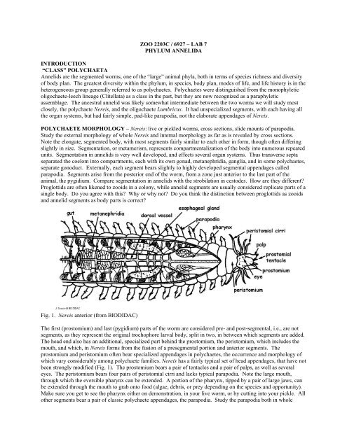
Lab 6: Annelida
Lab 6: Annelida
Lab 6: Annelida

Create successful ePaper yourself
Turn your PDF publications into a flip-book with our unique Google optimized e-Paper software.
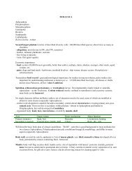
families recognized, we cover several locally common and important ones in this lab, as well as two others that usedto be considered separate phyla. You are expected to know the bolded ones. As you examine these animals, payspecial attention to the head structures on each: do they have prostomial tentacles, palps, peristomial cirri? Arethese simple or specialized? Do they have an eversible pharynx or jaws? How about the parapodia – are theysimple or elaborate; what are the chaetae like? These are the main characters that differentiate polychaete families.The relationships of polychaete families to each other are starting to be understood, but much remains to be learned.It is clear though that the ancestral polychaete was a fairly unspecialized, free-living worm with relatively simpleparapodia, and that three highly specialized lineages, that in the past were recognized as higher taxa (phyla andclass) – the Clitellata, Pogonophora, and Echiura – are highly modified members of the phylum. Recent work evensuggests that the phylum Sipuncula may turn out to be derived annelids as well.Family Nereididae – NereisSee above. Nereidids are “classic” errant (i.e. mobile, not sessile) polychaetes. Nereis vary greatly in their feedingmodes, both among as well as within species. Some are purely herbivorous, others eat animal prey (e.g.amphipods), deposit feed and even suspension feed (with a mucus bag!).Family Onuphidae – DiopatraDiopatra is a locally common (you have/will see it on field trips) errant polychaete that nevertheless constructs aparchment tube; the tube is buried in sediment except for its anterior end which projects a couple of centimetersabove the sediment surface. The emergent part of the tube is decorated with a variety of marine debris attached to itand is conspicuous in the field. Diopatra are omnivorous, feeding on live prey, dead animals, plants, andmicroorganisms. They have a pair of well developed pharyngeal jaws that can be everted to grab food. Theiranterior end has very well developed dorsal gills. Why do you think they have such well-developed gills? What arethe gills morphologically derived from?Family Glyceridae – Glycera - blood wormsGlycerids are characterized by a large, eversible proboscis armed with 4 jaws, each with a duct that delivers thesecretion of an associated poison gland. They readily evert their long proboscis when handled, and use it both forburrowing and predation. You can see the withdrawn proboscis, especially the jaws, through the body wall, but canalso elicit living worms to extend it by handling them. Glycerids are carnivores that live in elaborate burrowsystems in soft sediments, with multiple surface openings. They sense the vibration of their prey, and lunge outfrom their burrows to capture them. They have a characteristic conical prostomium with 4 short prostomialtentacles. The common name for glycerids, blood worms, refers to the presence of respiratory pigment in theircoelom, because, unusually among polychaetes, they lack a blood vascular system.Fig. 4. Head end and everted pharynx of glycerid (from Brusca X2 Invertebrates).Family Amphinomidae – fire wormsFire worms get their names from the pain they can inflict when handled. They have specialized, calcareous,harpoon-shaped chaetae. When disturbed, animals will arch their back to spread, display, and deploy these bundles
of fiber-glass like chaetae. The chaetae will readily penetrate and break off in your skin, and cause a lasting,burning and itchy sensation because of the discharge of associated neurotoxins. Amphinomids generally have arectangular cross section, and possess a caruncle – a mid-dorsal projection from the prostomium that extendsposteriorly across the front end of the animal. Many of the species are carnivorous (some are important coralpredators); they tend to be colorful and spectacular worms.Family Polynoidae – scale wormsThis large (and several related) families are characterized by paired dorsal scales (elytrae) that arch over, and areheld above the dorsum. How are the scales attached? What does the head end of the worm look like? What kind oflife style would you expect from a worm with a head like polynoids have?Family Aphroditidae – Aprhodite - sea miceSea mice are stout, large (to 20 cm) elliptical worms with wide segments, and a characteristic “hairy” dorsalcovering made of felt-like notochaetae (i.e. chaetae of the notopodium). These silky fibers project medially from thetop of the notopodium, and can completely cover the dorsum. Below the felt layer, aphroditids have a series ofpaired scales (elytra), like polynoids. Aphroditids are slow-moving, roving carnivores that live on muddy to sandybottoms. Sea mice have a large, eversible pharynx armed with two pairs of jaws.Family Arenicolidae – Arenicola - lug wormsArenicolids are large, deposit feeding polychaetes that live in U-shaped burrows in soft sediments. They swallowsediment, digest the organics from it, and defecate the remainder in coiled casts on the sediment surface. Like manyburrowing polychaetes, they lack much elaboration on their anterior end, but have a short, simple, eversible pharynx.Fig. 5. Life position of arenicolid (left) and pectinariid (right). Both are deposit feeders that live head down insediment. (from Brusca X2 Invertebrates).Family Pectinariidae – PectinariaBecause of the shape of their conical tubes, pectinariids are often referred to as ice cream cone worms. The neatlybuilt tubes are made of sand grains glued together edge to edge like bricks. Pectinariids are highly selective in theirchoice of sand grains to build their home – not the regularity in size, shape, and even the mineral of these grains.Despite living in tubes, pectinariids are mobile, living head down in sediment, with the narrow tip of their tubeprotruding above the sediment surface. They move about with specialized, large, golden chaetae (called paleae) thatextend from their anterior end; these can also close the tube like an operculum. Ice cream cone worms are selective,
subsurface deposit feeders. They are unusual among polychaetes in having a constant number of segments: 22 in allspecies!Family Chaetopteridae – ChaetopterusChaetopterus is one of the most highly specialized polychaetes. In addition to segmentation, it has a tagmatizedbody. Tagmatization refers to the specialization of different groups of segments for distinct functions. Arthropodsare tagmatized: the head, thorax, and abdomen of insects are tagmata, each comprised of several segments. Thelocal Chaetopterus variopedatus lives in a U-shaped parchment tube buried in sand, with both ends projecting abovethe sediment surface. It draws water into its tube with 3 specialized, dorsal, paddle-like flaps that are derived fromthe parapodia of segments 14-16. The water is passed through a mucus bag that is continuously secreted by a pair oflarge, dorsal, notopodial lobes on segment 12. The bag is collected by a dorsal cup on segment 13 that rolls themucus and captured food into a ball; this ball is periodically passed to the mouth via a dorsal food groove. The tubeof Chaetopterus provides a convenient home for several commensal animals. Thus most tubes have a pair of smallcrabs living in them. Note that the tube has a wide middle and two constricted ends. As the animal grows, the tubewould become tight – how do you think does the animal deals with this and makes its home bigger?Fig. 6. Chaetopterus variopedatus close up of anterior end (left) and life position in tube (right) (from Brusca X2Invertebrates).Family Terebellidae – spaghetti wormsSpaghetti worms are aptly named for their spaghetti-like feeding tentacles. These worms usually live in tubes builtfrom sediment, and extend their tentacles over the substratum in all directions from the tube opening. Tentaclesslowly “walk out” on their own, using muscular contractions and cilia, but can retract rapidly into the tube whendisturbed. Terebellids are selective deposit feeders on grains picked up by the tentacles. In pickles the tentacleslook like just a clump of curled spaghetti, but in living worms they are extended to several times the length of thebody – make sure you see undisturbed living worms on demonstration! Note also the well developed, reddish gillsthat look like a messy hair-do at the anterior end of the worm, quite close to the feeding tentacles.Family Sabellidae – feather duster wormsStudy the live sabellid in the display tanks, noting the spectacular plume of ciliated radioles used for suspensionfeeding. Add carmin to visualize water flow through this fan. Sabellids secrete a leathery tube from a collar-likeregion on the body. Animals can withdraw extremely rapidly into their tube when disturbed, because they have agiant nerve fibers (speed of nervous conduction is related to the diameter of the nerve; many animals haveindependently evolved giant nerve fibers for rapid conduction needed for escape behaviors). Examine a pickledexample to see the rest of the body, taken out of the tube. An unusual feature of sabellids is the inversion of
parapodia between the short, anterior, thoracic region and the long, posterior anterior region: note that the distinctneuropodia and notopodia are inverted at this junction. At the same junction the fecal groove (that transports fecesfrom the posterior anus to the anterior opening of the tube) switches around from a mid-ventral to a mid-dorsaltrack. How do you think this condition came about?Fig. 7. Sabellid brachiolar crown (left) and close-up of single brachiole showing morphology and particle transport(right) (from Brusca X2 Invertebrates).Family SerpulidaeSerpulids resemble sabellids in their well developed feeding fan, and suspension feed in a similar manner; howeverthey have a calcareous, rather than parchment tube. Many serpulids have a specialized operculum that closes theirtubes once they withdraw. The feeding fans of serpulids can be quite elaborate – see photos of “Christmas treeworms”, which live symbiotically in reef corals, and have a double stack of tree-like fans.Family SabellariidaeSabellariids are tube-dwelling polychaetes that construct their homes by gluing together sediment grains, andsuspension feed with ciliated tentacles. Sabellariids solved their sanitation problem quite differently from sabellids:the posterior end of their body is reduced to a thin, tail-like tube, which can be extended out of the tube opening, sortof like a perverted snorkel, for defecation. The larvae of at least some sabellariids are chemically attracted to andpreferentially settle on conspecifics; as a result these worms often live in large aggregations. Large reefs built by thesabellariid Phragmatopoma are common in some locations, including the southeast coast of Florida.Family Siboglinidae (former phylum Pogonophora) – picklesThe “Phylum Pogonophora” has a long and twisted history. Discovered over a hundred years ago, these gutless,thin, elongate worms were thought at various times to be deuterostomes or protostomes. Recent work places themfirmly within polychaetes; their unusual morphology is the result of rapid evolutionary change associated with theacquisition of a novel life-style: chemosymbiosis. All pogonophorans have chemoautotrophic bacteria living in alarge “trophosome” a mid-body tissue mass derived from the gut, but lacking a lumen. The animal provides thebacteria with H 2 S, which the bacteria use as a chemical energy source for fixing organic compounds, which theworm then uses. Siboglinids have one to many tentacles that extend from a large parchment tube. At the posteriorend of the body they have a small segmented portion, the opisthosoma, that hints at their polychaete origins.
Fig. 8. Schematic diagram of a pogonophoran (from Brusca X2 Invertebrates).Echiura (several families; formerly a phylum)Echiurans are unsegmented, coelomate worms, with a fairly simple morphology. They are largely infaunal in softsediments (extending to the deepest ocean depths, where they can be common), although several species nestle increvices. Recent evidence suggests that echiurans are highly derived polychaetes that have lost segmentation, butwhich retain a couple of chaetae in most species. They are selective deposit feeders that use their proboscis togather surface sediments. In some echiurans (family Echiuridae) the proboscis is chunky and short and both sexesare large. In the Bonellidae the proboscis is spaghetti-like, with a T-shaped end, and extends to many times thelength of the body. Bonellids have dwarf, degenerate males, which are not much more than bags of testes, and livewithin the female nephridium-gonoduct complex. Sex determination in bonellids is environmental: larvae that settleon females become males; those settling freely develop into females. Study the worms provided, noting theproboscis and body; find the chaetae in echiurids.Fig. 9. Three genera of echiurans: A. Echiurus, B. Listriolobus, C. Bonellia (left), echiuran anatomy (right) (fromBrusca X2 Invertebrates).CLITELLATA – OligochaetesOligochaetes are characterized by segments that have but two small clumps of chaetae and lack parapodia; they alsolack the various head appendages found in most polychaetes. They differ substantially in their reproductive biology
as well. Species are hermaphroditic, only a small number of segments are reproductive, and they have a clitellum, aseries of specialized segments, that secrete a cocoon for the eggs. The exercise on earthworm anatomy will runintroduce their reproductive specialization as well as their general morphology.OLIGOCHAETE MORPHOLOGY – Lumbricus dissection.The TA will anaesthetize your earthworm in a weak solution of alcohol. Lay out the worm in a dissecting pen, andpin it out in a couple of places. Note the simple anterior, lacking appendages, and the two bundles of short chaetae,but not parapodia, extending laterally from each segment. Chaetae help grip the soil during burrowing. Make ashallow, but long, longitudinal incision through the mid dorsal line along the anterior third of the worm. Carefullyseparate, and pin out the body wall, so as to reveal the internal anatomy. The gut includes an anterior pharynx, wellsupplied by external muscles, followed by an elongate esophagus, then a slightly distended crop, followed by agizzard, then an intestine. The dorsal vessel is clearly visible on top of the gut; remember it is set within the dorsalmesentery in a retroperitoneal position. Large circumesophageal vessels emerge from the dorsal vessel in theesophageal region and encircle the esophagus. Calciferous glands also lie on the esophagus in a dorsolateralposition; these are involved in Ca ++ regulation, taking up CaCO 3 out of ingested soil and precipitating it out inclumps of calcite so that it does not interfere with digestion. Note the paired metanephridia in each segment. Thefunnel of each metanephridium opens in one segment, the nephridioduct than traverses the posterior septum, so thatthe bulk of it, together with the nephridiopore opening to the outside, lie in the following segment.Fig. 10. Earthworm anatomy (top); schematic diagram of earthworm reproductive segments (below) (fromBIODIDAC).
The reproductive system of oligochaetes is highly specialized and conserved across the group, it extends fromsegment 9 to 15. Examine the diagrammatic summary above first, then try to locate the various structures in yourdissected animal. The male system includes a pair of small testes ventral to the large seminal vesicles, which storethe sperm produced. Sperm are then released into the coelom and collected by seminal funnels (similar in structureto metamenephridia). Two pairs of seminal funnels, which lie in male segments 10 & 11 are drained by commonsperm ducts that exit through male gonopores on segment 15. The female system is comprised of a pair of ovaries,and ovisacs (where eggs are stored prior to fertilization) and an oviducal funnel (homologous to the seminal funnel)on segment 13, that leads through a short oviduct to the female gonopore on segment 14. Segments 9 and 10 alsohave seminal receptacles that have a separate opening, and store sperm received during mating. The clitellum liesposterior to these reproductive segments, its position varying among, but consistent within species. The clitellum isa series of specialized segments with a highly glandular epithelium, and is present in all clitellates. Mating leads toreciprocal sperm transfer from the male gonopore to the partner’s seminal receptacle. Subsequent to mating, theworm creates a cocoon for its offspring by the clitellum. The cocoon is passed anteriorly, receives the eggs from thefemale gonopore, then the stored sperm, then it slips off over the head and is sealed.CLITELLATA – HIRUDINEALeeches – external morphology. Live and whole mount.Leeches are derived from within oligochaetes, making the latter paraphyletic. They are specialized in a number ofways. First, they develop anterior and posterior suckers that allow them to attach, as well as to move by inchwormtypelocomotion. Watch this locomotion in live animals if available. Second, unlike almost all polychaetes andoligochaetes, leeches have a fixed number of segments: most (i.e., “higher”) leeches have 34. Each segment issubdivided externally into several rings, or annuli, while several or the anterior and posterior segments are fused intothe anterior and posterior suckers. Finally, the coelom of leeches is reduced to a set of circulatory vessels thatresemble the BVS.Fig. 11. External morphology of a higher leach (Hirudo medicionalis) (from Brusca X2 Invertebrates).