ieee transactions on biomedical engineering, vol. 46, no. 10
ieee transactions on biomedical engineering, vol. 46, no. 10
ieee transactions on biomedical engineering, vol. 46, no. 10

You also want an ePaper? Increase the reach of your titles
YUMPU automatically turns print PDFs into web optimized ePapers that Google loves.
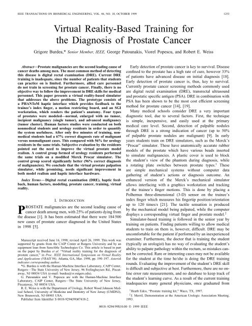
IEEE TRANSACTIONS ON BIOMEDICAL ENGINEERING, VOL. <strong>46</strong>, NO. <strong>10</strong>, OCTOBER 1999 1253Virtual Reality-Based Training forthe Diag<strong>no</strong>sis of Prostate CancerGrigore Burdea,* Senior Member, IEEE, George Patounakis, Viorel Popescu, and Robert E. WeissAbstract—Prostate malignancies are the sec<strong>on</strong>d leading cause ofcancer deaths am<strong>on</strong>g men. The most comm<strong>on</strong> method of detectingthis disease is digital rectal examinati<strong>on</strong> (DRE). Current DREtraining is inadequate, since the number of patients that studentscan practice <strong>on</strong> is limited. Furthermore, allied care pers<strong>on</strong>neldo <strong>no</strong>t train in screening for prostate cancer. Finally, there is <strong>no</strong>objective way to follow the improvement in DRE skills for medicalpers<strong>on</strong>nel. This paper presents a virtual reality-based simulatorthat addresses the above problems. The prototype c<strong>on</strong>sists ofa PHANToM haptic interface which provides feedback to thetrainee’s index finger, a moti<strong>on</strong> restricting board, and an SGIworkstati<strong>on</strong>, which renders the patient’s anatomy. Four typesof prostates were modeled—<strong>no</strong>rmal, enlarged with <strong>no</strong> tumor,incipient malignancy (single tumor), and advanced malignancy(tumor cluster). Human factors studies were c<strong>on</strong>ducted <strong>on</strong> bothn<strong>on</strong>medical students and urology residents in order to quantifythe system usefulness. After <strong>on</strong>ly five minutes of training, n<strong>on</strong>medicalstudents had a 67% correct diag<strong>no</strong>sis rate of malignantversus n<strong>on</strong>malignant cases. This compared with 56% for urologyresidents in the same trials. Subjective evaluati<strong>on</strong> by the residentspointed out the need to improve the virtual prostate modelrealism. A c<strong>on</strong>trol group formed of urology residents performedthe same trials <strong>on</strong> a modified Merck Procar simulator. Thec<strong>on</strong>trol group scored significantly better (96% correct diag<strong>no</strong>sisof malignancies). We c<strong>on</strong>clude that the virtual prostate palpati<strong>on</strong>simulator, while promising, needs significant improvement inboth model realism and haptic interface hardware.Index Terms— Digital rectal examinati<strong>on</strong> (DRE), haptic feedback,human factors, modeling, prostate cancer, training, virtualreality.I. INTRODUCTIONPROSTATE malignancies are the sec<strong>on</strong>d leading cause ofcancer death am<strong>on</strong>g men, with 25% of patients dying fromthe disease [1]. It has been estimated that there were 184 500new cases of prostate cancer diag<strong>no</strong>sed in the United Statesin 1998 [5].Manuscript received June 16, 1998; revised April 26, 1999. This work wassupported by grants from the CAIP Center at Rutgers University and by anequipment loan from SensAble Tech<strong>no</strong>logies Co. This article is based in part<strong>on</strong> the paper by Burdea et al. “Virtual reality training for the diag<strong>no</strong>sis ofprostate cancer,” in Proc. IEEE Internati<strong>on</strong>al Symposium <strong>on</strong> Virtual Realityand Applicati<strong>on</strong>s (VRAIS’98), Atlanta, GA, Mar. 1998, pp. 190–197. Asteriskindicates corresp<strong>on</strong>ding author.*G. Burdea is with the Human-Machine Interface Laboratory, CAIP Center,Rutgers—The State University of New Jersey, 96 Frelinghuysen Rd., Piscataway,NJ 08854 USA (e-mail: burdea@vr.rutgers.edu).G. Patounakis and V. Popescu are with the Human-Machine InterfaceLaboratory, CAIP Center, Rutgers—The State University of New Jersey,Piscataway, NJ 08854 USA.R. E. Weiss is with the Department of Urology, Robert Wood Johns<strong>on</strong> MedicalSchool, University of Medicine and Dentistry of New Jersey (UMDNJ),New Brunswick, NJ 08903 USA.Publisher Item Identifier S 0018-9294(99)07418-2.Early detecti<strong>on</strong> of prostate cancer is key to survival. Diseasec<strong>on</strong>fined to the prostate has a high rate of cure, however 33%of patients have advanced disease <strong>on</strong> initial diag<strong>no</strong>sis [18].Early detecti<strong>on</strong> of prostate cancer is, thus, key to survival.Currently prostate cancer screening methods comm<strong>on</strong>ly usedare digital rectal examinati<strong>on</strong> (DRE), transrectal ultrasoundand prostatic specific antigen (PSA). DRE in combinati<strong>on</strong> withPSA has been shown to be the most cost efficient screeningmethod for prostate cancer [14], [19].Many medical schools c<strong>on</strong>sider DRE a very importantdiag<strong>no</strong>stic tool, due to several factors. First, the techniqueis simple, inexpensive, and easily used at the primarymedical care level. Sec<strong>on</strong>d, detecti<strong>on</strong> of palpable <strong>no</strong>dulesthrough DRE is a str<strong>on</strong>g indicati<strong>on</strong> of cancer (up to 50%of palpable prostate <strong>no</strong>dules are malignant) [9]. In earlyyears students train <strong>on</strong> DRE simulators, such as the Merck’s“Procar” simulator. These have anatomically accurate rubbermodels of the prostate which have various beads insertedto simulate malignancies. A plastic cover is used to blockthe student’s view of the phantom during diag<strong>no</strong>sis, whilea rotating plate switches between prostate types. 1 Theseare simple mechanical systems without computer datagathering of student’s acti<strong>on</strong>s or diag<strong>no</strong>sis outcome. Anenhanced versi<strong>on</strong> of the Merck’s mechanical simulatorallows interfacing with a graphics workstati<strong>on</strong> and trackingof the trainee’s finger moti<strong>on</strong>s. This is d<strong>on</strong>e by placing aPolhemus three-dimensi<strong>on</strong>al (3-D) sensor <strong>on</strong> the trainee’sindex finger which measures his fingertip positi<strong>on</strong>/orientati<strong>on</strong>up to 120 times/s [21]. The tactile sensati<strong>on</strong> is producedby the mechanical model being palpated, while the computerdisplays a corresp<strong>on</strong>ding virtual finger and prostate model. 2Simulator-based training is followed in the senior year bytraining <strong>on</strong> patients. Finding patients willing to allow medicalstudents to train <strong>on</strong> them is, however, difficult. DRE may beuncomfortable for the patient if performed by an inexperiencedexaminer. Furthermore, the doctor that is training the student(typically an urologist) has <strong>no</strong> way of evaluating the student’sability to palpate pathology within the rectum, so mistakes can<strong>no</strong>tbe corrected. Rare or interesting cases may <strong>no</strong>t be availablefor the student at the time he/she is doing the DRE trainingrounds. Evaluating the improvement of the student’s DRE skillis difficult and subjective at best. Furthermore, there are <strong>no</strong> <strong>on</strong>lineerror rate measurements, and <strong>no</strong> database to keep track ofthe student’s learning curve. As a result of the current traininginadequacies many general physicians, <strong>on</strong>ce graduated from1 Heath Edco, “Prostate training kit,” Waco, TX, 1997.2 J. Merril, Dem<strong>on</strong>strati<strong>on</strong> at the American Urologic Associati<strong>on</strong> Meeting,1993.0018–9294/99$<strong>10</strong>.00 © 1999 IEEE
BURDEA et al.: DIAGNOSIS OF PROSTATE CANCER 1255Fig. 2. Simulati<strong>on</strong> hardware setup [4].loop, which renders organ models in the area of interest <strong>on</strong>the SGI High-Impact workstati<strong>on</strong>. When simulating humananatomy <strong>on</strong>e has the choice of generic, or patient-specificmodels obtained from computed tomography or magneticres<strong>on</strong>ance imaging data. In this proof-of-c<strong>on</strong>cept researchgeneric models were used, as they are easily available fromvarious vendors.A 3-D model was purchased from Viewpoint DataLabs[26] c<strong>on</strong>sisting of an anatomically accurate adult male urinarytract and intestines. In order to allow realtime rendering <strong>on</strong>our SGI medium-range computer the model was subsequentlysimplified by eliminating the area above the waist and belowthe knees, as well as the kidneys and the intestine abovethe rectal regi<strong>on</strong>. The model was then shaded using OpenGLgraphics libraries [20] and rendered at about 18 frames/swithout <strong>no</strong>ticeable latencies. In previous studies it was shownthat this frame rate is sufficient for virtual object manipulati<strong>on</strong>tasks [22].A low-resoluti<strong>on</strong> model of a right virtual hand was alsopurchased from Viewpoint DataLabs. Subsequently it wasc<strong>on</strong>figured in a fist gesture with the index extended at a fixedangle. This was needed since the PHANToM does <strong>no</strong>t readhand gestures, and <strong>on</strong>ly measures the 3-D positi<strong>on</strong> of thetrainee’s index fingertip. This data was mapped to the indexof the virtual hand, mimicking the moti<strong>on</strong> of the user’s indexand haptic interface. Once the trainee’s hand enters the rectalregi<strong>on</strong> of the virtual patient, and the prostate is in view, <strong>on</strong>lythe index finger is rendered, which further reduces the modelgraphics complexity.The prostate and its malignancies were modeled by generatingthe vertices of a hemisphere and then c<strong>on</strong>necting them intofour-sided polyg<strong>on</strong>s. The prostate surface median groove wascreated by depressing all the vertices that lie <strong>on</strong> this l<strong>on</strong>gitudinalline. After the polyg<strong>on</strong>s have been c<strong>on</strong>structed, the <strong>no</strong>rmalsto the surfaces and vertices were calculated for graphics andhaptics shading. A sample of the simulator graphics for anenlarged prostate is shown in Fig. 3 [4]. The complexity of themodel for the prostate was 200 vertices, while each malignancywas c<strong>on</strong>structed with an additi<strong>on</strong>al <strong>10</strong>0 vertices.The prostate model (but <strong>no</strong>t its malignancies) could bedeformed graphically where compressed by the virtual fin-Fig. 3. Interior view of an enlarged benign prostate [4].gertip. This was accomplished by detecting the “collisi<strong>on</strong>”between the surface vertex and the vertex of the fingertip.The magnitude of deformati<strong>on</strong> was determined by the distancethe finger penetrates the surface, with the surroundingvertices being deformed less, as the distance from the pointof collisi<strong>on</strong> increased. This gave the illusi<strong>on</strong> that the prostatewas graphically compressed by the fingertip.Graphics computati<strong>on</strong>s were <strong>on</strong>ly <strong>on</strong>e part of the workstati<strong>on</strong>load. The other important comp<strong>on</strong>ent of the softwareenvir<strong>on</strong>ment is the physical modeling, including c<strong>on</strong>tact detecti<strong>on</strong>and force feedback computati<strong>on</strong>. The graphics and hapticloops run c<strong>on</strong>currently but asynchr<strong>on</strong>ously, as the haptics loopc<strong>on</strong>trolling the PHANToM has a much larger bandwidth (about<strong>10</strong>00 Hz).The physical modeling task takes advantage of the GHOSThaptic library used to run the PHANToM arm [25]. In developingthe prostate palpati<strong>on</strong> simulati<strong>on</strong>, speed had to beweighted against force feedback realism. These two opposingrequirements called for optimizati<strong>on</strong> of the collisi<strong>on</strong> detecti<strong>on</strong>process, without which the model complexity would havebeen <strong>on</strong>ly <strong>10</strong>0 vertices (and <strong>no</strong>t 200–500 vertices as describedabove).The optimizati<strong>on</strong> takes advantage of the hemisphericalshape of the prostate surface. It assumes that when projected<strong>on</strong>to a plane the prostate top surface has the shadow ofc<strong>on</strong>centric circles. This assumpti<strong>on</strong> transforms the previoustwo-dimensi<strong>on</strong>al search for collisi<strong>on</strong> locati<strong>on</strong> into two <strong>on</strong>edimensi<strong>on</strong>alsearches. This results in a reducti<strong>on</strong> of the collisi<strong>on</strong>detecti<strong>on</strong> computati<strong>on</strong> time by an order of magnitude.As illustrated in Fig. 4 [4], the search first finds the closestc<strong>on</strong>centric circle to the fingertip vertex, then searches for theclosest vertex <strong>on</strong> that circle.Once the vertices of the polyg<strong>on</strong> the fingertip is in c<strong>on</strong>tactwith have been determined, the palpati<strong>on</strong> force vector isdetermined by the following formula:where represents the tissue stiffness. This parameter wasdetermined experimentally, based <strong>on</strong> the subjective evaluati<strong>on</strong>of our urologist coauthor, such that the virtual models felt
1256 IEEE TRANSACTIONS ON BIOMEDICAL ENGINEERING, VOL. <strong>46</strong>, NO. <strong>10</strong>, OCTOBER 1999Fig. 4. Vertex search optimizati<strong>on</strong> [4].Fig. 5. Interior view of a prostate with a single tumor [4].“real.” For example, the malignancies had a that was 80%larger than that of the stiffness of the surrounding tissue.In the above formula is the penetrati<strong>on</strong> distance al<strong>on</strong>g the<strong>no</strong>rmal defined at the surface pointand is the unity step functi<strong>on</strong> (forces are applied <strong>on</strong>ly whenthe surface is deformed by the fingertip).The functi<strong>on</strong> models a n<strong>on</strong>linear deformati<strong>on</strong>, instead ofthe customary Hooke’s law ( ). The simulati<strong>on</strong> usesan arctan functi<strong>on</strong> for the deformati<strong>on</strong> model, with the slopeof c<strong>on</strong>trolled according to the desired object deformati<strong>on</strong>model. A<strong>no</strong>ther useful aspect of the arctan functi<strong>on</strong> is that itproduces bounded forces, avoiding a premature exit due toGHOST safeguards (overload c<strong>on</strong>diti<strong>on</strong>). The <strong>no</strong>rmal vectorat the point of c<strong>on</strong>tact <strong>on</strong> the surface is determined in away similar to the Ph<strong>on</strong>g shading routine used in computergraphics [8]. In our simulati<strong>on</strong> force shading is implementedby calculating a weighted average of the vertex <strong>no</strong>rmals closestto the point of c<strong>on</strong>tact. The <strong>no</strong>rmal vector obtained from thiscalculati<strong>on</strong> is used to determine the directi<strong>on</strong> of the forcethe surface exerts <strong>on</strong> the finger and it is also used as theprojecti<strong>on</strong> vector for the calculati<strong>on</strong> of the distance the fingerpenetrated the surface. The magnitude of the force calculatedis subsequently averaged with the last force magnitude to geta smoother transiti<strong>on</strong> between subsequent forces. This reducesthe effect of mechanical vibrati<strong>on</strong>s, or “buzzing,” that wouldresult from unsmoothed force transiti<strong>on</strong>s.III. TRAINING SIMULATIONThe simulati<strong>on</strong> can be run in “training mode” and “examinati<strong>on</strong>mode.” The training mode allows for visual feedbackof the patient’s anatomy. It starts by showing the male patientbent in the usual DRE positi<strong>on</strong>. The trainee then positi<strong>on</strong>s thevirtual finger <strong>on</strong> the screen such that it lines up with the rectalarea. As the trainee pushes through the anus, the inertia effectproduced by GHOST provides the feel of resistance. Once theFig. 6. Interior view of a transparent prostate with an advanced malignantcluster [4].finger has penetrated the rectum, the inertia effect stops andthe graphics switches to an interior view of the patient.For training purposes the patient’s interior view showsa porti<strong>on</strong> of the rectum rendered in “wire frame” so thetrainee can see the intestine wall, the top of the prostate, thebladder, and an index finger. If the trainee pushes the virtualfinger against the rectum wall, he feels a small resistance tothe moti<strong>on</strong>. When the trainee starts palpating the prostate,the prostate model deforms and forces are fed back by thePHANToM, as described previously. The trainee can feel themedian groove which helps orient him hapticly before startingthe palpati<strong>on</strong> process.The simulator can display <strong>on</strong>e of four models that areavailable for palpati<strong>on</strong>. These are: 1) <strong>no</strong>rmal prostate, 2)benign enlarged prostate (prostatic hypertrophy), 3) earlysingle malignant <strong>no</strong>dule (illustrated in Fig. 5), and 4) lateinvasive cluster tumors (illustrated in Fig. 6). The tumorlocati<strong>on</strong>s are in turn randomized to four lobes of the prostatetop surface, which results in 12 cases of virtual patients totrain <strong>on</strong>. More could be created, such as rare cases, or patientspecificcases, but this was bey<strong>on</strong>d the scope of the currentresearch.
BURDEA et al.: DIAGNOSIS OF PROSTATE CANCER 1257The trainee has the opti<strong>on</strong> of seeing through the prostate surfaceby turning the model to wire frame rendering (see Fig. 6).This forms a mental mapping of the anatomy/pathology withthe haptic feedback the trainee receives when palpating thearea of interest. After the trainee feels c<strong>on</strong>fident with thecurrent case (model being palpated), he can switch to a<strong>no</strong>thercase with a simple keystroke.Once the trainee has palpated each type of 12 prostate cases,the simulati<strong>on</strong> is switched to the “examinati<strong>on</strong> mode.” Duringthis mode the trainee has to diag<strong>no</strong>se each case presentedwithout seeing the prostate <strong>on</strong> the screen, similar to realDRE’s. The screen displays <strong>on</strong>ly a red sphere during “rest”and a green sphere during “palpate” periods. In examinati<strong>on</strong>mode, all cases are randomized by prostate type and by tumorlocati<strong>on</strong>. The trainee inputs his diag<strong>no</strong>sis by pressing a keythat corresp<strong>on</strong>ds to the prostate type being examined. Theresp<strong>on</strong>se time for diag<strong>no</strong>sis, as well as diag<strong>no</strong>sis errors, arerecorded transparently for each trainee’s resp<strong>on</strong>se.During the examinati<strong>on</strong> each of the four different prostatetypes are presented three times in random order. After thetrainee gives his diag<strong>no</strong>sis, the trainee has a five-sec<strong>on</strong>d breakuntil the next case is presented. The trainee gets a three-minutebreak after every four cases presented. Once the examinati<strong>on</strong>is completed, the results can be viewed by reading a text filewhich lists what was recorded during the test. The trainee’sacti<strong>on</strong>s can also be recorded for play-back at a later time. Thisfile records the trajectory traced by the virtual finger duringexaminati<strong>on</strong> and the case presented at that time. This graphicsplayback provides a way to analyze the trainee’s palpati<strong>on</strong>technique, and what can be d<strong>on</strong>e to improve and correct anyerrors. The same software can be used to record and play backexpert examinati<strong>on</strong>s to show the trainee exactly how DRE’sshould be d<strong>on</strong>e. To play-back a recorded file, the simulati<strong>on</strong>simply changes the source of its input from the PHANToMto the data file.IV. HUMAN FACTOR STUDIESThe VR-based DRE training system has some clear advantagesover current training methods, however, it is essential toquantify its usefulness as a learning envir<strong>on</strong>ment for medicalstudents. This required human factors studies to quantifyobjective variables such as the trainee’s learning curve overrepeated cases. Other variables that were important to bemeasured include the diag<strong>no</strong>sis accuracy, the time taken to diag<strong>no</strong>seeach case, and the case that proved hardest to diag<strong>no</strong>se.In order to measure these variables we performed a series ofthree studies. These studies in<strong>vol</strong>ved two experimental groupsand a c<strong>on</strong>trol group.A. Experimental ProtocolThe first study used 22 n<strong>on</strong>medical students as subjects,16 male, and six female. All subjects were <strong>vol</strong>unteers, andwere <strong>no</strong>t compensated for participati<strong>on</strong> in the project. Eachsubject was given an overview of how to palpate the prostatecorrectly using rubber models of the prostate. These models aremanufactured by Health Edco (Waco, TX) and differ from themodels <strong>no</strong>rmally used by the Merck’s Procar simulator. Theywere told to first identify the median groove and evaluate thesize of the prostate. The subjects were then told to scan theprostate surface in small increments looking for <strong>no</strong>dules thatsignify tumors. Subsequently, each subject had five minutesof training using the PHANToM arm without the moti<strong>on</strong>restricti<strong>on</strong> board. During this time the wire-frame model ofthe prostate was made visible <strong>on</strong> the screen. Subjects werereminded of the correct way of palpating a prostate andpresented with all four prostate types (<strong>no</strong>rmal, enlarged, singlemalignancy, and cluster malignancy) at least twice. Duringtraining, subjects were allowed to c<strong>on</strong>centrate <strong>on</strong> the casesthey felt were difficult.Once the five-minute training period ended, the simulati<strong>on</strong>would blank the screen and display a red sphere signifyingthe initiati<strong>on</strong> of the test. From this point <strong>on</strong>, subjects were<strong>no</strong>t instructed <strong>on</strong> palpati<strong>on</strong> technique, as the simulati<strong>on</strong> was inthe “examinati<strong>on</strong> mode.” Each subject was presented with 12random cases with a three-minute break between every fourcases. They were <strong>no</strong>t given feedback <strong>on</strong> their performance untilafter the experiment was finished. The data recorded for eachsubject was the length of time to make a diag<strong>no</strong>sis (sec<strong>on</strong>ds),the diag<strong>no</strong>sis the subject chose, and the correct diag<strong>no</strong>sis forthat case. No subjective assessment was d<strong>on</strong>e by the subjectsin the first experimental group.Subsequently, a sec<strong>on</strong>d experimental group was selectedfrom the urology residents at Robert Wood Johns<strong>on</strong> MedicalSchool. These subjects were also <strong>vol</strong>unteers, so they were<strong>no</strong>t compensated for participati<strong>on</strong> in the project. Due to thec<strong>on</strong>straints of medical practice the sec<strong>on</strong>d group was muchsmaller, c<strong>on</strong>sisting of <strong>on</strong>ly four subjects. Furthermore thesesubjects were in a high stress/high fatigue state, having justcompleted 24-h rounds in the hospital emergency room.The protocol used <strong>on</strong> the sec<strong>on</strong>d group was similar tothat used <strong>on</strong> the first group, namely the same objectivevariables were measured and stored for analysis. The <strong>on</strong>eexcepti<strong>on</strong>, however, was that the residents had to use themoti<strong>on</strong> restricting board, which made the test harder, butsomewhat more realistic. Additi<strong>on</strong>ally, these subjects wereasked to subjectively assess the simulator, based <strong>on</strong> their priork<strong>no</strong>wledge of how real <strong>no</strong>rmal and malignant prostates feelduring DRE.The third (and last) subject group acted as a c<strong>on</strong>trol. Subjectsin this group were other <strong>vol</strong>unteer urology residents fromthe R. W. Johns<strong>on</strong> Medical School. The protocol in theircase in<strong>vol</strong>ved palpati<strong>on</strong> of rubber models as part of a Procarsimulator. The Procar was modified by adding the samemoti<strong>on</strong> restricti<strong>on</strong> board that was used with the PHANToMarm, however, the prostates were <strong>no</strong>w rubber models, ratherthan virtual <strong>on</strong>es. The examinati<strong>on</strong> time, rest periods, andcase randomizati<strong>on</strong> were the same as in the previous twoexperiments. Timing was measured with a stopwatch andresp<strong>on</strong>ses given orally (as opposed to using a keyboard asthe two previous groups did).B. Experimental ResultsTable I compares the overall diag<strong>no</strong>sis accuracy of thesubjects in our study. The n<strong>on</strong>medical students had c<strong>on</strong>-
1258 IEEE TRANSACTIONS ON BIOMEDICAL ENGINEERING, VOL. <strong>46</strong>, NO. <strong>10</strong>, OCTOBER 1999TABLE ICORRECT DIAGNOSIS RATES (ADAPTED FROM [4])SubjectcategoryExact Diag<strong>no</strong>sis(four cases)Benign/MalignantDiag<strong>no</strong>sisN<strong>on</strong>medicalStudents <strong>46</strong>% 67%Urology Residents 33% 56%C<strong>on</strong>trol Group 92% 96%sistently better diag<strong>no</strong>sis accuracy than medical residents(<strong>46</strong>% correct versus 33% correct) when using the PHANToMsimulator. This may be explained by the use of the moti<strong>on</strong>restricti<strong>on</strong> board by the residents, which increased the taskdifficulty. A<strong>no</strong>ther factor that impacted the performance ofthe sec<strong>on</strong>d group was fatigue when taking the test. A similartrend was observed when comparing the subject’s ability todistinguish the malignant cases (incipient or advanced) fromn<strong>on</strong>malignant cases (<strong>no</strong>rmal or enlarged prostates). Here, againthe performance of the first group was better (67% correctversus 56% correct). The c<strong>on</strong>trol group using the Procar didsignificantly better than both groups with exact diag<strong>no</strong>sticrates at 92% and detecti<strong>on</strong> of malignancies at 96%. Thismay be explained in part by the richer tactile informati<strong>on</strong>provided by the rubber models, compared to the simplifiedvirtual models used in the trials. Furthermore, the task for thec<strong>on</strong>trol group was easier since the locati<strong>on</strong> of the malignancywas <strong>no</strong>t randomized. There was <strong>on</strong>ly <strong>on</strong>e incipient and <strong>on</strong>eadvanced malignant rubber models in the Procar, versus foureach in the PHANToM simulator.Table II details the subjects’ diag<strong>no</strong>sis accuracy as a functi<strong>on</strong>of individual case type (benign, enlarged, single tumor,and cluster of tumors). The n<strong>on</strong>medical students were mostaccurate in detecting single tumors (58% correct diag<strong>no</strong>sis),and had the worst performance when detecting a cluster oftumors (34% correct diag<strong>no</strong>sis). The urology residents in thesec<strong>on</strong>d group of subjects were similarly most accurate indetecting single tumors (50% correct diag<strong>no</strong>sis), with theirworst performance when diag<strong>no</strong>sing benign prostates (25%accurate). This poor performance may be explained by theresidents’ subjective evaluati<strong>on</strong>. Here, they complained thatthe <strong>no</strong>rmal prostate model did <strong>no</strong>t feel sufficiently realistic,based <strong>on</strong> their prior mental model, and the median groovewas <strong>no</strong>t sufficiently delineated. The c<strong>on</strong>trol group had <strong>10</strong>0%accuracy for <strong>no</strong>rmal prostates. Their subjective evaluati<strong>on</strong>indicated that the median groove in the EDCO rubber modelswas too delineated, making it too easy to find. Similarly, resultsshow a much larger correct diag<strong>no</strong>sis rate for the c<strong>on</strong>trolgroup in the other three cases (benign enlarged, incipient, andadvanced malignancies). The correct diag<strong>no</strong>sis rates were inthis case 92%, 92%, and 83%, respectively.The above experimental results gave a measure of c<strong>on</strong>fidencein the simulator ability to model various prostate cases,even though the sample populati<strong>on</strong> was small. If the hapticmodels were poor, then all percentages in Table II would havebeen 25%, meaning chance probability. A<strong>no</strong>ther importantvariable is the learning curve, namely the increase in diag<strong>no</strong>sisaccuracy over repeating trials of the same case. As illustratedin Fig. 7 [4], both experimental groups using the PHANToMsimulator learned during the trials. However, the n<strong>on</strong>medicalstudents had a steeper learning curve between the first andsec<strong>on</strong>d trials, as they had <strong>no</strong> prior k<strong>no</strong>wledge or experiencewith DRE’s. The learning curve of the subjects in the sec<strong>on</strong>dgroup was different, with the steepest slope between the sec<strong>on</strong>dand third trials. The residents had to first adapt to the simulator,and its limited realism. Once they learned the simulator, theyimproved performance in the third trial. There was <strong>no</strong> learningfor the c<strong>on</strong>trol group, a clear indicati<strong>on</strong> that the task was tooeasy for them. These c<strong>on</strong>trols all had prior experience with theProcar, the models did <strong>no</strong>t have the same randomizati<strong>on</strong> as thePHANToM simulator had, and the rubber models providedmore tactile realism.Fig. 7 also shows the variati<strong>on</strong> in the standard deviati<strong>on</strong>in the correct diag<strong>no</strong>sis percentage over repeated trials. Therewas a small reducti<strong>on</strong> in the standard deviati<strong>on</strong> of n<strong>on</strong>medicalstudents’ resp<strong>on</strong>ses, which meant that as subjects learned,they behaved more uniformly as a group. A similar, butmore dramatic, trend was displayed by urology residents inthe sec<strong>on</strong>d group. These subjects had a very large standarddeviati<strong>on</strong> of the correct diag<strong>no</strong>sis percentage in their first trial.This large variability may be attributed in part to the lack ofsufficient realism of the virtual prostate cases, because of thelimited computing power of the workstati<strong>on</strong> used for testing.The large variability may also have been due in part to fatigue,as discussed previously. Subjects in the c<strong>on</strong>trol group havebetter uniformity of resp<strong>on</strong>se (smaller standard deviati<strong>on</strong>) thaneither of the experimental groups. Again, this is an indicati<strong>on</strong>of a familiarity with the task.Table III shows the overall time taken by each subject groupto diag<strong>no</strong>se individual cases. The n<strong>on</strong>medical students werec<strong>on</strong>sistently slower to diag<strong>no</strong>se than the residents, which is in<strong>no</strong> doubt a result of prior experience. Students took l<strong>on</strong>gestto diag<strong>no</strong>se enlarged prostates (60 s) and the least amount oftime to detect single tumors (48 s). The residents had similarlythe shortest time overall in detecting incipient tumors (39 s).Their difficulty was in diag<strong>no</strong>sing <strong>no</strong>rmal prostates (53 s),again due to the model geometry which was <strong>no</strong>t what theyhad expected. The c<strong>on</strong>trol group was three to five times fasterthan the residents in the sec<strong>on</strong>d group. They took the leastamount of time to detect a <strong>no</strong>rmal prostate (9 s) and the mosttime to diag<strong>no</strong>se advanced malignancies (16 s <strong>on</strong> average).Residents which were subjects in the sec<strong>on</strong>d and thirdgroups were asked subjective questi<strong>on</strong>s as to the usefulnessand realism of the simulati<strong>on</strong>, as well as perceived problemsor difficulties they may have had. The sec<strong>on</strong>d group resp<strong>on</strong>seswere optimistic in terms of eventual usefulness of the PHAN-ToM system as a teaching aid, provided more realism in themodeling was achieved. One resident had shorter fingers whichprevented him from fully palpating the prostate, which wesuspect may have happened also in actual DRE’s. Subjectsin the third (c<strong>on</strong>trol) group were asked to subjectively ratethe realism of the rubber models they palpated, as well asthe task difficulty when using the modified Procar with theadded moti<strong>on</strong> restricti<strong>on</strong> board. They felt that the model for the<strong>no</strong>rmal prostate was too soft and too delineated. Furthermore,they rated the board as too restrictive in terms of finger moti<strong>on</strong>when compared to the anal orifice.
BURDEA et al.: DIAGNOSIS OF PROSTATE CANCER 1259SubjectcategoryTABLE IICORRECT DIAGNOSIS OF INDIVIDUAL PROSTATE CASES (ADAPTED FROM [4])Benign<strong>no</strong>rmal sizeBenignEnlargedMalignant(incipient)Malignant(advanced)N<strong>on</strong>medical Students 48% 43% 58% 35%Urology Residents 25% 25% 50% 33%C<strong>on</strong>trol Group <strong>10</strong>0% 92% 92% 83%Fig. 7. Subjects’ learning curves: 0<strong>10</strong><strong>10</strong>n<strong>on</strong>medical students; — urology residents; and - - - c<strong>on</strong>trol group (adapted from [4]).SubjectcategoryTABLE IIIAVERAGE TIME TO DIAGNOSE FOR ALL CASES (ADAPTED FROM [4])Benign<strong>no</strong>rmal sizeBenignEnlargedMalignant(incipient)Malignant(advanced)N<strong>on</strong>medical Students 55 s 60 s 48 s 54 sUrology Residents 53 s 45 s 39 s 48 sC<strong>on</strong>trol Group 9 s <strong>10</strong> s 13 s 16 sV. CONCLUSIONS AND FUTURE WORKThe present research was aimed to determine if VR-basedsimulators are useful as a means of improving training inprostate palpati<strong>on</strong> through DRE. The prototype differs fromcurrent teaching methods since it uses virtual as opposedto mechanical (rubber) phantoms of the prostate in variousdisease stages. Diag<strong>no</strong>sis <strong>on</strong> 12 virtual patients was d<strong>on</strong>eusing a PHANToM haptic interface providing realtime forcefeedback to the trainee’s index finger. Initial human factorstests were performed <strong>on</strong> both n<strong>on</strong>medical students and urologyresidents. Both groups were able to palpate and correctlydiag<strong>no</strong>se the virtual cases presented to them, with accuracyrates above chance levels. Learning was evident in both groupsover repeated trials. Both groups, however, scored less than thec<strong>on</strong>trol group which palpated rubber models in a Procar. Therubber models were more realistic than the simplified virtual<strong>on</strong>es, and the tactile sensati<strong>on</strong> richer than that provided by thePHANToM simulator.The experimental results are encouraging with regard to thefeasibility of a VR-based DRE simulator. At the same time itbecame clear that improvements are needed in both simulatorhardware and software. Future research should develop awider variety of more realistic virtual prostate models, based<strong>on</strong> real patient data. Improvements in the haptic interfaceare needed to be able to measure the index orientati<strong>on</strong> andpossibly add torques to the present translati<strong>on</strong>al-<strong>on</strong>ly forcefeedback. Funding is needed to perform a l<strong>on</strong>g-range study ofthe efficacy of the simulator in an actual teaching curriculum.Diag<strong>no</strong>sis accuracy (and time to diag<strong>no</strong>se) should c<strong>on</strong>tinueto be the objective variables to be measured. However, thesubjects should be <strong>on</strong>ly medical students, some doing training<strong>on</strong> Procars and some <strong>on</strong> the new simulator. The true measureof success will be the overall accuracy levels obtained <strong>on</strong>ceboth groups palpate real patients.A promising device—the Mechanical Imaging probe—isbeing developed by Artann Laboratories (New Brunswick,
1260 IEEE TRANSACTIONS ON BIOMEDICAL ENGINEERING, VOL. <strong>46</strong>, NO. <strong>10</strong>, OCTOBER 1999NJ) in collaborati<strong>on</strong> with physicians at Robert Wood Johns<strong>on</strong>Medical Center. The probe has microsensors <strong>on</strong> its tip whichrecord stress and strain patterns over the prostate surface. Itcreates a 3-D computer image which correlates with DRE.This device will give an objective, reproducible image whichwill increase the realism of the haptic simulati<strong>on</strong> [6].ACKNOWLEDGMENTThe authors would like to thank M. Friedman M.D. ofGeorgetown University, Washingt<strong>on</strong>, DC, and the urology residentsat Robert Wood Johns<strong>on</strong> Medical School who d<strong>on</strong>atedtheir time for this study.REFERENCES[1] C. Bohring and T. Squires, “Cancer statistics,” CA Cancer J. Clin., <strong>vol</strong>.43, pp. 7–26, 1993.[2] G. Burdea, Force and Touch Feedback for Virtual Reality. New York:Wiley, 1996.[3] G. Burdea and P. Coiffet, Virtual Reality Tech<strong>no</strong>logy. New York:Wiley, Inc., 1994.[4] G. Burdea, G. Patounakis, V. Popescu, and R. Weiss, “Virtual realitytraining for the diag<strong>no</strong>sis of prostate cancer,” in Proc. IEEE Int. Symp.Virtual Reality and Applicati<strong>on</strong>s (VRAIS’98), Atlanta, GA, Mar. 1998,pp. 190–197.[5] CA Cancer J. Clin., <strong>vol</strong>. 48, p. <strong>10</strong>, 1998.[6] R. E. Weiss, A. P. Sarvazyan, P. Niemczyk, W. S. Ward, and K. B.Cummings, “Mechanical imaging: A <strong>no</strong>vel device for prostate imaging,”Surgical Forum, <strong>vol</strong>. XLIX, pp. 684–685, 1998.[7] Digital Image Co., “Virtual reality prostate exam,” Canada, 1997.[On–line]. Available: http://209.29.38.6/vr/vr-prostate.html.[8] J. Foley, A. van Dam, S. Feiner, and J. Hughes, Computer Graphics:Principles and Practice, 2nd ed. Reading, MA: Addis<strong>on</strong>-Wesley, 1990.[9] L. Gomella and J. Stephanelli, “Malignancies of the prostate,” PracticalOncol., pp. 325, 1993.[<strong>10</strong>] T. Hennigan, J. Franks, D. Hocken, and T. Allen-Mersh, “Rectalexaminati<strong>on</strong> in general practice,” Br. Med. J., <strong>vol</strong>. 30, <strong>no</strong>. 1, pp.478–480, 1990.[11] G. Higgins, G. Merrill, L. Hettinger, C. Kaufmann, H. Champi<strong>on</strong>,and R. Satava, “New simulati<strong>on</strong> tech<strong>no</strong>logies for surgical trainingand certificati<strong>on</strong>: Current status and future projecti<strong>on</strong>s,” Presence-Teleoperators Virtual Envir<strong>on</strong>., <strong>vol</strong>. 6, <strong>no</strong>. 2, pp. 160–172, Apr. 1997.[12] D. Irby, F. Lippert, and D. Schand, “Psychomotor skills for the generalprofessi<strong>on</strong>al educati<strong>on</strong> of the physician,” Teaching Learning Med., <strong>vol</strong>.3, pp. 2–5, 1991.[13] D. Kaufman and W. Bell, “Teaching and assessing clinical skills usingvirtual reality,” Studies in Health Tech<strong>no</strong>l. Informatics: Med. MeetsVirtual Reality, <strong>vol</strong>. 39, pp. <strong>46</strong>7–472, 1997.[14] P. Littrup, A. Goodman, and C. Mettlin, “The benefit and cost ofprostatic cancer early detecti<strong>on</strong>,” CA Cancer J. Clinicians, <strong>vol</strong>. 43, pp.134–149, 1993.[15] T. Massie and J. Salisbury, “The PHANToM haptic interface: A devicefor probing virtual objects,” in Proc. ASME Winter Annu. Meeting Symp.Haptic Interfaces for Virtual Envir<strong>on</strong>ment and Teleoperator Systems, NY,1994, <strong>vol</strong>. DSC-55-1, pp. 295–300.[16] K. McGovern and R. Johnst<strong>on</strong>, “The role of computer-based simulati<strong>on</strong>for training surge<strong>on</strong>s,” Studies in Health Tech<strong>no</strong>logy and Informatics:Medicine Meets Virtual Reality: Health Care in the Informati<strong>on</strong> Age,<strong>vol</strong>. 29, pp. 342–345, 1996.[17] G. Merril, R. Raju, and J. Merril, “Changing the focus of surgicaltraining,” VR World, pp. 56–61, Mar./Apr. 1995.[18] C. Mettlin, G. J<strong>on</strong>es, and G. Murphy, “Trends in prostate care inthe United States, 1974–1990: Observati<strong>on</strong>s from the Patient CareEvaluati<strong>on</strong> Studies of the American College of Surge<strong>on</strong>s Commissi<strong>on</strong><strong>on</strong> cancer,” CA Cancer J. Clin., <strong>vol</strong>. 43, pp. 83–91, 1993.[19] C. Mettlin, G. J<strong>on</strong>es, H. Averette, S. Gusberg, and G. Murphy, “Definingand updating the American Cancer Society guidelines for the cancerrelatedcheck-up: Prostate and endometrial cancers,” CA Cancer J. Clin.,<strong>vol</strong>. 43, pp. 42–<strong>46</strong>, 1993.[20] OpenGL Programming Guide: The Official Guide to Learning OpenGL,Release 1. Reading, MA: Addis<strong>on</strong>-Wesley, 1996.[21] Polhemus Navigati<strong>on</strong> Sciences Divisi<strong>on</strong>, Fastrack User’s Manual.Colchester, VT: McD<strong>on</strong>nell Douglas Electr<strong>on</strong>ics Co., 1992.[22] P. Richard, G. Birebent, P. Coiffet, G. Burdea, D. Gomez, and N.Langrana, “Effect of frame rate and force feedback <strong>on</strong> virtual objectmanipulati<strong>on</strong>,” Presence, <strong>vol</strong>. 5, <strong>no</strong>. 1, pp. 95–<strong>10</strong>8, 1996.[23] R. Satava, “Medicine 2001: The king is dead,” Studies in HealthTech<strong>no</strong>l. Informatics: Interactive Tech<strong>no</strong>logy and the New Paradigm forHealthcare, <strong>vol</strong>. 18, pp. 334–339, 1995.[24] R. Satava and S. J<strong>on</strong>es, “Virtual envir<strong>on</strong>ments for medical training andeducati<strong>on</strong>,” Presence, <strong>vol</strong>. 6, <strong>no</strong>. 2, pp. 139–1<strong>46</strong>, 1997.[25] GHOST User’s Manual, SensAble Tech<strong>no</strong>logies, Cambridge MA, 1997.[26] Viewpoint DataLabs Internati<strong>on</strong>al, Viewpoint Catalog, 3rd ed., Orem,UT, 1994.Grigore Burdea (S’87–M’87–SM’90) received thePh.D. degree from New York University, New York,NY, in 1987.He is presently an Associate Professor of ComputerEngineering at Rutgers University, Piscataway,NJ. He authored Virtual Reality Tech<strong>no</strong>logy,and Force and Touch Feedback for Virtual Reality,(New York: Wiley, 1994 and 1996, respectively). Healso coedited Computer-Aided Surgery (Cambridge,MA: MIT Press, 1996).Dr. Burdea will chair the upcoming IEEE VirtualReality 2000 c<strong>on</strong>ference.George Patounakis is a Junior in the Electricaland Computer Engineering Department at RutgersUniversity, Piscataway, NJ.He has been in<strong>vol</strong>ved in three-dimensi<strong>on</strong>al graphicsfor virtual reality medical applicati<strong>on</strong>s as well asfor entertainment applicati<strong>on</strong>s.George Popescu received the B.E. and M.E. degreesfrom the University Politehnica, Bucharest,Romania, in 1993. He is currently a Ph.D. degreecandidate in the Department of Electrical andComputer Engineering at Rutgers University, Piscataway,NJ.His research in<strong>vol</strong>ves virtual reality applicati<strong>on</strong>sin orthopedic rehabilitati<strong>on</strong>.Robert E. Weiss received the M.D. degree fromNew York University Medical School, New York,NY, and performed surgical and urologic trainingat Mount Sinai Medical Center, NY.He is presently an Assistant Professor of Urologyat Robert Wood Johns<strong>on</strong> Medical Center, NewBrunswick, NJ. He has authored numerous journalarticles <strong>on</strong> urologic cancer and has a particularinterest in prostate cancer detecti<strong>on</strong> and treatment.He is also the Director of the Annual ProstateCancer Screening Program at Robert Wood Johns<strong>on</strong>Medical Center.Dr. Weiss is a Fellow of the American College of Surge<strong>on</strong>s and a memberof the Society of Surgical Oncology and Society for Urologic Basic Research.