Monitor - International Varanid Interest Group
Monitor - International Varanid Interest Group
Monitor - International Varanid Interest Group
- No tags were found...

Create successful ePaper yourself
Turn your PDF publications into a flip-book with our unique Google optimized e-Paper software.
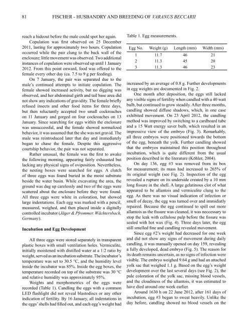
81FISCHER - HUSBANDRY AND BREEDING OF VARANUS BECCARIIreach a hideout before the male could spot her again.Copulation was first observed on 25 December2011, lasting for approximately two hours. Copulationoccurred while the pair clung to the back wall of theenclosure; little movement was observed. Two additionalinstances of copulation were observed up until 1 January2012. From this point onward, food was offered to thefemale every other day (ca. 7.5 to 9 g per feeding).On 7 January, the pair was separated due to themale’s continued attempts to initiate copulation. Thefemale showed increased activity, but no digging wasobserved, and her abdominal girth and tail base area didnot show any indications of gravidity. The female brieflyrefused insects and other food items for three days,but then reluctantly accepted two small cockroacheson 11 January and gorged on four cockroaches on 13January. Since searching for eggs within the enclosurewas unsuccessful, and the female showed normalizedbehavior, it was assumed that the she was not gravid. Themale was reintroduced later that day and immediatelybegan to chase the female. Despite this aggressivecourtship behavior, the pair was not separated.Rather unusual, the female was the first to awakethe following morning, appearing fairly exhausted butlacking any physical signs of oviposition. Nevertheless,the nesting boxes were searched for eggs. A clutchof three eggs was found buried in the moist substratebeside the water basin. While excavating the area, theground was dug up carelessly and two of the eggs werescattered about the enclosure before they were found.All three eggs were white in coloration, but showedlarge indentations. Each egg was marked with a pencil,measured, weighed, and then placed inside a digitallycontrolled incubator (Jäger & Pfrommer. Wächtersbach,Germany).Incubation and Egg DevelopmentAll three eggs were stored separately in transparentplastic boxes with small ventilation holes. Vermiculite,initially moistened with distilled water at a 1:2 ratio byweight, served as an incubation substrate. The incubator’stemperature was set to 30.5 °C, and the humidity levelinside the incubator was 85%. Inside the egg boxes, thetemperature recorded on top of the substrate was 30 °Cand relative humidity was approximately 95%.Weights and morphometrics of the eggs wererecorded (Table 1). Candling the eggs with a commonLED flashlight did not reveal blastodiscs or any otherindication of fertility. By 16 January, all indentations inthe eggs’ shells had filled out, and each egg’s weight hadTable 1. Egg measurements.Egg No. Weight (g) Length (mm) Width (mm)1 11.7 46 212 11.3 45 203 11.3 46 23increased by an average of 0.8 g. Further developmentsin egg weights are documented in Fig. 2.One month after deposition, the eggs still lackedany visible signs of fertility when candled with a 40 wattbulb, but continued to grow steadily. After three months,candling showed diffuse shadows, which, in one caseexhibited movement. On 23 April 2012, the candlingmethod was improved by switching to a cardboard tubeand a 15 Watt energy saver bulb, which resulted in animpressive view of the embryo (Fig. 3). Remarkably,all three embryos were positioned towards the bottomof the egg, beneath the yolk. Further candling showedthat the embryos maintained this position throughoutincubation, which is quite different from the usualposition described in the literature (Köhler, 2004).On day 136, egg #3 was removed from its boxfor measurement; its mass had increased to 265% ofits original weight (see Fig. 2). Inspection of the eggrevealed a rupture on its underside created by a 10 mmlong fissure in the shell. A large gelatinous clot of whatappeared to be allantois and vermiculite clung to theegg. As there was no visual indication of infection orsmell of decay, the egg was turned over and immidiatlyrepaired. Because the egg continued to spill out moreallantois as the fissure was cleaned, it was neccessary tostop the leak with cellulose pulp before the fissure wassealed with hot wax (Fig. 4). Three days later, the eggstill smelled fine and candling revealed movement.Since egg #2’s weight had decreased for one weekand did not show any signs of movement during dailycandling, it was manually opened on day 159, revealinga fully developed, dead embryo (Fig. 5). The reason forits death remains uncertain, as no signs of infection werevisible. The embryo weighed 9.64 g and had an attachedyolk sac that weighed 1.1 g. Based on the egg’s weightdevelopment over the last several days (see Fig. 2), thepale coloration of the yolk sac, missing blood vessels,and the cloudiness of the allantois, it was estimated tohave died around one week earlier.Around 1630 h on 22 June 2012, after 161 days ofincubation, egg #3 began to sweat heavily. Unlike theday before, candling showed no blood vessels on the