
You also want an ePaper? Increase the reach of your titles
YUMPU automatically turns print PDFs into web optimized ePapers that Google loves.
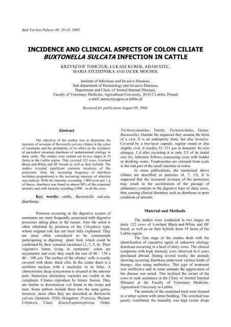
Bull Vet Inst Pulawy 49, 29-33, 2005<strong>IN</strong>CIDENCE AND CL<strong>IN</strong>ICAL ASPECTS OF COLON CILIATE<strong>BUXTONELLA</strong> <strong>SULCATA</strong> <strong><strong>IN</strong>FECTION</strong> <strong>IN</strong> <strong>CATTLE</strong>KRZYSZTOF TOMCZUK, ŁUKASZ KUREK, ADAM STEC,MARIA STUDZIŃSKA AND JACEK MOCHOLInstitute of Infectious and Invasive Diseases,Sub-department of Parasitology and Invasive Diseases,Department and Clinic of Animal Internal Diseases,Faculty of Veterinary Medicine, Agricultural University, 20-612 Lublin, Polande-mail: parazyt@agros.ar.lublin.plReceived for publication August 09, 2004.AbstractThe objective of the studies was to determine theintensity of invasion of Buxtonella sulcata ciliates in the colonof ruminants and the probability of its effect on the incidenceof periodical recurrent diarrhoea of undetermined etiology indairy cattle. The studies were carried out in two stages at 19farms in the Lublin region. They covered 122 cows, LowlandBlack-and-White and HF breeds as well as their hybrids. Thestudies revealed significant common incidence of thisprotozoon. Also, the increasing frequency of diarrhoeaincidence proportional to the increasing intensity of infectionwas noticed. With the intensity exceeding 1 000 cysts per 1 gof faeces, diarrhoea was found in almost 90% of the examinedanimals, and with intensity exeeding 2 000 – in all the cows.Key words: cattle, Buxtonella sulcata,diarrhoea.Protozoa occurring in the digestive system ofruminants are most frequently associated with digestiveprocesses taking place in the rumen. Also the colon isoften inhabited by protozoa of the Ciliophora type,whose original role has not been fully explained. Theyare most often considered to be commensalsparticipating in digesting plant feed, which could beconfirmed by their common incidence (2, 3, 5, 6). Theirvegetative forms living in ruminants’ colon areasymmetric and oval, they reach the size of 60 – 138 x46 – 100 µm. The surface of the ciliates’ cells is evenlycovered with short, thick cilia. In the centre there is areniform nucleus with a nucleolus in its hilus. Acharacteristic deep syncystoma is situated at the anteriorpole. Numerous alimentary vacuoles are visible in thecytoplasm. Ciliates reproduce by binary fission. Theyare similar to Balantidium coli found in the swine andman. Some authors include them into the same genus,however, more often they are described as Buxtonellasulcata (Jameson 1926) (Kingdom: Protozoa, Phylum:Ciliphora, Class: Kinetofragminophorea, Order:Trichostromatidae, Family: Pyenotrichidae, Genus:Buxtonella). Outside the organism they assume the formof a cyst. It is an endosporic form, but also invasive.Covered by a two-layer capsule, regular round or elseslightly oval, it reaches 52–131 µm in diameter. Its sizechanges; 3 d after excreting it is only 2/3 of its initialsize (6). Infection follows consuming cysts with fodderor drinking water. Trophozoites are released from cystsin the end part of the small intestine or colon.In some publications, the mentioned aboveciliates are described as parasites (4, 7, 13). It issupposed that the increased invasion of the protozoonmay result in the acceleration of the passage ofalimentary contents in the digestive tract of dairy cows,thus causing clinical disorders such as diarrhoea or poorcondition of animals.Material and MethodsThe studies were conducted in two stages ondairy 122 cows of Lowland Black-and-White and HFbreed, as well as on their hybrids from 19 farms of theLublin region.The first stage of the studies dealt with theidentification of causative agent of unknown etiologydiarrhoea occurring in a herd of dairy cows. The clinicalsymptoms with high intensity were observed in 6 cowspurchased abroad. During several weeks, the animalsshowing recurring diarrhoea underwent various kinds oftherapy, also using antibiotics. This type of treatmentwas ineffective and in some animals the aggravation ofthe disease was noted. This inclined the owner of thecows to seek assistance at the Clinic of Animal InternalDiseases at the Faculty of Veterinary Medicine,Agricultural University in Lublin.The animals of the examined herd were housedin a tether system with straw bedding. The cowshed waspoorly ventillated, the humidity was high (water drops
30on the walls) and the lighting was inadequate. Manurewas hand-removed. The cows did not have access to anoutside run or pasture. The feeding system was based onthe supply of free amount of hay, hay-silage as well aslittle amount of sugar beet pulp, and the mixture ofcereal and bran was given at the amount adapted to milkproductivity. Moreover, protein and mineral supplementcontaining in its composition mainly macroelements wasused. The animals had constant access to water.Blood from the external jugular vein and faecesfrom the rectum were sampled from the cows withadvanced diarrhoea.After having sampled the material, the therapyof the whole herd was started. The first part of thetherapy consisted an individual treatment of cowsshowing the most advanced clinical symptoms. Asymptomatic therapy was applied, i.e. parenteral supplyof hydrating fluids (PWE, 5% and 20% glucose), Bgroup vitamins, especially vitamin B 1, vitamin C, andpreparations per os containing glucogenic compounds(sodium proprionate). The other course of action aimedat improving zoohygienic conditions in a cowshed,introducing high biological value protein into the fodderand also balancing mineral–vitamin supplement.Moreover, the basic element of the efficiency of thetreatment ordered was to turn out the animals to theoutside run out pasture every day.The second stage of the studies comprised thedetermination of the intensity and extensity ofBuxtonella sulcata incidence in various herds the cows.In order to show the pathogenic effect of the parasite onthe condition of the alimentary tract of ruminants,monitoring examination of cattle from different herdswas performed to determine the incidence of thisprotozoon as well as the type of faeces (diarrhoeal orphysiological). The faeces for coproscopical studies wassampled from 116 cows from 18 farms. The faeces weretaken directly from the rectum of animals withsymptoms of diarrhoea and at random – from healthyanimals.To determine the intensity of the incidence ofB. sulcata, at both stages of the studies, the sampleswere examined by the method of decantation, usingMcMaster chamber (5), with the following our(Tomczuk) modification. Five gram samples of faeceswere strained through a strainer dipped in a 400 mlbeaker filled with water. After 20 min of decantation, apart of water was poured out, then the beaker was onceagain filled with water up to the volume of 400 ml. Theprocess of decantation was repeated 3–4 times until thetransparent sediment was obtained. Next, the sedimentwas transferred to smaller beakers and suspended in 30ml of water. The beakers were placed on a magneticstirrer and, while homogenising, the solution wastransferred with a pipette into both parts of McMasterchamber. The cysts were counted under a microscope inboth parts of McMaster chamber, and the number ofcysts in 1 g (CPG) of faeces was calculated from theformula:number ofCPG =cystsin bothparts of5McMasterchamber× 100ResultsAt the first stage, in the majority of cowsexamined clinically, except the ones in poor or badcondition, only pale pink colouring of the muscousmembrane of the conjunctival sac was found. Theremaining clinical parameters and also routinelyaccessible lymphatic nodes did not show deviationsfrom physiological norms. In a specific clinical study ofcows which showed diarrhoea, a low or medium level ofdehydration, changeable appetite and thirst as well as thedisorders in chewing and rumen motor activity werefound. Some animals had difficulty in raising.Coproscopical examination of faeces from these cows,sampled at the intensification of diarrhoeal symptoms,showed from 600 to 1 300 B. sulcata cysts in 1 g offaeces (Table 1) and from 14 to 21 eosinophils in 1 mlof blood in haematological examination.As the result of the treatment, after a weektherapyno cases of diarrhoea were observed, and aftertwo weeks – a marked improvement of the condition ofthe cows was noted.Coproscopical examination of faeces revealedfrom 100 to 500 B. sulcata cysts in 1 g faeces of thecows which earlier showed diarrhoea symptoms (Table1).Table 1Number of Buxtonella sulcata cysts (CPG) and faeces consistency in a herd of dairy cows before and after treatmentNo. of CPG Diarrhoeal CPGsample before treatment stool after treatmentDiarrhoeal stool1 1 300 + 500 -2 800 + 100 -3 1 200 + 460 -4 920 + 160 -5 600 + 100 -6 800 + 320 -
31Table 2B. sulcata invasion intensity and extensity (CPG) and faeces consistency in the examined herdsHerdABCDEFGHIJNo. ofsampleDiarrhoealstoolCPGDiarrhoealNo. ofHerdstoolsampleCPG1 240 - 1 260 +2 400 - 2 120 -3 0 - 3 360 -4 900 - 4 180 +5 120 + K 5 100 -6 60 +6 400 -1 60 - 1 1 380 +2 80 - 2 160 -3 100 - 3 80 -4 40 - 4 1 280 +5 3 760 + L 5 0 -6 140 +6 0 -1 1 420 + 1 2 960 +2 1 200 + 2 80 +3 1 180 + 3 140 -4 1 340 + 4 1 060 -5 440 - Ł 5 220 -6 1 120 +6 160 -1 1 600 + 1 260 -2 40 - 2 120 +3 40 - 3 60 -4 900 + 4 180 -5 1 800 +M5 100 +6 40 -6 140 +1 160 - 1 340 -2 20 + 2 20 -3 180 - 3 120 -4 260 - 4 240 -5 20 + 5 440 -6 60 +N6 180 -1 180 -7 280 -2 1 400 + 1 0 -3 180 - 2 60 -4 140 - 3 60 -5 1 440 + 4 20 -6 340 -O5 20 -1 0 + 6 40 -2 0 + 7 20 -3 0 -8 80 -4 0 + 1 2 860 +5 0 + 2 1 260 -6 0 - 3 60 -1 860 + 4 1 120 +2 200 - P 5 20 -3 220 - 6 340 -4 1 140 +7 20 -5 420 - 1 260 -6 1 080 + 2 100 -1 220 + 3 80 -2 160 - 4 200 -3 680 + 5 120 -4 1 060 + 6 400 -5 3 260 + 7 60 -6 400 - 8 440 -1 0 + 9 120 -2 0 - R 10 160 -3 0 - 11 60 -4 0 -12 80 -
33after returning the homeostasis of the digestive tract bythe normalization of feeding system and administeringprobiotics. Only the cases complicated by pathogenicbacterial flora will, at the further phase, need the generaltreatment with the use of adequately selected antibiotics.References1. Abubaker M.I., Nayel M.N., Fadlala M.E., AbdelrachmanA.O., Abuobeida S.A., Elgabara Y.M.: Prevalence ofgastrointestinal parasites in young camels in Bahrain. RevElev Méd Vét Pays Trop 2000, 53, 267–273.2. Bauer C.: Vorkommen und Beschreibung der Zysten desZäkumziliaten Buxtonella sulcata (Jameson 1926) im Kotvon Küen in Norddeutschland. Berl Munch TierarztlWschr 1983, 96, 371–374.3. Fox M.T., Jacobs D.E.: Patterns of infection withBuxtonella sulcata in British cattle. Res Vet Sci 1986, 41,90–92.4. Hong K.O., Youn H.J.: Incidence of Buxtonella sulcatafrom cattle in Kyonggi-do. Korean J Parasitol 1995, 33,135–138.5. Melhorn H., Duwel D., Raether W.: Diagnose undTherapie der Parasiten von Haus-, Nutz-, und Heimtieren.Gustav Fischer Verlag, Stuttgard 1993.6. Rommel M., Eckert J., Kutzer E., Korting W., SchneiderT.: Veterinarmedizinische Parasitologie. Parey Buchverlag,Berlin 2000.7. Urman H.D., Kellky G.W.: Buxtonella sulcata. A ciliateassociated with ulcerative colitis in a cow and prevalenceof infection in Nebraska cattle. Iowa State Univ Vet 1964,27, 118–122.8. Skotarczak B.: Bacterial flora in acute and symptom–freebalantidiosis. Acta Parasitol 1997, 42, 230–233.9. Skotarczak B.: Wpływ niektórych czynnikówzewnętrznych na encystacje i ekscystacje Balantidiumcoli. Wiad Parazytol 1984, 30, 565–573.10. Skotarczak B., Zieliński R.: Wzrost populacjiBalantidium coli w warunkach in vivo i in vitro. WiadParazytol 1997, 43, 171–178.11. Skotarczak B., Zieliński R.: Wpływ wybranychczynników biotopu Balantidium coli na przebiegbalantidiozy świń. Wiad Parazytol 1997, 43, 399–403.12. Tafti A.K., Maleki M., Oryan A.: Pathological study ofintestines and mesenteric lymph nodes of camels(Camelus dromedarius) slaughtered in Iran. J. CamelPract Res. 2001, 8, 209–213.13. Wacker K., Roffeis M., Conraths F.J.: Cow-calf herds inEastern Germany: status quo of some parasite species andcomparison of chemoprophylaxis and pasturemanagement in control of gastrointestinal nematodes. JVet Med B 1999, 46, 475–483.14. Ziomko I., Karamon J.: Występowanie a chorobotwórczośćorzęska Balantidium coli u prosiąt. Magazyn Wet2003, 84, 27– 8.