Laparoscopic -Assisted Transanal Endorectal Pull-Through for ...
Laparoscopic -Assisted Transanal Endorectal Pull-Through for ...
Laparoscopic -Assisted Transanal Endorectal Pull-Through for ...
- No tags were found...

You also want an ePaper? Increase the reach of your titles
YUMPU automatically turns print PDFs into web optimized ePapers that Google loves.
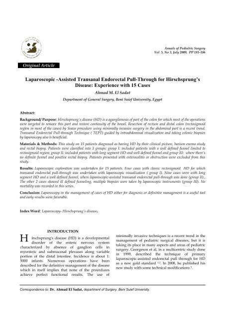
Annals of Pediatric SurgeryVol 5, No 3, July 2009, PP 181-186Original Article<strong>Laparoscopic</strong> -<strong>Assisted</strong> <strong>Transanal</strong> <strong>Endorectal</strong> <strong>Pull</strong>-<strong>Through</strong> <strong>for</strong> Hirschsprung’sDisease: Experience with 15 CasesAhmad M. El SadatDepartment of General Surgery, Beni Suief University, EgyptAbstract:Background/ Purpose: Hirschsprung's disease (HD) is a aganglionosis of part of the colon <strong>for</strong> which most of the operationswere targeted to remove this part and restore continuity of the bowel. Resection of rectum and distal colon (rectosigmoidregion in most of the cases) by Soave procedure using minimally invasive surgery in the abdominal part is a recent trend.<strong>Transanal</strong> <strong>Endorectal</strong> <strong>Pull</strong>-through Technique ( TEPT) guided by intraabdominal visualization and taking colonic biopsiesby laparoscopy also is beneficial.Materials & Methods: This study on 15 patients diagnosed as having HD by their clinical picture, barium enema studyand rectal biopsy. Patients were classified into 3 groups: group I: included patients with a well defined funnel limited torectosigmoid region; group II: included patients with long segment HD and well defined funnel and group III: where there’sno definite funnel and positive rectal biopsy. Patients presented with enterocolitis or obstruction were excluded from thisstudy.Results: <strong>Laparoscopic</strong> exploration was undertaken <strong>for</strong> 15 patients. Four cases with classic rectosigmoid HD <strong>for</strong> whichtransanal endorectal pull-through was undertaken with laparoscopic visualization ( group I). Nine cases were with longsegment HD and a well defined funnel, where laparoscopic-assisted transanal endorectal pull-through was done (group II).,The other 2 cases showed ill defined funneling, multiple biopsies were taken by laparoscopic instruments (group III). Nomortality was recorded in this series.Conclusion: Laparoscopy in the management of cases of HD either <strong>for</strong> diagnosis or definitive management is a useful tooland early results seem favorable.Index Word: Laparoscopy- Hirschsprung’s disease.INTRODUCTIONirschsprung's disease (HD) is a developmentalHdisorder of the enteric nervous systemcharacterized by absence of ganglion cells inmyenteric and submucosal plexuses along variableportion of the distal intestine. Incidence is about 1:5000 infants. Numerous operations have beendescribed <strong>for</strong> the definitive management of the diseasewhich in itself implies that none of the proceduresachieve perfect functional results. The use ofminimally invasive techniques is a recent trend in themanagement of pediatric surgical diseases, but it istaking its place in many aspects and areas of pediatricsurgery. Georgeson et al, in a multicentric study donein 1999, described the technique of primarylaparoscopic-assisted endorectal pull through <strong>for</strong> HDas a new gold standard 1,2 . In 2008, he published hisnew study with some technical modifications 3 .Correspondence to: Dr. Ahmad El Sadat, department of Surgery. Beni Suief University.
El Sadat A.PATIENTS AND METHODSFifteen patients with HD underwent surgery withlaparoscopic assisted endorectal pull throughtechnique during a time interval between November2006 and November 2008.Diagnosis was made by history, physical examinationand investigations. Detailed history was taken,including time of first passage of meconium, periodsof constipation, and history suggestive of intestinalobstruction or enterocolitis (those were followed uptill resolving of the condition). Clinical examinationincluded: general condition, presence of associatedanomalies, abdominal examination <strong>for</strong> distention andsigns of obstruction, rectal examination to detectabsence of the rectal ampulla and the impacted stools.Investigatory work up was done including routinelaboratory investigations, barium enema and rectalbiopsy.According to the findings of barium enema and rectalbiopsy, patients were classified into 3 groups:Group I: patients with rectosigmoid aganglionicsegment and well defined funnel.Group II: patients with long aganglionic segment andwell defined funnel.Group III: patients with ill defined funnel and positiverectal biopsy.Clinical Picture of HD + Positive rectal biopsy +Barium enema<strong>Laparoscopic</strong> visualization of the abdomenDefinite funnel Definite funnel no Definite funnelat the recto- above rectosigmoidzone sigmoid zoneTEPT with <strong>Laparoscopic</strong> <strong>Laparoscopic</strong><strong>Laparoscopic</strong> -<strong>Assisted</strong> biopsiesVisualization TEPTPreoperative decompression of the bowel <strong>for</strong> 2 dayswith saline irrigation through rectal tube negotiatedthrough the aganglionic part was done. The patientswere given nothing by mouth 12 hours preoperatively,only water was permitted with I.V fluids. Oralantibiotics were given as chemical preparation 24 hrspreoperatively in the <strong>for</strong>m of erythromycin, neomycinand metronidazole. At induction of anesthesia, 3 rdgeneration cephalosporin and I.V metronidazole weregiven.Infants are positioned transversely at the end of theoperating table with arm boards placed parallel to thelong axis to increase width of table while olderchildren were placed in a supine position (LloydDavis). The working surgeon stood at the patient’shead while working with the laparoscope in theabdominopelvic cavity. Pneumoperitoneum wasobtained using an open technique through theumbilicus with pressure of 10-12 mm water. A 5 mm,30° scope was introduced through a port placed belowthe liver edge in the right mid clavicular line. One 5mm port was placed in the right lower quadrant,another in the suprapubic region and another onesometimes in the left lower quadrant. The exactposition of the ports in relation to anterior superioriliac spine varied according to the age of the patient.The transition zone was visualized and localizedbe<strong>for</strong>e any attempt was made to devascularize ormobilize the colon. Rectosigmoid funnel (group I) wasan indication <strong>for</strong> beginning the transanal dissectionand the working surgeon begins 1-2 cm above dentateline creating submucosal plane by sharp and bluntdissection until the colorectum turned inside –outindicating that the transanal dissection had advancedto the level of the internal perirectal plane. The rectalsleeve is then opened posteriorly to join thesubmucosal plane with the extramural plane and thencarried circumferentially. At this step, the laparoscopicand perineal dissection planes were joined and thecolon was pulled few centimeters above the funnel toavoid the area of hypoganglionosis. The rectal musclecuff was made unfolded by inserting fingercircumferentially with pushing the cuff upwards andthen splitting it posteriorly to allow <strong>for</strong> neorectumreservoir. One layer anastomosis with interruptedabsorbable sutures between the anal mucosa above thedentate and full thickness of neorectum was thenmade. <strong>Laparoscopic</strong> visualization was done <strong>for</strong>untwisting the descended colon and to exclude anybleeder or collection inside.Annals of Pediatric Surgery 182
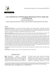
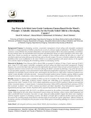
El Sadat A.If a long aganglionic segment was encountered onlaparoscopic visualization (group II), the sigmoidcolon was mobilized preserving proximity to themarginal artery with division of lateral fusion fasciaand sometimes freeing the splenic flexure until thecolon above the funneling is long enough to reachdeep into pelvis without tension. In those cases wherethere was no definite funneling by laparoscopicvisualization (group III), seromuscular biopsies weretaken by Metzenbaum endoscopic scissors and the siteof the biopsies were closed by various types of sutureswith various colors: (Prolene-blue, Maxon-green, PDSviolet,Undyed-White) to facilitate the pull-thoughsurgery at another step.Pneumoperitoneum was evacuated at the end of theprocedure with the camera inside to facilitate perfectclosure of port sites to prevent the occurrence of portsite hernia.An intra peritoneal open system tube drain was left inthe pelvis to be removed after 24 hoursNo nasogastric suction was needed. oral feeding wasallowed when bowel motions returned. Antibioticswere continued parenterally during the period of thehospital stay. After discharge, oral metronidazole wascontinued <strong>for</strong> three weeks to avoid enterocolitis in thepost operative period..The patient was seen 2 weeks after discharge andcareful rectal examination was done. Daily anorectaldilation is initiated if tightness is encounteredFig 1. : Barium enemaRESULTSFifteen patients were operated upon, Eleven males &four females. Age of patients ranged from 3 monthsand 5 years.Group I included 4 cases (27%). Group II included 9cases (60%), while group III included 2 cases (13%).Operating time varied according to the length of theaganglionic segment, average time <strong>for</strong> classicrectosigmoid aganglionosis (group I ) was 1.5 hour, incases where the funnel was extending higher (groupII) the average time was 2.5 hours, and those <strong>for</strong> whichmultiple biopsies were taken (group III) the averagetime was about one hour.Fig. 2: Creation of a window in the sigmoid colonFig. 3: color difference between normal and devascularizedcolon183 Vol .5, No 3, July 2009
El Sadat A.In Eleven cases (73%) oral intake started 48 hours afteroperation , in three cases (20%) it was delayed to 72 hrand in one case (6.7%) it was after 96 hours. The meanhospital stay was 5 days (range 4-7 days).Early complications (within 3 months postoperatively)occurred in 11 cases: enterocolitis in 3cases (20%) that responded to conservativemeasurement, fever in another 2 cases (13%) that wasnot explained to be due to enterocolitis, but wasprobably due to upper respiratory tract infection inone case, and urinary tract infection in the other case.Paralytic ileus occurred in 2 cases = (13.3) andresponded to NPO & intestinal prokinetic agents.Perianal excoriation occurred in 4 cases (26.4) thatresponded to zinc oxide ointment applications &constipating agents. There was no cases ofanastomotic leak .Late complications (after 3 months) occurred in 3patients: 2 cases (13.3%) developed anastomoticstricture that responded to regular dilatations, and onecases had 2 attacks of enterocolitis at 6 and 9 monthspost operatively that needed hospital admission andthe attacks had subsided under conservative measuresin the <strong>for</strong>m of parenteral antibiotics, intestinalantiseptics, I.V fluids and NPO.There were no complications peculiar to the use oflaparoscopy in this patients. Some patients hadhypercarbia intra operatively that responded tomodest hyperventilation. No inadvertent injury tointra abdominal structure. No port site hernia orwound infection. Estimation of fecal incontinenceneeds longer period of follow up but older childrenare showing good continence & regular bowel habitsin the late follow up. No mortalities occurred in thisseriesDISCUSSIONThe endorectal pull-through technique avoids injury tothe pelvic nerves by remaining within the muscularcuff of the aganglionic segment with lower incidenceof constipation, sexual dysfunction & micturationdisturbances 4 .Georgeson et al in 1995 had the initial experience inlaparoscopic assisted endorectal colon pull-through in12 children with good results 1 . In 1999, he publishedthe multi centric study on 80 patients 2 .Some modifications and technical points are valuablein describing the technique in his publication in 2008 3 ,as shifting from creating the pneumoperitoneum byverses needle to open transumbilical technique. In allcases in this study the open technique showed saferintroduction and reliability especially in those childrenwith distended colon.Portless technique was used in this work especially inthose sites where reentry is limited in grasping thecolon as the thin abdominal wall in children does nothinder working freely.It was described by some pediatric surgeons that therole of laparoscopy in the interventional surgery <strong>for</strong>Hirschsprung's disease is limited to those with longaganglionic segment based on contrast enema study 5,6 .In this study, the majority of cases were selected withlong segment & definite funnel to get the benefit ofintra abdominal dissection ( group II, 9 cases) and thework was extended to those with indefinite funnel onbarium study to obtain direct visualization & to takeseromuscular biopsies <strong>for</strong> planning of definite surgerywith minimal manipulation on viscera to avoid lateradhesionsIn the algorithm described in the study, the clinicalpicture of Hirschsprung's disease, positive rectalbiopsy & contrast enema were the basis of the selectionof the procedure to be taken and the laparoscopicvisualization at the beginning to confirm the site offunnel was used as diagnostic tool and then to assist inthe procedure.In cases where there was no definite funnel, biopsieswere taken to level the aganglionosis and to guide <strong>for</strong>the further procedure. In one of the 2 cases of groupIII, there was total colonic aganglionosis that neededtotal colectomy with ileal pull through in a later stepby open approach. Undertaking the whole procedureof colectomy in this cases needs good laparoscopicsurgical expertise and has a high conversion rate.A point to be mentioned is the ability to do colonicdissection more easily in younger age group. Themesentery of the colon can be pulled through withouttwist and with less manipulation through the cuff.Younger group are more challenging in theiranesthesia with more incidence of hypothermiaespecially when there's port leak.When the endorectal dissection is facilitated bylaparoscopic mobilization of the rectosigmoid colon,there's less risk of weakening the patient's internalsphincter by over dilatation 2 .Annals of Pediatric Surgery 184
El Sadat A.Children over five years of age and adults with a latediagnosis are not considered <strong>for</strong> a single stage repairby the laparoscopic assistance because of the hugelydilated chronically obstructed bowel proximal to theaganglionic colon 3 . However the role of laparoscopicassisted stoma <strong>for</strong>mation can be considered in furtherstudies.Exclusion criteria from this study were activeenterocolitis &obstructed patients, and those withdeteriorated general health or life threatening generalcondition 1,3 .The shortest length of resected aganglionic segmentwas 18 cm while the longest was 38 cm. In otherstudies the length of the aganglionic part was relatedto the conversion to laparotomy 3,7,8 .The postoperative hospital stay ranged between 4 to 7days (mean = 5 days). Georgeson et al in hismulticenteric study reported a mean of 4.8 days. Hereported that earlier in his study he used to keep thepatients <strong>for</strong> 7 days and then later they were dischargedon the 2 nd or 3 rd days 3 . In an EPSA study, meanhospital stay was 4.8 days 7 .Complication rate in this study was comparable toother studies. The incidence of enterocolitis was 20%which is nearly more than double the incidence in theGeorgeson et al multicenteric study (7.5%) 3 and littlemore than in EPSA multi centric study (17.8 %) 7 andin wang et al 8 on 61 patients was 21% but <strong>for</strong>tunatelyall cases responded to conservative measures.Anastomotic stricture occurred in 13% responded toregular dilatations.Conversion to conventional open procedure wasreported in 2 cases (2.5%) in Georgeson multi centricstudy but not reported with Wang et al or in this study7,8.No colonic per<strong>for</strong>ation or anastomotic leak occurred inthis study. Per<strong>for</strong>ations occurred in one case (1.6%) inWang et al and no per<strong>for</strong>ations with Georgeson et al.Leak occurred in 2 cases in Georgeson’s (2.5%) and 2patients (3.3%) in Wang et al that was managed bydiverting colostomy 7,8 .No mortality was reported in the follow up period.Continence to stools needs longer period of follow-upespecially in younger age group. On those childrenabove 3 years, continence results were satisfactory inthe follow up period and reported to be good by someauthors 9 . The procedure is cost-effective and acceptedby the parents. 10 .CONCLUSIONFor most patients with Hirschsprung's disease,laparoscopic-assisted transanal endorectal pullthroughcan be per<strong>for</strong>med in a single stage.Laparoscopy gives beneficial aspects regardingdiagnosis & management of the condition. The idea ofminimal invasive surgery seems satisfactory to theparents and ameliorates the impact of the decision ofsurgery on them.The use of laparoscopy has many advantages being aone stage procedure with less post operative stayespecially in long segment cases, less post operativepain, early post operative recovery of intestinalmotility, minimal intra operative manipulation andsmall access wounds that decrease post operativemorbidity regarding adhesions and cosmeticdisfiguring.Additional advantages of the laparoscopic assist <strong>for</strong>transanal pull-through include the flexibility af<strong>for</strong>dedto per<strong>for</strong>m almost all procedures <strong>for</strong> correctingHirschsprung’s disease.It can allow a delay in the definitive surgery <strong>for</strong>permanent histologic confirmation if rapid frozensection indicates a long aganglionic segment or if rapidfrozen section is indeterminate as to the position oftransition zone.REFERENCES1. Georgeson KE, Fuefner MM, Hardin WD. Primarylaparoscopic pull-through <strong>for</strong> Hirschsprung’s disease ininfants and children. J pediatr surg 30:1017-1021. 19952. Gerogeson KE, Cohen RD, Hebra A, et al. Primarylaparoscopic endorectal colon pull-through <strong>for</strong>Hirschsprung’s disease: a new gold standard. Ann Surg229:678-682. 19993. Gerogeson KE and Muensterer OJ. Laparoscpic-<strong>Assisted</strong> <strong>Transanal</strong> <strong>Pull</strong>-through <strong>for</strong> Hirschsprung’s Disease,Holschnider AM and Prem Puri (editors), Hirschsprung’sdisease and Allied disorders, 3rd edition, Ch.23, 323-333,Springer. 20084. Albanese CT., Jennings RW, Smith B. et al. Perinealone stage pull-through <strong>for</strong> Hirschsprung’s disease J. Pediatr.Surg. 34:377-380. 19995. Hadidi A. <strong>Transanal</strong> endorectal pull-through <strong>for</strong>Hirschsprung’s disease, experience with 68 patients. JPediatr Surg 38:1337-1340. 2003185 Vol .5, No 3, July 2009
El Sadat A.6. Langer JC, Minkes RK, Mazziot MV, et al. <strong>Transanal</strong>one-stage Soave procedure <strong>for</strong> infants with Hirschsprung’sdisease. J Pediatr Surg 34:148-151. 19997. EL Halaby EA, Hasish A, El Barbary MM et al.<strong>Transanal</strong> one stage <strong>Endorectal</strong> pull-through <strong>for</strong>Hirschsprung’s disease: A multi centric study. J. Pediatr.Surg: 39 (3): 345-351. 20048. Wang NL, Lee Hc, Yeh Ml, et al. Experience withprimary laparoscopy-assisted endorectal pull-through <strong>for</strong>Hirschsprung’s disease. Pediatr Surg Int 20:118-122. 20049. Naho Fujiwaraa, Kazuhiro Kaneyama . Comparativestudy of laparoscopic-assisted pull-through and open pullthrough<strong>for</strong> Hirschsprung’s disease with special reference topostoperative faecal incontinence J Pediatr. Surg. 42:2071-2074. 200710. Bufo AJ, Chen MK, Shah R, et al. Analysis of the costsof surgery <strong>for</strong> Hirschsprung’s disease: one stage laparoscopicpull-through versus two-stage Duhamel procedure. ClinPediatr 38:593-596. 1999.Annals of Pediatric Surgery 186