Protocols for field and laboratory rodent studies - HAL
Protocols for field and laboratory rodent studies - HAL
Protocols for field and laboratory rodent studies - HAL

You also want an ePaper? Increase the reach of your titles
YUMPU automatically turns print PDFs into web optimized ePapers that Google loves.
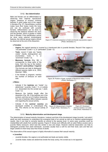
<strong>Protocols</strong> <strong>for</strong> <strong>field</strong> <strong>and</strong> <strong>laboratory</strong> <strong>rodent</strong> <strong>studies</strong>3.1.2. Sex determinationMale <strong>and</strong> female can be differentiated byobserving their external reproductiveorgans: presence of scrotum coveringtesticles in adult males <strong>and</strong> of the vaginain females (Fig. 37). Nevertheless, juvenilemales have their testes inside the body<strong>and</strong> can be confused with juvenilefemales. Differentiation can be done byobserving the distance between the anus<strong>and</strong> the genitals, which is greater in malesthan in females. If the identification cannotbe done using external morphologicalfeatures, the internal sexual organs shouldbe observed at the end of the dissection.Figure 37: External comparison of male <strong>and</strong> female reproductive organs(Photo: Herbreteau V.)● Females:- Vagina: the vaginal opening is covered by a translucent skin in juvenile females. Record if the vagina is“not per<strong>for</strong>ated” (Code = 1), or “per<strong>for</strong>ated” (Code = 2).- Teats: record if teats are “barelyvisible” (hardly distinguished <strong>and</strong>enumerated, Code = 1) or“prominent” (Code = 2).- Mammary <strong>for</strong>mula (Fig. 38): itcorresponds to three parts of thebody, from the top to the bottom:pectoral + post-axillary + inguinalThis <strong>for</strong>mula can help to distinguishdifferent species (Aplin et al., 2003;Marshall, 1988).- If the female is pregnant, mentionthe number of embryos on eachside. Figure 38: Position of teats, example of B<strong>and</strong>icota indica (1+2+3)(Photo: Herbreteau V.)● Males:- Indicate if the testicles are “inside” (i.e.abdominal = juvenile, Code = 1), or outside(i.e. descended into the scrotal sac = adult,Code = 2).- Measure the testicle length after thedissection (Fig. 39). If the testicles are in thescrotum, pull one out of it be<strong>for</strong>e measuring.The size of testicles differs between species<strong>and</strong> regarding the sexual activity.- Indicate if the seminal vesicles aredeveloped (visible) or undeveloped (Fig.40). This in<strong>for</strong>ms on the sexual activity ofmales.Figure 39: Measurement of testiclelength (Photo: Herbreteau P.)Figure 40: Developedseminal vesicles in an adultmale (Photo: Herbreteau P.)223.1.3. Maturity determination <strong>and</strong> development stageThe determination of sexual maturity (immature / mature) <strong>and</strong> that of the development stage (juvenile / sub-adult /adult) are both necessary to underst<strong>and</strong> the ecological traits of an animal as well as to interpret epidemiologicalresults. Also it can help to correctly identify an animal to the species level. In some case, juveniles could bemisidentified with adults from another species. Figure 41 shows such difficulty with an adult Rattus exulans <strong>and</strong> ajuvenile Rattus tanezumi, which look like similar in size <strong>and</strong> shape. However external observation can help todistinguish juveniles from adult individuals. Juveniles usually have a soft fur <strong>and</strong> their skull, ears <strong>and</strong> feet arelarger (relative to their body size) than those of adults.The observation of the sexual organs is highly in<strong>for</strong>mative to assess their sexual maturity:● Juveniles:- Juvenile females: the vagina is not per<strong>for</strong>ated <strong>and</strong> teats are barely visible.- Juvenile males: testes are abdominal (inside the body cavity), the scrotal sac is not apparent.