Pulse Amplitude Modulation Fluorometry - Coral Reef Targeted ...
Pulse Amplitude Modulation Fluorometry - Coral Reef Targeted ...
Pulse Amplitude Modulation Fluorometry - Coral Reef Targeted ...

You also want an ePaper? Increase the reach of your titles
YUMPU automatically turns print PDFs into web optimized ePapers that Google loves.
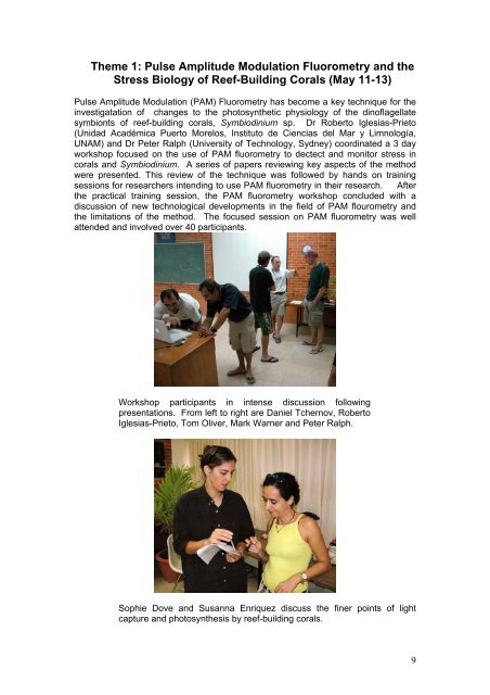
Theme 1: <strong>Pulse</strong> <strong>Amplitude</strong> <strong>Modulation</strong> <strong>Fluorometry</strong> and theStress Biology of <strong>Reef</strong>-Building <strong>Coral</strong>s (May 11-13)<strong>Pulse</strong> <strong>Amplitude</strong> <strong>Modulation</strong> (PAM) <strong>Fluorometry</strong> has become a key technique for theinvestigatation of changes to the photosynthetic physiology of the dinoflagellatesymbionts of reef-building corals, Symbiodinium sp. Dr Roberto Iglesias-Prieto(Unidad Académica Puerto Morelos, Instituto de Ciencias del Mar y Limnología,UNAM) and Dr Peter Ralph (University of Technology, Sydney) coordinated a 3 dayworkshop focused on the use of PAM fluorometry to dectect and monitor stress incorals and Symbiodinium. A series of papers reviewing key aspects of the methodwere presented. This review of the technique was followed by hands on trainingsessions for researchers intending to use PAM fluorometry in their research. Afterthe practical training session, the PAM fluorometry workshop concluded with adiscussion of new technological developments in the field of PAM flourometry andthe limitations of the method. The focused session on PAM fluorometry was wellattended and involved over 40 participants.Workshop participants in intense discussion followingpresentations. From left to right are Daniel Tchernov, RobertoIglesias-Prieto, Tom Oliver, Mark Warner and Peter Ralph.Sophie Dove and Susanna Enriquez discuss the finer points of lightcapture and photosynthesis by reef-building corals.9
Roberto Iglesias-PrietoThe Photosynthetic Apparatus ofSymbiodinium.Unidad Académica Puerto Morelos, Instituto de Ciencias del Mar y Limnología,Universidad Nacional Autónoma de México, Apartado Postal 1152, Cancún QR77500, México.Invertebrates symbiotic with dinoflagellates in the genus Symbiodinum are amongthe most important primary producers in coral reefs. In these environments they areresponsible not only for the high gross production, but also for the construction andmaintenance of the reef structure. Despite the importance of this symbiosis, ourknowledge about the biochemical organization of the photosynthetic apparatus ofdinoflagellates is still incomplete. This gap in our understanding is particularlyimportant in the context of the study of the photobiology of thermal stress. We havecommonly relied on the use of green plant models to make interpretations ofchlorophyll a fluorescence kinetic data from Symbiodinium. Although in some casesthese models can explain the experimental observations, caution should be takenbefore ascribing photosynthetic phenomenon to Symbiodinium that have not beencharacterized empirically in dinoflagellates, such as the induction of state transitionsand their putative role in photo-protection.ChromophoresThe functions of photosynthetic pigments are to capture photons, transfer excitationenergy to the reaction centers where primary photochemistry takes place, and, insome cases, provide photo-protection), The light harvesting function indinoflagellates is performed by Chl c 2 and peridinin, in addition to Chlorophyll a (Chla. Chlorophylls are porphyrin derivatives that form a cyclic tetrapyrrol with a chelatedMg atom ligated at the center of the macrocycle (Scheer 1991). The spectralcharacteristics of these molecules depend on the side groups attached to themacrocycle. In contrast with other chromophyte algae, dinoflagellates contain onlyChl c 2 , and their diagnostic carotenoid, peridinin. This carotenoid is capable oftransfering excitation energy to Chl awith efficiencies close to 100%, andtherefore plays a major role in lightcollection. All functionalphotosynthetic pigments are noncovalentlybound to specific proteinsforming Chl –protein complexes.The function of the protein moiety isto orient and space the chorophoresto assure the the excitation eneregyis efficiently transfered to thereaction centers. Functionally, Chlproteincomplexes are divided intolight harvesting complexes orantennae and their reaction centers.Fig 1. Schematic representation of thephotosynthetic apparatus of Symbiodinium.12
Iglesias-Prieto R (1996) Biochemical and spectroscopic properties of the lightharvestingapparatus of dinoflagellates. In: Chaudhry BR, Agrawal SB (eds)Cytology, Genetics & Molecular Biology of Algae. SPB Academic Publishing,Amsterdam, p 301-322Iglesias-Prieto R, Govind NS, Trench RK (1993) Isolation and Characterization ofthree membrane-bound chlorophyll-protein complexes from four dinoflagellatespecies. Philosophical Transactions of the Royal Society of London B. 340:381-392Iglesias-Prieto R, Trench RK (1997) Acclimation and adaptation to irradiance insymbiotic dinoflagellates. II. Responses of chlorophyll protein complexes todifferent light regimes. Marine Biology 130:23-33Scheer H (1991) Structure and ocurrence of chlorophylls. In: Scheer H (ed)Chlorphylls. CRC Press, Boca Raton, p 3-30Warner ME, Fitt WK, Schmidt GW (1999) Damage to photosystem II in symbioticdinoflagellates: a determinant of coral bleaching. Proceedings of the NationalAcademy of Sciences USA 96:8007-801214
Multiple scattering on coral skeleton enhanceslight absorption by symbiotic algaeSusana Enríquez, Eugenio R. Méndez 1 , & Roberto Iglesias-Prieto.Unidad Académica Puerto Morelos, Instituto de Ciencias del Mar y Limnología,Universidad Nacional Autónoma de México, Apartado Postal 1152, Cancún QR77500, México; 1. Departamento de Óptica, División de Física Aplicada, Centro deInvestigación Científica y Educación Superior de Ensenada, Km 107 carreteraTijuana-Ensenada, Ensenada BC 22860, México.New examination of the optical properties of intact coralsThe evolutionary success of scleractinian corals as reef-builders relied on theformation of mutualistic symbioses with photosynthetic dinoflagellates. Algalphotosynthesis provides nutritional advantages to scleractinians, as the translocationof photosynthates may account for their entire metabolic needs, while promotingrapid calcification. Therefore, symbiotic reef-building corals depend heavily on theefficiency with which they collect solar energy. In spite of the significant progress thatcoral photobiology has achieved over the last 20 years, the optical properties of intactcorals have not been directly assessed yet. A major obstacle for the study of theoptical properties of intact corals is the complexity of the coralline structure,consisting of coelenterate tissue, endosymbiotic algae and the intricate geometries ofthe aragonite coral skeleton. We provide in this work measurements of theabsorption spectra of intact corals for the first time. We selected the Caribbeanscleractinian Porites branneri, because the morphological characteristics of thisspecies permits the preparation of even and thin coral laminae (thickness = 3 ± 0.1mm) with homogeneous pigmentation suitable for spectroscopic analyses. During anatural bleaching event we collected specimens exhibiting a broad variation insymbiont and chlorophyll a content per unit of surface area (Chl a density), whichallowed us to assess the effect of such variability on the optical properties of P.branneri.Methodology employed hereAbsorption spectra of the coral laminae were recorded between 380 and 750 nm with1 nm resolution, with an Aminco DW2 (USA) spectrophotometer controlled by anOLIS (USA) data collection system. Skeleton laminae were used as reference. Thelight beams of the spectrophotometer were baffled with black tape apertures to matchthe exact dimensions of individual samples. Reflectance spectra of corals andskeletons were measured between 400 and 750 nm with 1 nm resolution using a4800S Lifetime spectrofluorometer (SLM-Aminco, USA) equipped with a red sensitivephoto-multiplier tube (R955, Hamamatsu, Japan). The use of thin laminae allowed usto obtain high quality absorption spectra of intact coral surfaces. As a result ofbleaching, we had available a series of absorption spectra of specimens whose Chl adensity varied from 3.3 mg m -2 to 102,1 mg m -2 . The observed 30-fold variation in Chla density resulted in an approximately 5-fold variation in coral absorptance.Measurements of light absorption on transmission mode are not only laborious, butcan be difficult to implement with corals of other morphologies. We propose as analternative technique for estimating coral absorption, the determination of reflectancespectra, since the inferred absorption spectrum compared well with those obtained intransmission mode.15
Variation in light absorption properties as a function of coral pigment contentDetermination of absorptance as a function of the variation in pigment content showedthat the light-harvesting capacity of P. branneri decreases abruptly only for Chl adensity below 20 mg m -2 , remaining practically constant for values above thisthreshold. These results differ from former estimations based on filtered coral slurries (1-4) .To quantify the variations in pigment light-absorption efficiency, we estimated thechanges in the chlorophyll a specific absorption coefficient (a*) as a function of Chl adensity. The analysis of this variation indicates that the values of a* estimated forintact corals are between 2 and 5 times higher than those estimated fromsuspensions of freshly isolated symbionts with similar pigment density. On the otherhand, the a* values obtained in our study for a suspension of freshly isolatedsymbionts are consistent with measurements of the absorption of filtered blastates (1-4) , and with values reported for phytoplankton (5) . The increase in the absorptionefficiency may be understood through simple physical considerations. In simplifiedform, a coral structure may be visualized as a thin layer of small, pigmented particles,above one dimensional surface of coral skeleton. As the illumination reaches thepigment layer a fraction of the incident light is absorbed (Φ abs (i) ). Part of the lighttransmitted through the layer is backscattered by the skeleton and passes again(Φ abs (s) ) as diffuse light through the layer of pigment, increasing thus the capacity oflight absorption by the particles. This theoretical model concludes that a flatscattering surface can enhance the absorption of the particle by a factor of up to 3(for a non absorbing surface, Φ abs = Φ abs(i)+ Φ abs(s)= (1+2R)Φ abs (i) ). The modelbecomes theoretically intractable when the particle is placed inside a concavity or isexposed to several reflective coral skeleton surfaces. Nevertheless, it concludes thatlight absorption by the particle could be amplified by a factor much higher that 3.The comparison done in the North Queensland Tropical Museum (Townsville,Australia) on 56 coral skeletons of Favide spp, and 18 spp of other massivetaxonomic families, reveled a large variability among species in the variation of themaximum enhancement factor. We found values from a minimum of 2.9 showed byCaulastrea curvata to a maximum of 8.3 showed by Cyphastrea japonica. It isnoteworthy that the three Porites spp from the Indo-Pacific examined showed similarvalues than the maximum value estimated in this work for Porites branneri. Weconcluded that coral skeletons are efficient bulk scatterers allowing to enhancing thecapacity of light absorption by algal pigments through the diffusive propagation oflight over longer optical paths. Multiple scattering by the coral skeleton providesdiffuse and homogeneous light fields for the symbionts reducing pigment selfshading.Biological and ecological implicationsThe biological and ecological implications of the optical properties of the intact coralstructure revealed by this study are diverse. We concluded that: a) the light fieldswithin coral tissue are not predictable from the water column light attenuationdescriptions, and are very dependent on pigment density; b) the study ofphotoacclimatization of different species of corals and the propagation of the thermalstress needs to be revised from this new perspective; c) the modulation of theinternal light field by the coral skeleton may be an important driving force in theevolution of symbiotic scleractinian corals; and d) determinations of the minimumquantum requirements for symbiotic scleractinian corals need to be re-assesed. Ourresults indicate that symbiotic corals are one of the most efficient solar energycollectors in nature. These organisms are capable of harvesting the same amount ofincident radiation as the leaf of a terrestrial plant with six times less pigment density.16
References1- Dubinsky, Z., Falkowski, P.G., Porter, J.W., and Muscatine L. 1984. Proc. R. Soc.Lond. B. 222: 203-214.2- Dubinsky, Z., Stambler, N., Ben-Zion, M., McClauskey L.R., Muscatine L., andFalkowski P.G. 1990. Proc. R. Soc. Lond. B. 239: 231-246.3- Wyman, K.D., Dubinsky, Z., Porter, J.W., and Falskowski, P.G. 1987. Mar. Biol.96: 283-292.4- Lesser, M.P., Mazel, C., Phinney, D. and Yentsch, C.S. 2000. Limnol. Oceanogr.45: 76-86.5- Morel, A., and Bricaud A. 1981. Deep Sea Res. 28: 1375-1393.6- Enríquez, S., Méndez, E.R., and Iglesias-Prieto R. 2005. Limnol. Oceanogr. 50:1025-1032.17
Mark E. Warner 1An overview of the interpretation and currentuse of chlorophyll fluorescence to understandcoral bleaching1. University of Delaware, College of Marine Studies, 700 Pilottown Rd. Lewes,Delaware, 19958, USA.As the thermal sensitivity of some zooxanthellae is seen as one of the primarycauses of coral bleaching, many laboratories have undertaken intensive studies inthe photobiology of these symbiotic dinoflagellates to better understand howphotosynthetic processes may be affected by excessive thermal and light exposure.Several methods of measuring active chlorophyll fluorescence to infer photosyntheticfunction are becoming widely used in coral biology and for assessing photosystemstress during experimental or natural bleaching in particular. The saturation pulsemethod commonly used with the pulse amplitude modulation (PAM) fluorometer wasintroduced to the field of coral biology almost a decade ago, and has become acommon tool for many reef biologists investigating coral bleaching. While the methodand the data that it can quickly generate has proven quite beneficial in extending ourfundamental understanding of the photobiology of zooxanthellae symbioses, it is notwithout certain caveats that should be addressed in light of current efforts to betterunderstand the biochemical and cellular pathways involved in coral bleaching.Importance of experimental design and key parametersWhile core proteins and pigments within the reaction centers of photosystem I and II(PSI & PSII hereafter) are highly conserved across all known photosyntheticeukaryotes, there are many differences at the level of light harvesting complexstructure and function which can and do affect commonly used fluorescenceparameters. It is prudent to note that the much of the theoretical groundwork thatforms the basis of using PAM fluorometry extends from work with higher plants whichcan represent a significant departure from the physiological variability one is likely toencounter in working with algal groups (including the dinoflagellates) that extend fromthe red algal lineage. A second important point is that there are many differences incurrent designs of coral bleaching experiments utilizing PAM fluorometry such thatresults should be evaluated in light of the scale of the design itself. It is important toview current results in relation to the ecological relevance of the experimental designversus the degree of physiological reduction. The field of chlorophyll fluorescencecontains a semantic minefield of terminology that can be an unnecessary source ofconfusion when comparing different bleaching experiments, and one should strive tofollow previously published guidelines for correct use of such terms (see Kromkampand Forster 2003). Some of the more common parameters in use today are the darkacclimated quantum yield of PSII (Fv/Fm), the effective quantum yield of PSII(∆F/Fm’), photochemical (qP) and nonphotochemical fluorescence quenching (qNand NPQ), and electron transport rate (ETR).Fv/Fm and ∆F/Fm’ are two of the most common parameters used for rapidlyassessing the status of PSII in zooxanthellae within corals, as they are easy tomeasure and have a long history of use in plant and phytoplankton biology fordetecting photoinhibition. <strong>Coral</strong>s exposed to thermal stress as well as those sampledduring natural bleaching events have shown a significant loss in Fv/Fm compared tocorals held in non-bleaching conditions which typically precedes detection of any loss18
in zooxanthellae density. Declines in Fv/Fm are correlated with the loss of PSII D1protein content in some cases (Warner et al. 1999; Lesser and Farrell 2004), therebyenforcing the idea of thermal stress exacerbating a pathway of cellular damage seenin light enhanced photoinhibition studies. However, more work is needed to fullyestablish if this correlation holds across different species of zooxanthellae. Forexample, some zooxanthellae could possibly show a loss in PSII activity whiledegradation of reaction center proteins is impeded. Likewise, an important point isthat one should be careful to delineate stress related loss of Fv/Fm with possibleseasonal declines in Fv/Fm that largely reflect photoacclimation as opposed tochronic cellular stress. Similarly, one can use the effective quantum yield (∆F/Fm’) orPSII capacity in the light acclimated state as a proxy for stress, so long as one hasan understanding of the typical patterns of diurnal decline and recovery due simply toincreased levels of light energy dissipation during daylight hours (i.e. enhancedquenching of the fluorescence signal due to non-photochemical pathways such asxanthophyll cycling) in their organism of interest. Recent work has shown thatanalysis of ∆F/Fm’ can also indicate when a bleaching experimental design may beinducing more artificial chronic stress than would be seen in the field (see Franklin etal. 2004, Fig.1, for a good example). A second point of caution is that ∆F/Fm’ isdependent on the previous light history of the alga and one should take care ininterpreting results for experiments that involve acute light shocks that may notreplicate physical conditions typically seen in the field. In this same vein, afluorescence induction curve conducted with a light intensity significantly higher thanthat used in the experimental treatment can yield large decreases in ∆F/Fm’ whichreflect an artefact of the light intensity used for the induction curve rather than how acoral may have processed light under the experimental conditions. Of the quenchingparameters typically measured, photochemical quenching (qP) is typically thehardest to measure under field conditions. This difficulty is due to the fact that theequations traditionally used to calculate this variable rely on accurate measurementof any quenching of the initial fluorescence signal (F o ), which can happen quitefrequently in zooxanthellae (personal observation). Proper assessment of qPrequires the ability to rapidly darken a sample and apply a pulse of far-red light to reoxidizePSII traps. Such protocols are not available on current submersibleinstrumentation (e.g. the diving PAM), and alternative equations developed withhigher plants to circumvent the need to know F o for qP calculation have proven to beless accurate for corals (personal observation).Other parameters, such as excitation pressure over PSII (sensu Iglesias-Prieto et al.2004) and NPQ have shown to provide a good proxy for potential thermal stress insome corals. The central idea to this point is that some thermally sensitivezooxanthellae may have less capacity to dissipate excess energy than others, andtheir homeostatic level of excitation pressure is higher than that of otherzooxanthellae that show greater thermal resistance. On the other hand, more work iscurrently underway to better understand how (or if) any compensatory electronturnover at PSII may also play a role in explaining how some corals with elevatedNPQ or excitation pressure can maintain PSII function during thermal stress.Lastly, electron transport rate or ETR has been used heavily to infer changes inphotosynthetic activity in general and during thermal and/or light stress. Whileplotting ETR vs. irradiance is a common practice one should take great caution ininterpreting such data using traditional photosynthesis to irradiance terminology andconcepts, as they are not synonymous in many cases. For example, one should notassume that maximal ETR is representative of maximal photosynthetic rate (P max ) asmeasured by other methods (e.g. respirometry). Comparisons of grossphotosynthesis and ETR in several groups of algae have shown that these twovariables co-vary only within a range of light intensities and that they can significantly19
depart from each other at higher levels of light (Geel et al. 1997). Such an effect ismost likely due to non-assimilatory electron flow through PSII such as that due toMehler activity, which can change between different algae or under different physicalconditions. Likewise, current evidence suggests that coral absorptance can changesubstantially during bleaching (Enriquez et al. 2005), thus rendering anymeasurement of “relative” ETR that does not account for such absorptance changeshighly suspect to gross error.ReferencesEnriquez S, Mendez ER, Iglesias-Prieto R (2005) Limnol Oceanogr 50: 1025-1032Franklin DJ, Hoegh-Guldberg P, Jones RJ, Berges JA (2004) Mar Ecol Prog Ser 272:117-130Geel C, Versluis W, Snel JFH (1997) Photosynth Res 51: 61-70Iglesias-Prieto R, Beltran VH, LaJeunesse TC, Reyes-Bonilla H, Thome PE (2004)Proc Royal Soc London 271: 1757-1763Kromkamp J, Forster RM (2003) Eur J Phycol 38: 103-112Lesser MP, Farrell JH (2004) <strong>Coral</strong> <strong>Reef</strong>s 23: 367-377Warner ME, Fitt WK, Schmidt GW (1999) Proc Nat Acad Sci 96: 8007–801220
Diel Cycling of Nitrogen Fixation in <strong>Coral</strong>s withSymbiotic CyanobacteriaMichael P. Lesser 1 , Luisa I. Falcón 2 , Aimé Rodríguez-Román 3 , SusanaEnríquez 3 , Ove Hoegh-Guldberg 4 , and Roberto Iglesias-Prieto 31 Department of Zoology and Center for Marine Biology, University of NewHampshire, Durham, New Hampshire 03824, USA; 2 Instituto de Ecología,Universidad Nacional Autónoma de México, Circuito Exterior s/n CiudadUniversitaria, CP 04510 México, D. F. México, 3 Unidad Académica Puerto Morelos,Instituto de Ciencias del Mar y Limnología, Universidad Nacional Autónoma deMéxico, Cancún QR 77500, México, 4 University of Queensland, Centre for MarineSciences, 4072, St. Lucia, Queensland, Australia<strong>Coral</strong>s in mutualistic symbiosis with endosymbiotic dinoflagellates (zooxanthellae)are essential components of the ecological diversity of tropical coral reefs.Zooxanthellate corals also exist in an environment where inorganic nitrogen limits thegrowth and abundance of zooxanthellae in hospite 1-3 . Many colonies of theCaribbean coral, Montastraea cavernosa, contain endosymbiotic cyanobacteria 4 .These cyanobacteria co-exist with the zooxanthellae and express the nitrogen fixingenzyme nitrogenase 4 . Here we show that the percentage of colonies containingsymbiotic cyanobacteria increases with increasing depth, and that measurements ofnitrogen fixation show a diel pattern with the highest rates of nitrogen fixation in theearly morning and evening. No nitrogen fixation was measurable in non-symbioticcon-specifics. The δ 15 N stable isotope data show a strong nitrogen fixation signal inthe zooxanthellae fraction of corals with cyanobacterial symbionts suggesting thatzooxanthellae use fixed nitrogen products. The timing of nitrogen fixation avoidsmaximum periods of photosynthesis to avoid severe hyperoxia, and nitrogen fixationdoes not occur when the coral experiences hypoxia or anoxia. These cyanobacteriarequire low oxygen tensions to support the respiratory processes that provide theenergy required to fix nitrogen. Nitrogen fixation in these corals provides animportant supplemental source of a limiting element for this novel microbialconsortium.Figure 1. Green-brown (left) and red morphs of Montastraea cavernosa under Bluelight and viewed through an orange filter set. Photo: O. Hoegh-Guldberg21
Figure 2 Percent abundance ofMontastraea cavernosa coloniescontainingendosymbioticcyanobacteria at different depthsaround Lee Stocking Island, Bahamas.Significant differences were observedwith depth using a contingency tableanalysis; Likelihood ratio, χ2 =18.17P=0.006, Pearson, χ2 =14.065P=0.03. Post-hoc multiple comparisontesting showed that the population oforange M. cavernosa were significantlygreater deeper (≥15 m) compared toshallow depths (≤ 12 m).The presence of cyanobacterial symbionts that can fix nitrogen inzooxanthellate corals represents not just a novel microbial consortium ofphotosynthetic eukaryotes and prokaryotes, but one that challenges the longtermparadigm that nitrogen is limiting in corals. Our observations indicate thatseveral species of scleractinian corals in the Caribbean and on the GreatBarrier <strong>Reef</strong> have individuals harboring stable, non-pathogenic populations ofendosymbiotic cyanobacteria. If the occurrence of this consortium in otherspecies of scleractinian corals residing in oligotrophic tropical waters is morecommon than previously believed, it could provide an important supplementalsource of a limiting element for zooxanthellate corals. These inputs of newnitrogen from endosymbiotic cyanobacteria in corals not only has importantimplications for our current understanding of the role of nitrogen as a limitingand regulatory element in these associations, but also requires that we reexaminethe role corals play in the nitrogen budgets of coral reefs. Becausecorals release large quantities of dissolved organic material containing highconcentrations of both organic and inorganic sources of nitrogen the implicationof corals as nitrogen fixes consortiums for the biogeochemical fluxes of nitrogenin carbonate sands and pore waters of coral reefs are potentially very large.22
Figure 3. a) Nitrogen fixation (acetylene reduction) brown/green (N=3) versus orange(N=3) colonies of M. cavernosa. There were significant effects of colony type(ANOVA: P
Figure 4. a) The relationship between time (min) and depth (m) during which nitrogenfixation can take place in Montastraea cavernosa with endosymbiotic cyanobacteria.The open circles represent predictions using the data from the fitted P-I curve. Theclosed squares are the simulation results using the empirical respiration rates, andthe closed circles represent the simulation results under conditions where we allowednitrogen fixation to take place at the same irradiances experienced in situ by ourcoral samples. b) Using the percent abundance data of Montastraea cavernosa withendosymbiotic cyanobacteria and the simulation data of the percentage of daylightirradiances that are below the compensation point (± 10%) there is a significantfunctional relationship (ANOVA: P
Figure 1. Effect of transplanting Montipora monasteriata between high (unshaded) and low(shaded) light fields on host pigments. Absorption spectrum of (A) pocilloporin (B) putativegreen GFP. Transplantation of blue and tan morph from high to low PFD (C,D); of green, redand brown morphs from low to high PFD (E,F). *, p
Imaging-PAM: Operation and PossibilitiesRoss Hill 1 and Karin E. Ulstrup 11 Institute for Water and Environmental Resource Management and Department ofEnvironmental Sciences, University of Technology, Sydney, Westbourne St, GoreHill, NSW 2065, Australia.High resolution imaging of variable chlorophyll a fluorescence emissions was used toidentify 2-dimensional heterogeneity of photosynthetic activity across the surface ofcorals. In comparison to earlier studies of fluorescence analysis (Ralph et al. 2002),the Imaging-PAM enables greater accuracy by allowing different tissues to be betterdefined and by providing many more data points within a given time (Hill et al. 2004;Ralph et al. 2005). The resolution of the instrument provides detail down to 100 µmand the area imaged can be controlled by the user. The standard Imaging-PAMmeasures an area of 3.5 x 4.5 cm and the Maxi-Imaging-PAM measures 10 x 13 cm.A micro-head attachment is also available for more detailed, fine scale investigations.An added component to the apparatus is a 96 well plate (imaged under the Maxi-Imaging-PAM) which has applications for ecotoxicological studies. This instrumentcontains a ring of blue, red and near-infra-red (NIR) LED’s and a CCD camera forfluorescence detection. A new feature provided by this instrument enables themeasurement of absorptivity = 1-(Red/NIR).Photosynthetic responses of coral tissue to lightImages of fluorescence emission indicated that the photosynthetic activity ofcoenosarc and polyp tissues responded differently to changing light and dielfluctuations in Acropora nobilis, Goniastrea australiensis, and Pavona decussata.Fig. 1 shows variable chlorophyll a fluorescence images of A. nobilis under 295 µmolphotons m -2 s -1 .Absorp. F t F m ’ EQY PS NPQ32 mmFig. 1: Chl a fluorescence images of A. nobilis showing PAR absorptivity, F t , F m ’, effectivequantum yield (EQY), relative photosynthesis rate (PS) and non-photochemical quenching(NPQ). Colour scale shown.Diel fluctuations in F v /F m revealed that different tissue types showed varying degreesof downregulation/photoinhibition spatially and temporally. Upon exposure toexperimentally controlled high light conditions (1000 µmol photons m -2 s -1 ),downregulation of photosynthesis occurred as well as higher NPQ within the polypsof G. australiensis and on the polyp walls and coenosarc of A. nobilis.Studying coral bleaching with the Imaging-PAMIn Pocillopora damicornis, A. nobilis and Cyphastrea serailia the Imaging-PAM wasused to map the impact of bleaching stress. The effect of bleaching conditions (33°C29
and 280 µmol photons m -2 s -1 ) was studied over a period of 8 h. Marked changes influorescence parameters were observed for all three species. Although a decline inEQY was observed, P. damicornis showed no visual signs of bleaching on theImaging-PAM after this time. In A. nobilis and C. serailia, visual signs of bleachingover the 8 h period were accompanied by marked changes in F t (light-adaptedfluorescence), NPQ and EQY. These changes were most noticeable over the first 5h. The most sensitive species was A. nobilis, which after 8 h at 33°C had reached anEQY value of almost zero across its whole surface (Fig. 2). Differential bleachingresponses between polyps and coenosarc tissue were found in P. damicornis, butnot in A. nobilis and C. serailia. Spatial variability of photosynthetic performance fromthe tip to the distal parts was revealed in one species of branching coral, A. nobilis.Control 1 h 2 h 3 h 4 h 5 h 6 h 7 h 8 hFig. 2: EQYof A. nobilisover 8 h ofexposure tobleachingconditions.Colour scaleshown.Photosynthetic performance of zooxanthellae within animal tissue affected byPorites Ulcerative White Spot (PUWS) SyndromeThe Imaging-PAM was also used to map thephotosynthetic gradient across syndromelesions. In this case, Porites Ulcerative WhiteSpot (PUWS) Syndrome was imaged (Fig. 3).Several variable chlorophyll a fluorescenceparameters (F o , F m and EQY at highirradiances (596 µmol photons m -2 s -1 )showed distinct gradients across thesyndrome lesion. This suggests that in thecase of PUWS, photosynthetic performanceof zooxanthellae is affected and thatfluorometry may be a useful tool to assess thehealth of the symbionts associated with coralsyndromes in general.F v/F mF o F m EQY (596)EQY (5)F v/F m30Fig. 3: Photograph and variable chlorophyll afluorescence of Porites sp. showing F v /F m , F o , F m ,EQY at 596 and 5 µmol photons m -2 s -1 .Conclusions and future directionsThe Imaging-PAM allows for a range of photosynthetic parameters to be measuredacross a 2-dimentional photosynthetic surface and also provides the means tomeasure absorptivity. The results of these experiments indicate that stress-inducedphotosynthetic responses are rarely continuous across a coral surface and thatvariations exist between the various tissue types. As a result, it is emphasised that itis unwise to extrapolate single-point measurements to a whole colony.
Seasonal fluctuations in the physiology ofStylophora pistillataGidon Winters 1 , Sven Beer 1* and Yossi Loya 21 Department of Plant Sciences, Tel Aviv University, Tel Aviv 69978, Israel;Department of Zoology, Tel Aviv University, Tel Aviv 69978, IsraelSeasonal fluctuations in the maximal quantum yield of photosynthetic electron flowthrough photosystem II (F v /F m ) as measured in situ were found to occur naturally inzooxanthellae of non-bleaching colonies of the branching coral Stylophora pistillatagrowing at 5, 10 and 20m. These fluctuations correlated stronger with changes inirradiance than changes in seawater temperature. Seasonal fluctuations were alsofound in the chlorophyll a density, which was due mostly to seasonal changes inzooxanthellae density. Results show that during the summer of a “non-bleachingyear”, corals will loose 80% of their zooxanthellae (in comparison with zooxanthellaedensities measured during the winter) the equivalent of zooxanthellae loss measuredin the Caribbean during the 1998 mass bleaching event (Warner et al. 2002).Underwater photographs taken monthly reveal the dramatic colour changes corals gothrough even during “non-bleaching” years. These results shed some light on theissue of what is “real” bleaching as apposed to seasonal changes. Possibly, what istermed mass bleaching should be seen as taking this normal (seasonal) paling ofcoral colour one notch forwards. For future PAM fluorometry based studies, it issuggested that, in order to correlate F v /F m values with anthropologically causedstresses, (a) F v /F m measurements be performed in situ under natural conditions and(b) natural seasonal fluctuations in F v /F m be taken into account when using thisparameter for diagnosing coral bleaching. It is further suggested that high irradiancesmay cause decreased F v /F m values at least as much as, if not more than, hightemperatures.FiguresFig.1. a) Study site and b) experimental set up: Specially made plastic holder for theDiving-PAM’s probe, allowing for repetitive measurements to be performed keepingthe same angle (69o) and distance (1 cm) between the sample and the PAM’s probe32
0.750.70.650.60.555m10m20m0.5Jan-04Feb-04Mar-04Optimal quantum yield of PSII (Fv/Fm)Apr-04May-04Jun-04Jul-04Aug-04Sep-04Oct-04Nov-04Dec-04Month (2004-5)Fig. 2. Seasonal variations in F v /F m in Stylophora pistillata (n=5, ±SE) growing at 5,10 and 20mMaximum diel global radiation (W m -2 )12001000800600400Jan/04Feb/04Mar/04Apr/04May/04Jun/04Jul/04Aug/04Sep/04Oct/04Nov/04Dec/04Jan/05Max diel global radiationMax diel water temperature2928272625242322Maximum diel seawater temperature ( o C)20021Month (2004)Fig. 3. Seasonal variations in global diel maximum global radiation (left y-axis) anddiel maximum seawater temperature (right y-axis) for the year 2004.33
Optimal quantum yield of PSII (Fv/Fm)0.660.640.620.60.580.560.54ar=0.1620 21 22 23 24 25 26 27 28Monthly average water temperature ( o C)Optimal quantum yield of PSII (Fv/Fm)0.660.640.620.60.580.56br=0.850.54500 600 700 800 900 1000Monthly average maximum diel radiation (W m -2 )Fig. 4 Correlations between monthly F v /F m measurements of Stylophora pistillata(n=5) growing at 5m and monthly average of a) seawater temperature ( o C) and b)diel maximum global radiation (W m -2 )34
32.521.510.50Jun-04Jul-04Aug-04Sep-04Oct-04Nov-04zooxanthellae density(10 6 cells/ cm 2 )Dec-04Jan-05Feb-05Mar-05Apr-05May-05Jun-05Month (2004-5)Fig. 5. Seasonal dynamics in zooxanthellae density of Stylophora pistillata (n=5-6,± SE) growing at 5mFig. 6. Photographs following the same branch of Stylophora pistillata growing at 5mthrough out the year.35
The cellular mechanism of coral bleachingDaniel Tchernov 1,3 , L Haramaty 1 , T. S. Bibby 1 , Max Y. Gorbunov 1 ,andPaul G. Falkowski 1,21 Environmental Biophysics and Molecular Ecology Program, Institute of Marine andCoastal Sciences, Rutgers University, 71 Dudley Road New Brunswick, NJ08901,USA;§ Department of Geological Sciences, Rutgers University, WrightGeological Laboratory, 610 Taylor Road, Piscataway, NJ 08854, USA; ¥ TheInteruniversity Institute of Eilat, P.O.B 469, Eilat 88103, IsraelThe phenomenon of mass coral bleaching has developed into a major concernbecause of the coral reef’s key ecological role as a major habitat for the most diversecommunity in the marine realm and its key economic function in numerouseconomies. <strong>Coral</strong> bleaching is induced by positive anomalous temperatures insurface waters of 1.5 to 2 ºC. However, not all reefs or corals within a reef areequally susceptible to elevated temperature stress. Here we wish to report thatthylakoid membrane (TM) lipid composition in the algal symbiont plays a key role indetermining the symbiosis’ susceptibility to thermal stress. However, there seems tobe no correlation between phylogenetic assignment of the symbiotic algal type andTM lipid composition. In addition, we show that apoptosis, triggered by the productionof reactive oxygen species (ROS) in the symbiotic algae (zooxanthellae) that residewithin host animal cells, can induce an apoptotic caspase cascade in the host animalthat leads to expulsion of the algae and can also lead to death of animal. Thebleaching process can be experimentally manipulated by the addition of extracellularROS and caspase inhibitors. This mechanistic explanation is of major importance inenabling a comprehensive understanding of bleaching on a biochemical andmolecular level. Our findings might also reflect on the interpretation of ecological andevolutionary processes that are currently observed in coral reefs world wide.Tolerant26 o CTolerant32 o CA meltdownof biologicalmembranesSensitive26 o CSensitive32 o CSensitiveS. pistillata26 o CA meltdownof biologicalmembranesSensitiveS. pistillata32 o C36