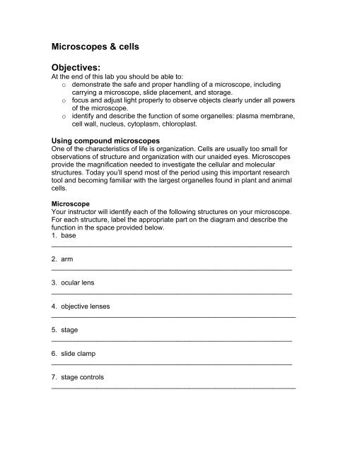
Microscopes & cells Objectives:
Microscopes & cells Objectives:
Microscopes & cells Objectives:

You also want an ePaper? Increase the reach of your titles
YUMPU automatically turns print PDFs into web optimized ePapers that Google loves.
7. coarse focusing knob_______________________________________________________________8. fine focusing knob________________________________________________________________
Using stainsStains are used to improve contrast. This improves viewing with a microscope.The next sections use two different stains. The first stain, methylene blue, stainsmembranes. The second stain, iodine, stains starch.Methylene blue and cheek <strong>cells</strong> Gently scrape the inside of your cheek with the tip of a toothpick andsmear it on your slide. Add a small drop of methylene blue. Add a cover slip. Start on low power and progress up to high power Focus on a single cell. Try to identify the plasma membrane, cytoplasm, and nucleus. Notice that animal <strong>cells</strong> do not have acell wall. Sketch a single cell and label organelles. Find a group of <strong>cells</strong> clumped together. Sketch this clump of <strong>cells</strong>.Single cheek cell under high powerwith organelles labelledClump of cheek <strong>cells</strong> under high power26. Describe the spatial relationship between individual <strong>cells</strong> in thisgroup of cheek <strong>cells</strong>: side by side, or overlapping? __________________27. What might be the advantage to how cheek <strong>cells</strong> are in relationship to eachother?______________________________________________________________28. Describe the spatial relationship between individual <strong>cells</strong> in theElodea leaf:______________________________________________________________