have demonstrated that identified neurones of the rostral andcaudal components of the ITN have fundamentally differentfiring properties in vivo, somatodendritic architectures andconnectivity at the level of the striatum (Lacey et al., 2007).Using a multidisciplinary approach, we have demonstrated thatneurones of the rostral and caudal ITN provide functionallydistinct inputs to the striatum. We carried out recordings ofsingle CL and Pf neurones in anaesthetized rats and, afterphysiological characterization, we juxtacellularly labelled theneurones with a tracer, Neurobiotin. The juxtacellularrecording/labelling technique is powerful because it enablesone to selectively label the same neurone that you haveextracellularly recorded from and thus unambiguously identifyand morphologically characterize the recorded neurone(Pinault, 1996). Using this technique, we were able to preciselydefine the location of the recorded neurones within in the CL orthe Pf, and, more importantly, we could correlate thesomatodendritic characteristics, axonal projection patterns andsynaptic connectivity of a labelled single neurone with its in vivophysiological properties (Lacey et al., 2007). The results of ourstudies show that CL and Pf neurones are different at severalfunctional levels. First, CL neurones, but not Pf neurones,engage in burst firing modes during slow-wave (sleep) activity(Fig. 2), which is a stereotypical property of thalamic relayneurones (see above). Thus, CL and Pf neurones operate astypical and atypical thalamic neurones, respectively. In additionto differences in physiological properties of neurones in thesetwo nuclei, the morphological properties and synapticconnectivity were distinct (Fig. 3). We confirmed that CL and Pfneurones had strikingly different dendritic architectures; theformer exhibited dendrites typical of thalamic relay neurons(Fig. 3A), but the latter did not (Fig. 3C). The study also revealedthat the axons of identified CL and Pf neurones establishedsynaptic connections with different targets in the striatum:these neurones target, on average, dendritic spines anddendritic shafts, respectively (Fig. 3B and D). Importantly, Pfneurones, but not CL neurones, exhibit a clear heterogeneitywith respect to their postsynaptic targets in striatum. Thus,single Pf neurones can innervate both dendritic spines andshafts, or exclusively innervate dendritic shafts, or selectivelyinnervate dendritic spines. Furthermore, a single axon terminalof a Pf neurone could simultaneously establish synapses withboth dendritic spines and shafts (see cover of this issue). Theselective innervation of distinct domains of the somatodendriticmembrane of MSNs is likely to have different influences ontarget cell activity, although how and to what degree thisinfluences striatal output is not yet understood.The thalamostriatal pathway: a new outlookThe results of these studies have further clarified thefundamental physiological and anatomical attributes of therostral and caudal ITN neurones and their <strong>projections</strong> to thestriatum. Although the ITN are commonly considered asfunctionally homogeneous, our work has directly demonstratedthat the fundamental properties of CL and Pf neurones differand they provide different temporally-patterned inputs todistinct striatal targets. One important implication of these datais that the field should re-evaluate the dogma that dorsalthalamic relay neurones are homogeneous in their firingproperties, given that in our study Pf neurones do not expressthe typical operational principles of thalamic relay neurones.Moreover, because of the multiple differences in thephysiological and anatomical characteristics of CL and Pfneurones alone, the ITN should no longer be collated into abroad category of “the thalamostriatal pathway”, with acommon mode of operation, for the sake of conceptualconvenience. This new information has far-reachingimplications for our understanding of both the thalamocorticaland thalamostriatal pathways (and other subcortical brainareas), and future studies should perhaps be dedicated todissecting out other potential differences between the rostraland caudal components of the ITN and their <strong>projections</strong>.AcknowledgementsThe author wishes to extend her warmest thanks to her thesissupervisors, Professor Paul Bolam and Dr. Peter Magill, both ofwhom more than generously devoted their time and energy tosuccessfully getting her through the D.Phil. Her thanks also go toeverybody at the MRC Anatomical Neuropharmacology Unit,Oxford. The author would also like to extend special thanks to BenMicklem for help with figures and for designing the coverillustration. This work was funded by the Medical Research CouncilUK. During her D.Phil., the author was also supported by travelawards from The Guarantors of Brain and The Keble Association,and the Faith Ivens Travel Grant (Keble College, Oxford).ReferencesBolam JP, Booth PAC, Hanley JJ, Bevan MD (2000)Synaptic organisation of the basal ganglia. J Anatomy196:527-542.DeLong MR (1990) Primate models of movementdisorders of basal ganglia origin. Trends Neurosci13:281-285.Deschênes M, Bourassa J, Parent A (1995) Twodifferent types of thalamic fibers innervate the ratstriatum. Brain Res 701:288-292.Deschênes M, Bourassa J, Doan VD, Parent A (1996) Asingle-cell study of the axonal <strong>projections</strong> arising fromthe posterior intralaminar thalamic nuclei in the rat. EurJ Neurosci 8:329-343.Destexhe A, Sejnowski TJ (2003) Interactions betweenmembrane conductances underlying thalamocorticalslow-wave oscillations. Physiol Rev 83:1401-1453.Groenewegen HJ, Berendse HW (1994) The specificity ofthe 'nonspecific' midline and intralaminar thalamicnuclei. Trends Neurosci 17:52-57.Jones EG (1985) The Thalamus. New York: Plenum Press.Kimura M, Minamimoto T, Matsumoto N, Hori Y (2004)Monitoring and switching of cortico-basal ganglia loopfunctions by the thalamo-striatal system. Neurosci Res48:355-360.Lacey, C.J., Bolam J. P. and Magill P.J. (2007). Noveland distinct operational principles of intralaminarthalamic neurons and their striatal <strong>projections</strong>. J.Neurosci. 27: 4374-4384.Lisman JE (1997) Bursts as a unit of neural information:making unreliable synapses reliable. Trends Neurosci20:38-43.Llinás R, Ribary U, Contreras D, Pedroarena C (1998)The neuronal basis for consciousness. Philos Trans RSoc Lond B Biol Sci 353:1841-1849.Llinás RR, Steriade M (2006) Bursting of thalamicneurons and states of vigilance. J Neurophysiol95:3297-3308.Matsumoto N, Minamimoto T, Graybiel AM, Kimura M(2001) Neurons in the thalamic CM-Pf complex supplystriatal neurons with information about behaviorallysignificant sensory events. J Neurophysiol 85:960-976.McHaffie JG, Stanford TR, Stein BE, Coizet V,Redgrave P (2005) Subcortical loops through the basalganglia. Trends Neurosci 28:401-407.Paxinos G and Watson C (2007) The Rat Brain inStereotaxic Coordinates, 6th Ed, Elsevier AcademicPress, San Diego.Pinault D (1996) A novel single-cell staining procedureperformed in vivo under electrophysiological control:morpho-functional features of juxtacellularly labeledthalamic cells and other central neurons with biocytinor Neurobiotin. J Neurosci Methods 65:113-136.Scheibel ME, Scheibel AB (1967) Structuralorganization of nonspecific thalamic nuclei and theirprojection toward cortex. Brain Res 6:60-94.Sherman SM (2001) Tonic and burst firing: dual modesof thalamocortical relay. Trends Neurosci 24:122-126.Smith Y, Bevan MD, Shink E, Bolam JP (1998)Microcircuitry of the direct and indirect pathways ofthe basal ganglia. <strong>Neuroscience</strong> 86:353-387.Smith Y, Raju DV, Pare JF, Sidibe M (2004) Thethalamostriatal system: a highly specific network of thebasal ganglia circuitry. Trends Neurosci 27:520-527.Steriade M, McCormick DA, Sejnowski TJ (1993)Thalamocortical oscillations in the sleeping andaroused brain. Science 262:679-685.17
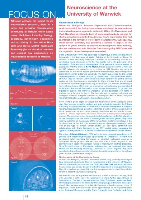
FOCUS ON...<strong>Neuroscience</strong> at theUniversity of WarwickAlthough perhaps not famed for its<strong>Neuroscience</strong> research, there is alarge and growing <strong>Neuroscience</strong>community at Warwick which spansmany disciplines including biology,sociology, psychology, economicsand art history. In this article MarkWall and Kevin Moffat (BiologicalSciences) give an historical overviewand current day perspective on<strong>Neuroscience</strong> research at Warwick.Figure 1Figure 2Figure 3Figure 4Figure 5<strong>Neuroscience</strong> in BiologyWithin the Biological Sciences Department (http://www2.warwick.ac.uk/fac/sci/bio) the first groups to have an interest in <strong>Neuroscience</strong>took a developmental approach. In the mid 1980s, Liz Oliver-Jones andHugh Woodland developed a bank of monoclonal antibody markers forstudying development in the frog, Xenopus leavis. In particular, they hadan interest in the formation of ectoderm (neurectoderm). Subsequently,Oliver-Jones’s laboratory has published a number of papers on theanalysis of genes involved in early neural development. More recently,she has collaborated with Nicholas Dale investigating NTPDases andthe regulation of eye development (see below).Cahir O’Kane (1988-1993) had developed the technique of enhancer-trapping inDrosophila in the laboratory of Walter Gehring at the University of Basel. AtWarwick, Cahir’s laboratory developed a number of enhancer-trap markers fordeveloping neural structures in the fly. This rapidly led to the publication of adescription of the embryonic development of the peripheral nervous system inDrosophila. With the arrival of Kevin Moffat, first as a post-doc in the O’Kane labin 1990, subsequently appointed as a Lecturer in 1994, Warwick was quick tocollaborate on the use of second generation enhancer-traps, first developed byBrand and Perrimon at Harvard University. This technique allowed for the controlof gene expression in marked cells during development. They quickly built a bankof “GAL4 lines” or “drivers” with defined expression control in the central nervoussystem of both the developing and adult fly. Many of these lines are still usedwidely in the fly neurobiology community, for example, GAL4 drivers expressing inthe mushroom bodies of the brain (an area in involved in olfactory processing) andin the giant fibre circuit (involved in visual escape behaviours). To go with theexpression system, the Warwick Drosophila groups developed new tools toanalyse neural function in the fly: The first attempts to analyse cell shape viatransgenic markers; toxigenic ablation via transgenic Ricin-A chain; toxigenicneuronal inactivation via transgenic tetanus toxin-A chain.Kevin Moffat’s group began to analyse the development of the Drosophila adultgiant fibre neurons, using the markers and tools he had developed in the O’Kanelaboratory. His group was able to describe the complete development of the giantneurons. More recently, his group have identified a number of new genes involvedin the development of adult neural circuitry: short-stop, ken and barbie, arouser(Figure 1 - Ribbed appearance of arouser expression in Drosopholia embryo) andslowmo. The development of the genetic tools has also led the Moffat laboratoryto use Drosophila for the study of neurologically important genes. They haverecently published on the analysis of the human torsin dystonia homologue in thefly, describing for the first time neurodegenerative phenotypes associated withloss of this type of protein. Similar studies are now being continued withcollaboration with Bruno Frenguelli on an ART funded project analysing thehyper-phosphorylation of tau in the well established Drosophila Alzheimer’s models.The arrival of Richard Baines in 2001 led to the introduction of a combination ofgenetic and electrophysiological approaches in Drosophila. Richard haddeveloped patch clamping techniques of identified embryonic neurons inDrosophila. The fly group at Warwick was thus able to make progress on thecontributions of specific transcription factors to motoneuron electrical properties.Since Richard’s move to Manchester, collaboration has continued with KevinMoffat and the Luschnig laboratory, University of Zurich. Together they havedescribed a translational control mechanism to regulate expression of the voltagegated sodium channel gene, paralytic.The formation of the <strong>Neuroscience</strong> GroupIn 2000, Ted Pridgeon, a retired Lincolnshire farmer living in nearby LeamingtonSpa, generously endowed a Chair of <strong>Neuroscience</strong> at the University of Warwick.The first and current occupant of this Chair, Nicholas Dale, started to establish<strong>Neuroscience</strong> as a serious discipline within the Department of Biological Sciences.Thus Nicholas Dale and Mark Wall, the first two appointments, joined Kevin Moffatto form a nascent <strong>Neuroscience</strong> grouping.The establishment of a graduate entry medical school at Warwick, initially jointlywith Leicester in 2001, gave the opportunity to make further appointments in<strong>Neuroscience</strong>. This led to the recruitment of David Spanswick, Kevin Lee, PeterStanfield (group leader of the Molecular Physiology group) and Richard Baines (seeabove). <strong>Neuroscience</strong> research at Warwick has now entered a second phase ofexpansion. Firstly, there have been recent appointments to the experimentalistswithin Biological Sciences –notably Bruno Frenguelli, Yuriy Pankratov and Sonia18