NAPPO DIAGNOSTIC PROTOCOLS DP 01 Citrus Tristeza Virus (CTV)
NAPPO DIAGNOSTIC PROTOCOLS DP 01 Citrus Tristeza Virus (CTV)
NAPPO DIAGNOSTIC PROTOCOLS DP 01 Citrus Tristeza Virus (CTV)
- No tags were found...

Create successful ePaper yourself
Turn your PDF publications into a flip-book with our unique Google optimized e-Paper software.
<strong>NAPPO</strong> <strong>DIAGNOSTIC</strong> <strong>PROTOCOLS</strong><strong>DP</strong> <strong>01</strong><strong>Citrus</strong> <strong>Tristeza</strong> <strong>Virus</strong> (<strong>CTV</strong>)The Secretariat of the North American Plant Protection Organization1431 Merivale Road, 3 rd Floor, Room 140Ottawa, Ontario, Canada, K1A 0Y9March 19, 2<strong>01</strong>3
ContentsPageReview.............................................................................................................................3Approval ..........................................................................................................................3Implementation ................................................................................................................3Amendment Record.........................................................................................................3Distribution.......................................................................................................................31. Pest information ....................................................................................................41.1 Transmission.........................................................................................................42. Taxonomic information..........................................................................................53. Detection and identification...................................................................................53.1 Host plants ............................................................................................................53.2 Signs and symptoms.............................................................................................54. Risk of pest spread ...............................................................................................65. Methods for detection and identification................................................................65.1 ELISA (Enzyme Linked Inmunosorbent Assay......................................................75.2. Immunoimpression- ELISA .................................................................................145.3 Biological techniques ..........................................................................................156. Records...............................................................................................................177. Contacts for additional information......................................................................178. References..........................................................................................................18<strong>DP</strong> <strong>01</strong><strong>Citrus</strong> <strong>Tristeza</strong> <strong>Virus</strong> (<strong>CTV</strong>)2
1. Pest information<strong>Citrus</strong> tristeza closterovirus (<strong>CTV</strong>) is the cause of one of many economically seriousdiseases of citrus. <strong>CTV</strong> probably originated in Asia and was disseminated to manycitrus producing countries by infected propagative material. Subsequent spread byaphid vectors caused epidemics in the 20th century. Millions of trees planted on sourrootstocks have been lost to tristeza decline in many countries, including the UnitedStates, Spain, and Brazil. Trees on tolerant or resistant rootstocks may be affected bystem pitting strains that can reduce yields and quality (Bar-Joseph et al. 1989; Garnseyet al. 1998; Lee and Bar-Joseph 2000).<strong>CTV</strong> probably occurs in all citrus growing areas worldwide, although its incidencevaries. <strong>CTV</strong> is usually detected in isolated citrus trees and still is not consideredepidemic in many countries such as Albania, Algeria, Cyprus, Egypt, France, Greece,Italy, Jordan, Lebanon, Libya, Morocco, Palestine, Portugal, Syria, Tunisia and Turkeyand in some Central and South America countries such as Belize, Chile, Ecuador, ElSalvador, Honduras, Mexico and Nicaragua. <strong>CTV</strong> is considered to cause the mostdamaging viral disease of citrus. In Argentina, with its occurrence in 1945, it caused thedeath of approximately 10 million trees, and in 1959 this number increased to 18 million.Brazil reported over 10 million of trees lost in 1958. In the sixties in Spain and in theeighties in Venezuela, <strong>CTV</strong> destroyed approximately 16 million of trees (Rocha-Peña etal. 1995).In Mexico, <strong>CTV</strong> was first detected in 1983 in the State of Tamaulipas, and in 1986 inVeracruz; both outbreaks were quickly eliminated. However, the virus was subsequentlyreported in other Mexican States.Since 1939, the disease has also been observed in California, United States.Observations made by Halma, Smoyer and Schwalm (1944, 1945) indicated that quickdecline reported in California was similar to the "tristeza" disease reported in Brazil.Fawcett and Wallace (1946) at the same time as Meneghini (1946), reported thattristeza in Brazil was caused by a virus. It can be concluded that this virulent strain oftristeza caused the death of at least 3 million citrus trees in California. Grant andSchneider (1951) reported for the first time the occurrence of <strong>CTV</strong> in Florida, althoughno doubt its presence in the state goes back many years before its identification. Thetristeza strains identified in the state of Florida are different than those present inCalifornia. In Texas, Olson and Sleeth (1954) and Olson and McDonald (1954) found‘Meyer’ lemon trees infected with tristeza. The ‘Meyer’ lemon was the first host reportedin the State of Arizona (Carpenter 1956).1.1 Transmission<strong>CTV</strong> can be transmitted by grafting and semi-persistently by one of several differentspecies of aphid including Toxoptera citricida (Kirkaldy), T. aurantii (Boyer deFonscolombe), Aphis gossypii (Glover), and A. spiraecola (Patch) (Moreno et al. 2008).Efficiency of aphid transmission can be affected by the donor and receptor host, by thevirus isolate and its ability to replicate in a given host, by the vector species and itsbiology, and by environmental and horticultural factors. The most efficient aphid vectoris T. citricida (the Brown <strong>Citrus</strong> Aphid). However, aphid transmission by other species isefficient enough to cause economically significant spread of <strong>CTV</strong> (Rocha-Peña et al.1995). T. citricida was detected in February 2000 in the northern part of the states ofQuintana Roo and Yucatán, México. It has now been detected in the states of Yucatan,<strong>DP</strong> <strong>01</strong><strong>Citrus</strong> <strong>Tristeza</strong> <strong>Virus</strong> (<strong>CTV</strong>)4
Quintana Roo, Veracruz, Tabasco, Chiapas, Oaxaca, Puebla, and Campeche inMexico. In the United States, T. citricida has been reported from Florida but not fromCalifornia, Arizona, or Texas. Extensive research has been done on the epidemiology of<strong>CTV</strong> in different citrus growing areas and how it is influenced by aphid, host and virusisolate factors (Gottwald et al. 1999). Techniques have been developed and tested formonitoring and predicting rates and patterns of spread (Hughes et al. 20<strong>01</strong>). In general,once natural infection rates reach 0.1% to 0.5%, infection rates increase rapidly andsuppression by tree removal becomes difficult. In areas with active natural spread, thelevel of actual infection will exceed that determined by testing since some recentinfection will not be detected. <strong>CTV</strong> has not been demonstrated to be seed-transmitted.Many species of citrus and specific related genera of Rutaceae (Aurantioideae subfamily)have been reported to be hosts of <strong>CTV</strong>. However, trifoliates (Poncirus trifoliata)and many of their hybrids are resistant to <strong>CTV</strong>.2. Taxonomic information<strong>CTV</strong> is a member of the family Closteroviridae and has the binomial Closterovirus citrustristeza virus. The pathogen itself is a flexible virion of about 2,000 X 11 nm and has anon-segmented, positive-sense, single stranded ribonucleic acid (RNA) genome ofapproximately 20 Kb. Studies on the molecular properties of <strong>CTV</strong> have revealed that itis a complex virus group. Three major genotype groups have been recognized alongwith some variations among these. Recombinants between different genotypes and thefact that single “pure” isolates are actually populations of RNA variants further increasethe complexity (Rubio et al. 20<strong>01</strong>).Associations have been established between biological reactions and various geneticmarkers, but at this point no specific molecular pathogenicity determinants for decline orstem pitting have been established (Nikolaeva et al., 1998, Hilf and Garnsey 2000,Moreno et al. 2008).3. Detection and identification3.1 Host plantsNatural hosts for <strong>CTV</strong> include mainly species of the genera <strong>Citrus</strong> and Fortunella, butinfection has been detected in genera related to the genus <strong>Citrus</strong>, that have beenexperimentally inoculated such as Aegle, Aeglopsis, Afraegle, Atalantia, Citropsis,Clausena, Eremocitrus, Hesperthusa, Merrillia, Microcitrus, Pamburus, Pleiospermiumand Swinglea. Experimental inoculations have also been achieved with non-citrusspecies such as Passiflora gracilis y Passiflora coerulea using the aphid vector (Morenoet al. 2008)3.2 SymptomsDifferent isolates of <strong>CTV</strong> vary greatly in the symptoms they produce in different citrushosts. Some are extremely mild and cause little injury, others cause medium damageeven in sensitive hosts. Some are severe in specific hosts and benign in others, andsome can cause very severe reactions in a wide range of hosts. Different isolates canhave a wide range of severity in some hosts and, conversely, the same isolate can varyin symptoms expressed in different hosts. Graft inoculation of a range of differentindicator plants is commonly used to develop a profile of the biological properties of<strong>CTV</strong> and to predict probable severity in commercial hosts (Garnsey et al. 2005).<strong>DP</strong> <strong>01</strong><strong>Citrus</strong> <strong>Tristeza</strong> <strong>Virus</strong> (<strong>CTV</strong>)5
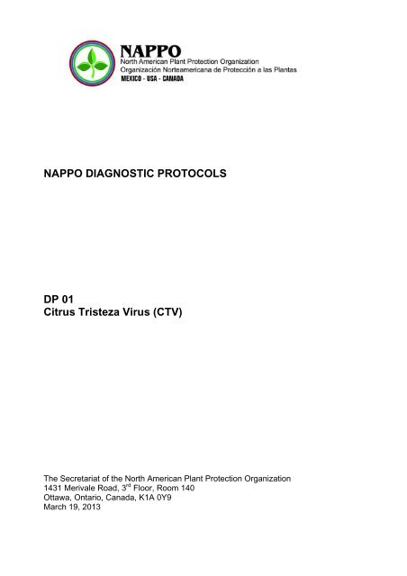
Common <strong>CTV</strong> symptoms include vein clearing, stunting, chlorosis, several types ofstem pitting, and seedling chlorosis (Moreno et al. 2008).<strong>CTV</strong> isolates and symptom phenologies can be categorized as follows: i) tristezadecline or quick decline strains which induce a collapse of citrus grown on sour orangerootstock; ii) mild (weak) strains that are symptomless in citrus grown on <strong>CTV</strong>-tolerantor resistant rootstocks; iii) seedling yellows strains which produce stunting and chlorosisin seedlings of orange, grapefruit, or Eureka lemon in a greenhouse virus index(generally considered to indicate severe strains); iv) stem pitting strains which inducemoderate to severe stem pitting in bark and adjacent wood in branches and trunks ofsweet orange, grapefruit, or mandarins regardless of rootstock.There are essentially two commercially important <strong>CTV</strong> diseases of citrus. One is theclassic tristeza or decline that is associated with a decline of trees grafted on sourorange rootstocks caused by a virus-induced phloem necrosis at the bud union (Figures1a, 1b). The classic decline disease has caused severe losses in many citrus growingareas, including California, and still threatens citrus industries having sour orange as themain rootstock. The <strong>CTV</strong>-induced decline can be avoided by use of decline-tolerant or –resistant rootstocks in lieu of sour orange; however, the use of these rootstocks mayresult in other diseases and horticultural problems (Moreno et al. 2008).The second major <strong>CTV</strong>-induced disease is stem pitting. In contrast to decline, stempitting can reduce the vigour and production of scions regardless of rootstock. Somerootstocks can be affected by stem pitting even when the scion is not. <strong>CTV</strong> stem pittingisolates has caused serious losses in many other citrus growing areas (Moreno andGarnsey 2005; Rocha-Peña et al. 1995).4. Risk of pest spread<strong>CTV</strong> may be spread to various citrus growing areas within the same country or to manycountries as a result of propagation by grafting and the subsquent transmission of thevirus in this fashion, added to spread by the vector.5. Methods for detection and identification<strong>CTV</strong> can be detected by biological indexing and various non-biological methodsincluding light and electron microscopy, serology and a variety of molecular-basedtechniques. The latter include several types of reverse transcription polymerase chainreaction (RT-PCR), including an immunocapture PCR with multiple molecular markers(MMM) (Hilf et al. 2005) and real time PCR (Ruiz-Ruiz et al. 2009), SSCP analysis(Rubio et al. 1996), oligoprobes (Narvaez et al. 2000) and RFLP analysis (Gillings et al.1993). The following techniques are approved by <strong>NAPPO</strong>: biological indexing, ELISA,and immunoprint ELISA.It is important to consider that sampling to detect <strong>CTV</strong> infected propagative materialmust be done during the citrus flushing season, coinciding with less hot months (whenthe monthly average temperature is around 27 °C and with little fluctuation during theyear). When moving plants within Mexico, sampling is done regardless of temperature.Each sample is made up of 10-15 cm shoots, of five trees, taken from the four treequadrants. If plants are too small and have no shoots, petioles can be used.<strong>DP</strong> <strong>01</strong><strong>Citrus</strong> <strong>Tristeza</strong> <strong>Virus</strong> (<strong>CTV</strong>)6
In California, where temperatures vary more than in Mexico, <strong>CTV</strong> incidence is very lowduring the winter and summer months (when temperatures are extreme) in most of theregions of California. The ideal season to detect <strong>CTV</strong> is between April and June or inOctober/November. This allows testing to be performed when incidence is high,providing more reliable diagnostics (Dodds et al. 1987).5.1 ELISA (Enzyme Linked Inmunosorbent AssayTo detect <strong>CTV</strong>, the Enzyme Linked Inmunosorbent Assay (ELISA) serological techniqueis used. This technique is based in the capability of certain proteins, known asantibodies, to recognize and link a specific antigen associated to the pathogen. This is areliable and quick technique to detect a plant pathogen virus. Some variants have beendeveloped, but so far the DAS-ELISA (Double Antibody Sandwich) direct method is themost commonly used (Salazar 1990). This technique consist of specific antibodiesabsorption to a polyestirene plate which later on the antigen is added to and then reactsto the antibodies adhered to the plate. Then, an enzymatic conjugate is added (secondantibody) to form the complex antibody-antigen-antibody.The specificity of monoclonal antibodies and the ease to trade them (since they arehomogenous and stable), in addition to the advantages offered by the technique suchas sensibility, low cost, speed, reliability and capability for mass usage, have favouredthe application of the ELISA technique in all citrus growing countries to detect <strong>CTV</strong>,using monoclonal antibodies capable to recognize any virus isolate without any reactionwith components of the host plant.5.1.1. ProcedureThe antibodies developed by Nikolaeva et al. (1995) and/or Lee et al. (2005) (primaryantibodies CREC 1052, CREC 27, CREC 28, CREC 29, CREC 30, or CREC 31 and thesecondary antibody G604) are commonly used in California. Agdia and BioRebacommercial kits for <strong>CTV</strong> are presently used in Mexico.The United States uses two different techniques, one by the <strong>Citrus</strong> Clonal ProtectionProgram (CCPP) using Gumpf antibodies and the other used by the GermplasmRepository (NCGRCD) which uses Lee et al. (2005) antibodies.5.1.1.1 Procedure of the <strong>Citrus</strong> Clonal Protection Program (CCPP) (Clark and Adams1977, modified for tristeza by J.M. Díaz of CCPP).A. Sample preparation1. Collect young branches from the four tree quadrants (or eight quadrants for treesat risk of field infection).2. Remove the bark from the branch, calculate approximately 1.0 g and cut in piecesof 1-2 mm.3. Prepare the samples to 1:10 (p/v) in extraction buffer, using the KLECO tissuepulverizer or the Ultra-Turaz T25 Tissumizer 1 .1 Manufacturing: Kleco Tissue Pulverizer, 14097 Ave 272,Visalia, CA 93292, 559-732-3785 or Tissuemizer,Ultra Turraz T25, Janke & Kunkel<strong>DP</strong> <strong>01</strong><strong>Citrus</strong> <strong>Tristeza</strong> <strong>Virus</strong> (<strong>CTV</strong>)7
Buffer PBSNaClKH 2 PO4Na 2 HPO4.7H 2 OKClNaN 3Adjustd the pH to 7.4, 1 L of H 2 O.8.0 g0.2 g2.17 g0.2 g0.2 gPBS-Tween Buffer: PBS Buffer + Tween20 2 ml/LExtraction Buffer: Buffer PBSTween + 20 g/L PVP40 polyvinylpyrrolidone (Sigma)4. Samples can be frozen and thaw before preparing the plate. This helps lower thebottom of the optical density (recommended but not required).B. Preparation of ELISA 2 plates1. Place 200 µl of γ-globulin in buffer coat in each well of the plate. Incubate for 4hours at 37 ºC. Normal concentration is 1-2 µg/ml. Ideal concentration must bedetermined by limiting dilution.Buffer coatNa 2 CO 3NaHCO 3NaN 3Adjust the pH at 9,6, in 1 L of H 2 O.1.59 g2.93 g0.2 g2. Wash the plate with PBS-Tween. Let it settle for 3 minutes. Repeat washing 3times, emptying the plate each time.C. Sample addition1. Place proportionally 200 µl of samples in each of two wells. Incubate at 6 ºCovernight or at 37 ºC for 4 hours.2. Wash the plate 3 times as in step B.2 and wash the plate 3 times with distilledwater, which helps lower the bottom of optical density.D. Addition of secondary and conjugated antibodies1. Place proportionally 200 µl of secondary antibody [according to Drs. A and OKarasev (Nikolaeva et al. 1995)] in each well to the ideal pre-determined dilution.Incubate at 37 ºC for 4 hours. For this step, use the conjugated buffer.Enzyme conjugate buffer: Buffer PBS-Tween + 20 g/L PVP40 polivinylpirrolidona +2 g/L ovalbumin (Sigma A-7030)2. Wash the plate 3 times as in step B.2.3. Place proportionally 200 µL of commercial conjugate (Sigma A-8025 Anti-RabbitIgI Alkaline Phosphatase Antibody, developed on goat), which usually has adilution of 1:8.000 to 1:10.000. Incubate at 37 ºC for 4 hours.E. Substratum addition (P-nitrophenyl phosphate disodium, Sigma S-0942, 5 mgtablets)1. Wash the plate 3 times as indicated in step B.2.2. Place proportionally 200 µl of recently prepared substrate at a concentration of 0,6-- 1,0 mg/ml of p-nitrophenyl phosphate in substrate buffer.2 Microtiter plates: Immulon IV Flat plates, Fisher #14245153 (50 plates/pack)<strong>DP</strong> <strong>01</strong><strong>Citrus</strong> <strong>Tristeza</strong> <strong>Virus</strong> (<strong>CTV</strong>)8
4. Pipet 200 μl of the supernatant into a dilution tube filled with 800 μl of coatingbuffer. Mix by pipetting the solution up and down a few times.Direct extraction using stem from new, tender flush tissue1. After step 4 (wash procedure), fill all sample wells of the plate proportionally with200 μl Aliquots of coating buffer containing 0.1 % Tween-20.2. Chop the stem in 1-2 mm long pieces using pruning shears.3. Drop the pieces of tissue directly into the wells filled with the buffer.4. Incubate overnight at 4 ºC.B. Preparation of controlFor BSD-ELISA usage of glycerol-prepared controls stored at –20 o C is preferred forstandardization purposes (Lee et al. 2005). A healthy (<strong>CTV</strong> -) and a positive (<strong>CTV</strong> +)control are included with the detection kit. These controls are ready to load into theELISA plate. 100 ul aliquots are loaded into wells that are pre-filled with 100 ul ofcoating buffer with 0.2% Tween 20.Alternatively, lyophilized, dried or fresh tissue can be used for negative and positivecontrols. In this case, use about 50 mg of tissue in 5 ml of coating buffer. Let the driedtissue hydrate as long as possible up to 24 hours before homogenizing. A good strategyis to hydrate the controls early in the morning, grind late in the afternoon, and extractovernight at 4 o C with the candidate samples.C. Preparation of ELISA plates1. Coat the ELISA plates with polyclonal antiserum made against purified <strong>CTV</strong>(preparation of IgG CREC27, CREC28, CREC31, CREC1051, or CREC 1052).Dilute the “CREC29 coating buffer” with the coating buffer to the concentrationindicated on the coating buffer tube (usually 1:5,000). Add 200 μl of the dilutedantiserum to each of the wells on the plate except the uncoated control well; twowells may be left as an uncoated control, usually wells A11 & A12. Incubate 1 to 3hours at room temperature, or overnight at 4 o C. (Note that when using overnightincubations, higher dilutions can be used for the coating antiserum. For example,if running the step at room temperature using a 1/5,000 dilution, or if running anovernight incubation in the cold, use a 1/10,000 dilution.)D. Wash1. Shake the solution from the plate and wash 3X under a stream of deionized water.2. Fill the plate with PBST, and let set for 5 minutes or longer (let set for at least 30minutes on the first wash).PBSTNaCl8.0 gKH 2 PO 40.2 gNa 2 HPO 41.15 g (anhydrous)KCl0.2 gAdjust to pH 7,4 using HCl , 1 L.Add 1 ml of Tween 203. Repeat steps 1 and 2 twice.4. Repeat step 1.5. Invert and tap on a clean paper towel to drain excess water immediately before thenext step. Make sure the plate does not dry out.<strong>DP</strong> <strong>01</strong><strong>Citrus</strong> <strong>Tristeza</strong> <strong>Virus</strong> (<strong>CTV</strong>)10
E. Samples1. Fill all wells of the ELISA plate with 100 μl of coating buffer containing 0.2%Tween-20.2. Add 100 μl aliquot of the sample per well, use two wells per sample. Mix thesamples with the buffer already in the well by pipetting up and down in the well afew times.3. Incubate overnight at 4 ºC.4. Wash the plate following the wash procedure.F. Secondary antibody1. Dilute the G-604 secondary antibody to a 1:20,000 dilution in conjugate buffer 4 .Conjugate buffer (500 ml)PBSTPVP-40(Bovine serum albumin, Fraction V).500 ml10.0 g1.0 g2. Add 100 μl of this solution to all wells in the plate.3. Incubate for 1 hour at 37 ºC, or overnight at 4 ºC.4. Wash the plate following the wash procedure.G. Conjugate1. Dilute Rabbit Anti-goat antibody conjugate with alkaline phosphatase to therecommended dilution on the tube (usually 1:30.000) in conjugate buffer.2. Add 100 μl aliquots of this solution to all wells in the plate.3. Incubate for 3 hours at 37 o C or overnight at 4ºC.4. Wash the plate following the wash procedure.H. Reaction1. Add 0.6 mg/ml of substrate (4-nitrophenyl phosphate hexahydrate) to coatingbuffer. Make sure the coating buffer is fresh, unless it was prepared using NaN 3 orit is not older than a few days and stored at 4ºC.2. Add 200 μl aliquots of this solution to each well.3. Incubate at room temperature.4. Read the plate after one or two hours, and/or on the next morning. Colour usuallydevelops very slowly. To calibrate it, set the plate reader to blank on the uncoatedwells. This will subtract the absorption of light caused by the plate and the buffer.No significant background reaction should develop during an overnightdevelopment of the plate. If this is the case, the secondary antibody should bediluted further.I. Evaluation1. Evaluate the controls first. The uncoated control should not give any reaction at allsince the reader is adjusted to zero on these wells,. Reaction in the uncoated wellsindicates improper washing of the plates after the conjugate step, improperpreparation of one of the buffers, or loss of the specificity of the antibodies againstthe antigen.4 Each lot of G-604 must be calibrated making a series of dilutions.<strong>DP</strong> <strong>01</strong><strong>Citrus</strong> <strong>Tristeza</strong> <strong>Virus</strong> (<strong>CTV</strong>)11
2. It is not unusual for the buffer only wells to show some reaction, sometimes evenmore than that of the healthy control (no antibodies).3. The healthy (negative) control wells should show none or very low reaction.4. A good plate will show OD405 values under 0.050 for the healthy and buffer onlywells, and values over 1.000 for the positive controls. In that case, a sample isconsidered positive if the average OD value is more than 0.100. Antibody dilutionsneed to be adjusted when higher values are obtained for the negative controls, orlower values obtained for the positive controls.Note: For all protocols, the final reaction volume in each step is the same. For this case, it is100 μl.5.1.1.3 Procedure used with the commercial Agdia kit1. Preparation of the samplea. Remove the bark from each shoot 5 of the sample collected. Weigh 0,2 g of barkand cut into pieces of 1-2 mm.b. Empty the tissue in a 50 ml tube and add 2 ml of extraction buffer.Extraction buffer solution (Macerated plant samples)Sodium sulfite (anhydrous)Polyvinylpyrrolidone (PVP) Molecular weight of 24 -40,000Sodium AzideGround Egg (chicken) albumin, Grade IITween-20Dissolve in 1Lt of Phosphate buffer solution 1X (PBST)Note: Adjust the pH to 7,4 (+/- 0,2). Store at 4 °C on refrigeration1.3 g20.0 g0.2 g2.0 g20.0 gc. Blend for 15 seconds using a tissue homogenizer.d. For better extraction, store at 4 °C overnight before placing the samples on theELISA plate.2. Preparation of ELISA platesa. Sensitize the polystyrene plate. Place 100 μl of the specific antibody dilution for<strong>CTV</strong> (Dilution 1:200 6 ) in each well plus the coating buffer solution taking intoaccount the sample distribution pattern, considering the samples and negative andpositive controls with their replicates.b. Incubate in wet chamber for 2 hours at 37 +/- 2 °C or overnight at 4 °C.Coating bufferSodium carbonate (anhydrous)Sodium bicarbonateSodium azideAdjust the pH at 9,6 (+/- 0,2). Store at 4 °CDissolve in 1 L of distilled water1.59 g2.93 g0.2 g5 Leaves main veins or petioles can also be used.6 Dilution commonly used. Follow the manufacturer’s instructions.<strong>DP</strong> <strong>01</strong><strong>Citrus</strong> <strong>Tristeza</strong> <strong>Virus</strong> (<strong>CTV</strong>)12
3. Plate washinga. Wash the plate 3 to 5 times 7 with PSTB-T 1XPhosphate (PBST) buffer solution (wash)Sodium chloride8.0 gDibasic sodium phosphate (anhydrous)1.15 gMonobasic potassium phosphate (anhydrous)0.2 gPotassium chloride0.2 gTween 200.5 g/mlDissolve in 1L of distilled waterAdjust the pH to 7,4 (+/- 0,2), washing solution can be prepared as aconcentrate first and then diluted. Prepare a concentration of 20X (20times concentrated).4. Sample additiona. Plant samples of interest are crushed or homogenized with extraction buffer. Add100 μl of the homogenized tissue to each well, with its replicates, in the previouslyselected wells.5. Control preparationa. In each of the wells associated with the negative controls, place either a 100 μl ofthe extraction solution by itself or commercial negative control. It is recommendednot to use the wells close to the edges since they can develop non-specificreactions (Salazar 1990).b. Dilute the positive control and place 100 μl of this in the wells.c. Incubate in wet chamber for 2 hours at 37 +/- 2°C or overnight at 4 °C.d. Wash the plate 6 to 10 times with PSTB-T 1X.6. Add the antibody + conjugatea. Mix the monoclonal antibody (Bottle A) with the enzymatic conjugate (Bottle B) inECI buffer in a 1:200 8 proportion for both antibodies. From this mix, 100 μl ispoured in each well and the plate is kept in a wet chamber for two hours 37+/- 2°C.b. Dissolve in 1000 ml of phosphate buffer solution 1X (PBST):Conjugate solution (ECI)Bovine serum albumin (BSA)Poly vinyl pyrrolidinone (PVP)Molecual weight 24-40,000Sodium azideAdjust the pH to 7,4 (+/- 0,2), store at 4°C.2.0 g10.0 g0.2 ge. Wash the plate 6 to 10 times.7. Addition of substrate.a. Prepare the substrate solution from the enzyme diluting the para-nitrophenylphosphate in extraction buffer in a proportion of 1mg/ml (usually one tablet/5 ml).7 Discard the plate contents in a sink, shake hard several times on paper towel. Fill each of the sensitized wells with the wash buffer, starting onthe first line, then the second line and so on, to cover all the needed wells, let stand for 1 minute and discard again the plate contents, shake wellover the paper towel.8 Dilution commonly used. Follow the manufacturer’s instructions.<strong>DP</strong> <strong>01</strong><strong>Citrus</strong> <strong>Tristeza</strong> <strong>Virus</strong> (<strong>CTV</strong>)13
. Place 100 μl in all the working wells, including the target and incubate under darkconditions from 15 to 45 minutes.Buffer solution for PNP (developing)Diethanolamine97.0 mlSodium azide0.2 gAdjust the final volume to 1 L. with sterile distilled water. Adjust thepH to 9,8 with hydrochloric acid, store at 4°C in refrigeration in amberbottle or wrap in aluminium to protect from light.8. Plate evaluationa. Turn on and program the ELISA plate reader.b. Note if there is colour in the wells. Change in colour is indicative of positive results.c. Read in the plate reader and print the readings.d. Analyze the results and record them in the record book for laboratory samples,writing the date of the response. Record the results in the database.ConsiderationsFor a diagnostic with reliable readings, the following controls must be included in theplate:• Uncovered wells• Extraction buffer• Healthy plant (negative control)• Sick plant (positive control)Infected plants with different <strong>CTV</strong> variants can be included 9 as positive controls. Anegative control corresponding to a healthy plant of the same species should also beincluded. In both cases (positive and negative controls) replicates should be included.Positive readings must be twice the value of the negative control. Negative results ofunknown samples must present absorbency values similar to the negative controls.If the samples are different than their replicate or have readings 1.5 times the value ofthe negative control, the diagnostic must be repeated. If the positive controls do notreact properly, the diagnostic must also be repeated.5.2. Immunoimpression- ELISAThe preparation of the plant extracts to be diagnosed is the most important limitingfactor to detect <strong>CTV</strong> with conventional ELISA. Therefore, some variants have beendeveloped such as immunoimpression-ELISA, which is a technique that uses capturemembranes that do not require sample crushing or homogenization, thus allowing theanalysis of thousands of samples in a simple and fast way (Cambra et al. 2000).The process consists of four basic and sequential stages:1. Collection and impression of membrane samples.2. Blocking the pressed membrane and reaction.3. Washing.4. Developing and reading the results.9 Two mild and two severe.<strong>DP</strong> <strong>01</strong><strong>Citrus</strong> <strong>Tristeza</strong> <strong>Virus</strong> (<strong>CTV</strong>)14
5.2.1 Procedure1. Make transversal or oblique cuts to young shoots 10 , leaves, pedicels, or pedunclesof newly ripe fruit with very sharp instruments.2. Firmly press the cross sectional tissue against the nitrocellulose membrane of 0.45mm pore, which is used as an immunoadsorbent. Make two impressions per shoot,cutting the ends from its base and apex. This will allow making 10 impressions permature tree. Positive and negative controls should be included.3. Let the membranes dry for a few minutes and keep them in a dry place andprotected from light until they are analyzed.4. Block the pores from the membrane (and the rest of the surface) with a bovineserum albumin (BSA) solution with a concentration of 1 % in distilled water. Themembranes must stay in the blocking solution for 1 hour at room temperature, or for16 hours if the operation is done at 4 ºC. Slightly shake the membrane to soak itand cover it completely with the blocking solution.5. Add a solution of specific monoclonal antibodies for <strong>CTV</strong>, marked with the alkalinephosphatase enzyme (0.1 mg/ml) to the membrane soaked with the albuminesolution.6. Incubate for 2 or 3 hours at room temperature with the solution completely coveringthe membrane. Once the reaction time has elapsed, discard the solution ofconjugated antibodies.7. Rinse the membrane 11 with washing buffer (PBS + 0.05% Tween 20)8. Add the substrate (BCIP-NBT Sigma Fast Tablets) 12 specific for the enzyme (10tablets in 100 ml of distilled water). Slightly cover the membranes and incubate atroom temperature until violet-purple precipitation start to show.9. When positive controls have shown some colour, stop the reaction by washing themembranes with running water. Precipitations will show after 3-7 minutes ofincubation at room temperature.10. Take the reading from the membranes once they are dry. In many cases, a readingat first sight is enough, but they should be observed with a magnifying glass ordissecting microscope.5.3 Biological techniquesBiological indexing is a technique that, under proper conditions, guarantees an accuratediagnosis. Biological indexing (bioindexing) for citrus pathogens is based on the use ofcitrus indicator plants free of pathogens, which react to the virus infection by expressingdiagnostic symptoms depending on the pathogen and isolate (Roistacher 1991). Eachsample (budwood to be tested) is inoculated into replicated indicator plants.Uninoculated indicators are used as healthy (negative) controls and indicatorsinoculated with tissue known to be infected with mild to severe isolates of the pathogenare used as positive controls.Biological indexing requires appropriate environmental conditions in a greenhouseenvironment and one month to a year to evaluate the material tested, depending uponthe pathogen.10 It is preferable to collect shoots of 10-15 cm from the youngest material available, from various areas around the tree, preferably from themedium-high section of the canopy, which is the area most likely to be visited by aphids.11 The washing stage is essential since it eliminates all the antibodies marked with alkaline phosphatase which have not reacted. That iway therewould only be traces of enzyme on the selections of printed plant material infested with <strong>CTV</strong>.12 Nitro blue tetrazolium and bromo chlorine indolyl phosphate (NBT+BCIP)<strong>DP</strong> <strong>01</strong><strong>Citrus</strong> <strong>Tristeza</strong> <strong>Virus</strong> (<strong>CTV</strong>)15
Bioindexing has been validated as reliably identifying citrus pathogens in mixedinfections, which are common in the case of citrus (Vidalakis et al. 2004).5.3.1. ProcedureThe procedures used for biological indexing for <strong>CTV</strong> are well established (Roistacher1991). For the biological indexing of <strong>CTV</strong> the preferred indicator plant is Mexican lime(<strong>Citrus</strong> aurantiifolia), the small-fruited acid lime. The Mexican lime seedlings must behealthy (i.e, no infections) with stems of 4-10 mm diameter and 100 cm height. Theseedlings of Mexican lime should be planted 3 per pot. Each pot will have 2 inoculatedseedlings and 1 non-inoculated. The uninoculated plant will serve as a healthy control.There should be between 4 and 8 replicates from each tree diagnosed and at least 2positive controls (one strong, one weak) for each test.From the plants to be tested, take budsticks from a minimum of four quadrants of eachtree. In the case of field trees where there is a risk of infection, take budsticks from eightquadrants. This is very important in the case of foundation trees maintained in the field.“Buds” are grafted two per indicator plant. It is important that the inoculum includephloem tissue and that the phloem of the inoculum and indicator are in good contactsince <strong>CTV</strong> is phloem-limited. If leaf disks are used, a minimum of five or six should beused per plant. The blades used to do the graft should be disinfected with a solution of10 % of commercial sodium hypochloride between samples. Immediately after theinoculation the plants should be cut back to a height of 20-25 cm.Once the plants are inoculated, symptom expression is favoured by maintainingtemperatures of 24 – 28 ºC (day) and 17 – 21 ºC (night) for 2 – 4 months. After twoweeks, the grafting tape should be removed and survival recorded. The side branchesof the seedlings should not be pruned for the first three flushes (more or less 8 weeks)in order to obtain the maximum quantity of leaves to observe for symptoms.The majority of inoculated plants will show symptoms within five weeks and almost allwithin eight weeks. The primary symptom of tristeza is vein clearing in young andmature leaves. This symptom is observed best in leaves held above so that sunlightshines through the leaf. The best time to observe these symptoms is when the leaf juststops increasing in size. In the case of severe strains of tristeza (seedling yellows), thevein clearing can develop into vein corking.Another characteristic symptom of tristeza in Mexican lime is leaf cupping. This is seenwhen conditions are very good for indexing. It can persist in mature leaves. On the otherhand, leaf cupping is also a symptom of vein enation in Mexican lime and so leafcupping by itself is not diagnostic for tristeza. After eight weeks, the bark can beremoved and stem pitting observed with certain strains of tristeza. However, stem pittingin Mexican lime is better evaluated by removing the side shoots after the third flush(about eight weeks) and training the plants to a single leader. Stem pitting can then beevaluated after four to six months after the initial inoculations.Indicator plants can also be used to biocharacterize isolates of tristeza (Garnsey et al.1987; Roistacher 1991). In addition to the intensity of the reaction in Mexican lime, theisolates can be evaluated for stem pitting (Figure 1g), seedling yellows (Figure 1h), orquick decline. All these symptoms indicate a severe strain that can produce economicconsequences.<strong>DP</strong> <strong>01</strong><strong>Citrus</strong> <strong>Tristeza</strong> <strong>Virus</strong> (<strong>CTV</strong>)16
Although various isolates of tristeza can cause stem pitting or seedling yellows invarious indicator plants, 'Duncan' grapefruit is preferred for this use. Sour orange canserve as an additional indicator for stem pitting and 'Madame Vinous' or 'Pineapple'sweet orange can show whether a strain causes stem pitting in sweet orange varieties.Thus, five indicators are needed to biocharacterize different isolates of tristeza: Mexicanlime (basic symptoms, stem pitting); sour orange (seedling yellows, stunting); sweetorange (stem pitting, stunting); 'Duncan' grapefruit (seedling yellows, stem pitting,stunting); and sweet orange budded on sour orange (phloem necrosis, stunting). Thegrapefruit and sour orange are grown three per pot and the sweet orange one per potfor these uses. Observation of decline on sour orange rootstocks has taken 2 – 3 yearsin the past. Recently, a more rapid method has been developed (Pina et al. 2005)where a reduction of growth of sour orange buds grafted onto ‘Madam Vinous’ sweetorange inoculated with a decline-inducing strain has been correlated with declineinducingisolates.6. RecordsA record of tested samples must be kept containing the following:• Code for the reference number of the sample.• Variety and origin of the sample.• Symptoms description (including pictures, as appropriate) or no symptoms.• Methods used in the diagnostic and the results obtained with each method, includingcontrols (pictures of the diagnostic gel o records of results for the ELISA testing, onwhich the diagnostic was based)• Name of the laboratory, and when appropriate, the names of the personsresponsible for the diagnostic.Records and evidences of the diagnostic results should be kept for at least one year.7. Contacts for additional informationCentro Nacional de Referencia Fitosanitaria. (CNRF). Dirección General de SanidadVegetal. Guillermo Pérez Valenzuela No. 127 Col. Del Carmen, Coyoacán, Del.Coyoacán, México, DF 04100.Instituto de Fitosanidad. Colegio de Postgraduados. Campus Montecillo Km. 35.5 Carr.México- Texcoco CP. 56230 Montecillo, Edo. de México. México.<strong>Citrus</strong> Clonal Protection Program, Department of Plant Pathology, University ofCalifornia, Riverside, CA 92521 USA.USDA-ARS. National Clonal Germplasm Repository for <strong>Citrus</strong> & Dates, 1060 MartinLuther King Blvd., Riverside, CA 92507, USA.<strong>DP</strong> <strong>01</strong><strong>Citrus</strong> <strong>Tristeza</strong> <strong>Virus</strong> (<strong>CTV</strong>)17
8. ReferencesBar-Joseph, M., R. Marcus and R.F. Lee. 1989. The continuous challenge of citrustristeza virus control. Annual Review of Phytopathology 27:291-316.Cambra, M., M.T. Gorris, M. P. Román, E. Terrada, E. Camarasa, S. M. Garnsey, E.Camarasa, A. Olmos and M. Colomer. 2000. Routine detection of <strong>Citrus</strong> <strong>Tristeza</strong> <strong>Virus</strong>by Direct Immunoprinting-ELISA Method Using Specific Monoclonal and RecombinantAntibodies. Fourteenth OICV Conference. Pp. 34-41.Carpenter, J.B. 1956. Identification of tristeza in Meyer lemon in Arizona. Plant DiseaseReporter 40:8.Clark, M.F. and A.N. Adams. 1977. Characteristics of the micro-plate method ofenzyme-linked immunosorbent assay for the detection of plant viruses. Journal ofGeneral Virology 34:475-483.Fawcett, H.S. and J.M. Wallace. 1946. Evidence of virus nature of citrus quick decline.California Citrograph 32:88-89.Garnsey, S. M., T.R. Gottwald and R.K. Yokomi. 1998. Control Strategies for <strong>Citrus</strong><strong>Tristeza</strong> <strong>Virus</strong> (<strong>CTV</strong>). pp 639-658. In: Plant <strong>Virus</strong> Disease Control. A. Hadidi, R. K.Khetarpal and H. Koganezawa (eds.) APS Press, St. Paul, MN.Grant, T.J. and H. Schneider. 1951. Initial evidence of the presence of tristeza or quickdecline of citrus in Florida. Phytopathology 43:51-52.Gillings M.P., Broadbent, J. Indsto and R.F. Lee. 1993. Characterization of isolates andstrains of citrus tristeza closterovirus using restriction analysis of the coat protein geneamplified by the polymerase chain reaction. J Virol Methods 44:305–317Gottwald, T.R., G. Gibson, S.M. Garnsey and M. Irey. 1999. Examination of the effectof aphid vector population composition on the spatial dynamics of citrus tristeza virusspread by stochastic modeling. Phytopathology 89:603-608.Halma, F.F., K.M. Smoyer and H.W. Schwalm. 1944. Quick decline associated withsour rootstocks. California Citrograph 29:245.Halma, F.F., K.M. Smoyer and H.W. Schwalm. 1945. Rootstock in relation to quickdecline of citrus. California Citrograph 30:150-151.Hilf, M.E. and S.M. Garnsey. 2000. Characterization and classification of <strong>Citrus</strong> tristezavirus isolates by amplification of multiple molecular markers, Pp. 18-27. In: Proc. 14thConf. IOCV, IOCV, Riverside, CA.Hilf, M.E., V.A. Mavrodieva and S.M. Garnsey. 2005. Genetic marker analysis of aglobal collection of isolates of <strong>Citrus</strong> tristeza virus: Characterization and distribution of<strong>CTV</strong> genotypes and association with symptoms. Phytopathology 95: 909-917.Hughes, G., T.R. Gottwald and S.M. Garnsey. 20<strong>01</strong>. Development of methods andmodels and their application to disease problems in the perennial citrus crop system. In:<strong>DP</strong> <strong>01</strong><strong>Citrus</strong> <strong>Tristeza</strong> <strong>Virus</strong> (<strong>CTV</strong>)18
Jeger M.J. and Spence N.J. (eds). Biotic interactions in plant pathogen associations.CABI Publishing Wallingford, UK.Lee, R.F. and M. Bar-Joseph. 2000. <strong>Tristeza</strong>. Pp 61-63. En: Compendium of <strong>Citrus</strong>Diseases. 2nd edition. Timmer, L.W., S.M. Garnsey, and J.H. Graham (editors). St Paul(MN): American Phyotpathological Association.Lee, R.F., M. G. H. Dekkers and M. Bar-Joseph. 2005. Development of stable, uniformantigen controls for use in ELISA assays for <strong>Citrus</strong> tristeza virus. Pp 127-136 In: Proc.16 th Conf. IOCV. IOCV, Riverside.Meneghini, M. 1946. Sôbre a naturaza e transmissibilidade do doencia “tristeza” docitrus. O Biologico 15:115-118.Moreno P.S and S.M. Garnsey. 2<strong>01</strong>0. <strong>Citrus</strong> <strong>Tristeza</strong> Disease -A WorldwidePerspective. Pp.27-49 In: Karasev, A.V. y Hilf, M.E. (eds.). <strong>Citrus</strong> tristeza virus complexand tristeza diseases. American Phytopathological Society Press.Moreno P.S., Ambros, M.R. Albiach-Marti, J. Guerri and L. Pena. 2008. <strong>Citrus</strong> tristezavirus: a pathogen that changed the course of the citrus industry. Molecular PlantPathology 9: 251-268.Narvaez, G., B.S. Skander, M.A. Ayllon, L. Rubio, J. Guerri and P. Moreno. 2000. Anew procedure to differentiate citrus tristeza virus isolates by hybridization withdigoxigenin-labelled cDNA probes. J. Virol. Methods 85:83-92.Nikolaeva O.V., A.V. Karasev, S.M. Garnsey and R.F. Lee 1998. Serologicaldifferentiation of the citrus tristeza virus isolates causing stem pitting in sweet orange.Plant Dis. 82:1276-1280.Olson, E.O. and J.R. McDonald. 1954. <strong>Tristeza</strong> in satsuma varieties in Texas. PlantDisease Reporter 38:439-441.Olson, E.O. and B. Sleeth. 1954. <strong>Tristeza</strong> virus carried by some Meyer lemon trees inTexas. Proceedings of the Rio Grande Valley Horticultural Institute 8:84-88.Rocha-Peña, M.A. 1995. <strong>Citrus</strong> tristeza virus and its aphid vector Toxoptera citricida.Plant Disease 79:437-445.Roistacher, C.N. 1991. Graft-Transmissible Diseases of <strong>Citrus</strong>: Handbook For DetectionAnd Diagnosis. Rome: Food and Agricultural Organization of the United Nations.Rubio, L., M.A. Ayllón, J. Guerri, H. Pappu, C.L. Niblett and P. Moreno. 1996.Differentiation of citrus tristeza closterovirus (<strong>CTV</strong>) isolates by single-strandconformation polymorphism analysis of the coat protein gene. Ann. Appl. Biol. 129: 479-489.Rubio, L., M.A. Ayllón, P. Kong, A. Fernandez, M. Polek, J. Guerri, P. Moreno and B.W.Falk. 20<strong>01</strong>. Genetic variation of <strong>Citrus</strong> tristeza virus isolates from California and Spain:evidence for mixed infections and recombination. J. Virology 75:8054-8062<strong>DP</strong> <strong>01</strong><strong>Citrus</strong> <strong>Tristeza</strong> <strong>Virus</strong> (<strong>CTV</strong>)19
Ruiz-Ruiz, S., P. Moreno, J. Guerri and S. Ambrós. 2009. Discrimination between mildand severe <strong>Citrus</strong> tristeza virus isolates with a rapid and highly specific real-timereverse transcription-polymerase chain reaction method using TaqMan LNA probes.Phytopathology 99:307-315.Salazar, L. F. 1990. Metodología Para La Detección De <strong>Virus</strong> De Papa: Pasado,Presente Y Futuro. Revista Latinoamericana de la Papa. 3(1):1-12Vidalakis, G., S.M. Garnsey, J.A. Bash, G.D. Greer and D.J. Gumpf. 2004. Efficacy ofbioindexing for graft-transmissible citrus pathogens in mixed infections. Plant Disease88:1328- 1334.<strong>DP</strong> <strong>01</strong><strong>Citrus</strong> <strong>Tristeza</strong> <strong>Virus</strong> (<strong>CTV</strong>)20
a b cd e fghFigure 1. Symptoms of <strong>Citrus</strong> <strong>Tristeza</strong> Closterovirus, a) <strong>Tristeza</strong> decline of sweet orange grown on sourorange rootstock, b) and c) phloem necrosis of bud union of sweet orange – sour orange infected withdecline strain of <strong>CTV</strong>, d) vein clearing in leaves of Mexican lime indicator, e) vein corking in severe(seedling yellows) strain of <strong>CTV</strong> in Mexican lime indicator, f) Leaf cupping in leaves of Mexican limeindicator, g) stem pitting in Eureka lemon indicator, h) seedling yellows reaction in grapefruit indicator. (Allphotos by CN Roistacher)<strong>DP</strong> <strong>01</strong><strong>Citrus</strong> <strong>Tristeza</strong> <strong>Virus</strong> (<strong>CTV</strong>)21