EFFECT OF EXTRACELLULAR HYPERTONICITY AND ALKALOSIS ...
EFFECT OF EXTRACELLULAR HYPERTONICITY AND ALKALOSIS ...
EFFECT OF EXTRACELLULAR HYPERTONICITY AND ALKALOSIS ...
- No tags were found...

Create successful ePaper yourself
Turn your PDF publications into a flip-book with our unique Google optimized e-Paper software.
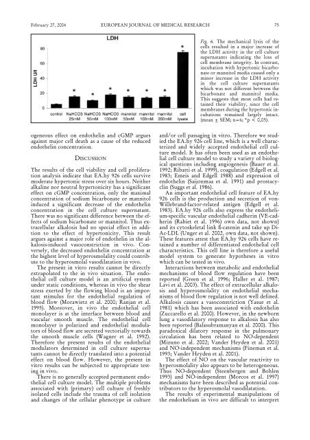
February 27, 2004 EUROPEAN JOURNAL <strong>OF</strong> MEDICAL RESEARCH75Fig. 6. The mechanical lysis of thecells resulted in a major increase ofthe LDH activity in the cell culturesupernatants indicating the loss ofcell membrane integrity. In contrast,incubation with hypertonic bicarbonateor mannitol media caused only aminor increase in the LDH activityin the cell culture supernatantswhich was not different between thebicarbonate and mannitol media.This suggests that most cells had retainedtheir viability, since the cellmembranes during the hypertonic incubationsremained largely intact.(mean ± SEM; n=6; *p < 0,05).ogeneous effect on endothelin and cGMP arguesagainst major cell death as a cause of the reducedendothelin concentration.DISCUSSIONThe results of the cell viability and cell proliferationanalysis indicate that EA.hy 926 cells survivemoderate hypertonic stress over six hours. Neitheralkaline nor neutral hypertonicity has a significanteffect on cGMP concentration, only the maximalconcentration of sodium bicarbonate or mannitolinduced a significant decrease of the endothelinconcentration in the cell culture supernatant.There was no significant difference between the effectsof sodium bicarbonate or mannitol. Thus extracellularalkalosis had no special effect in additionto the effect of hypertonicity. This resultargues against a major role of endothelin in the alkalosis-inducedvasoconstriction in vivo. Conversely,the decreased endothelin concentration atthe highest level of hyperosmolality could contributeto the hyperosmolal vasodilatation in vivo.The present in vitro results cannot be directlyextrapolated to the in vivo situation. The endothelialcell culture model is an artificial systemunder static conditions, whereas in vivo the shearstress exerted by the flowing blood is an importantstimulus for the endothelial regulation ofblood flow (Morawietz et al. 2000; Ranjan et al.1995). Moreover, in vivo the endothelial cellmonolayer is at the interface between blood andvascular smooth muscle. The endothelial cellmonolayer is polarized and endothelial modulatorsof blood flow are secreted vectorially towardsthe smooth muscle cells (Wagner et al. 1992).Therefore the present results of the endothelialmodulators determined in cell culture supernatantscannot be directly translated into a potentialeffect on blood flow. However, the present invitro results can be subjected to appropriate testingin vivo.There is no generally accepted permanent endothelialcell culture model. The multiple problemsassociated with (primary) cell culture of freshlyisolated cells include the trauma of cell isolationand changes of the cellular phenotype in cultureand/or cell passaging in vitro. Therefore we studiedthe EA.hy 926 cell line, which is a well characterizedand widely accepted endothelial cell culturemodel. It has often been used as an endothelialcell culture model to study a variety of biologicalquestions including angiogenesis (Bauer et al.1992; Ribatti et al. 1999), coagulation (Edgell et al.1983; Emeis and Edgell 1988) and expression ofendothelin (Saijonmaa et al. 1991) and prostacyclin(Suggs et al. 1986).An important endothelial cell feature of EA.hy926 cells is the production and secretion of von-Willebrand-factor-related antigen (Edgell et al.1983). EA.hy 926 cells also express the endothelium-specificvascular endothelial cadherin (VE-cadherin(Rabiet et al. 1996) own data, not shown)and its cytoskeletal link ß-catenin and take up Di-Ac-LDL (Unger et al. 2002, own data, not shown).These features attest that EA.hy 926 cells have retaineda number of differentiated endothelial cellcharacteristics. This cell line is therefore a usefulmodel system to generate hypotheses in vitrowhich can be tested in vivo.Interactions between metabolic and endothelialmechanisms of blood flow regulation have beenreported (Green et al. 1996; Haller et al. 1987;Lavi et al. 2003). The effect of extracellular alkalosisand hyperosmolality on endothelial mechanismsof blood flow regulation is not well defined.Alkalosis causes a vasoconstriction (Yasue et al.1981) which has been associated with endothelin(Zuccarello et al. 2000). However, in the newbornlung a vasodilatory response to alkalosis has alsobeen reported (Balasubramanyan et al. 2000). Thisparadoxical dilatory response in the pulmonarycirculation has been related to NO-dependent(Mizuno et al. 2002; Vander Heyden et al. 2001)and NO-independent mechanisms (Fineman et al.1993; Vander Heyden et al. 2001).The effect of NO on the vascular reactivity tohyperosmolality also appears to be heterogeneous.Thus NO-dependent (Steenbergen and Bohlen1993) and NO-independent (Morcos et al. 1997)mechanisms have been described as potential contributorsto the hyperosmolal vasodilatation.The results of experimental manipulations ofthe endothelium in vivo are difficult to interpret