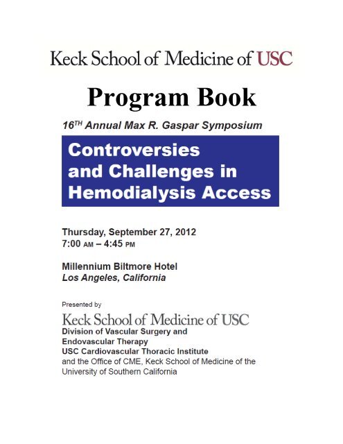
Program Book - Keck School of Medicine of USC - University of ...
Program Book - Keck School of Medicine of USC - University of ...
Program Book - Keck School of Medicine of USC - University of ...
- No tags were found...

Create successful ePaper yourself
Turn your PDF publications into a flip-book with our unique Google optimized e-Paper software.
<strong>Program</strong> <strong>Book</strong>
TABLE OF CONTENTSNeeds Assessment 2Course Description and Learning Objectives 3Acknowledgement <strong>of</strong> Commercial Support & Exhibitors 4<strong>Program</strong> Faculty & Disclosures 5<strong>Program</strong> Schedule and Contents 7Cultural and Linguistic Competency Policy & Resources 1501
Needs AssessmentOver 370,000 patients in the United States require hemodialysis to sustain life. 1 Growth in theincident hemodialysis population has been driven by a linear increase since 1980. 1 In 2009, therewere 106,333 new hemodialysis patients. 1 Kidney Disease Outcomes Quality Initiative (K-DOQI)Clinical Practice Guidelines for Hemodialysis Adequacy have stated since 2006 that patients shouldhave a functional permanent access at the initiation <strong>of</strong> dialysis therapy. 2 However, 81% <strong>of</strong> patientsstill dialyze through a catheter at initiation <strong>of</strong> hemodialysis. 3 This may be related to the fact thatmany patients have challenging anatomy that make traditional arteriovenous fistulas or graftsdifficult to create.Advancements in biomedical engineering have accelerated the technological development andapplication <strong>of</strong> vascular access grafts. With the explosion <strong>of</strong> endovascular technology in vascularsurgery, many new grafts have been developed which use a combination <strong>of</strong> an open andendovascular approach to treating patients with challenging anatomy. This has resulted in morenon-catheter based hemodialysis access options.In 2003, the Fistula First Breakthrough Initiative was created with a goal <strong>of</strong> achieving a 70%prevalent fistula rate. 4 While the prevalent fistula rate has nearly doubled from 32% in 2003, as <strong>of</strong>January 2012, the prevalent national fistula rate is still only 60%. 5 One contributing factor to thefailure to achieve the 70% goal is that improvements in overall management <strong>of</strong> end-stage renaldisease patients have decreased the annual mortality almost 18% from 2000 to 2008. 1 However,during that same period, the prevalence <strong>of</strong> patients wait-listed for a kidney transplant increasednearly 33%. 1 As a result, the survival <strong>of</strong> this patient population increases at a rate that outpaces theavailability <strong>of</strong> kidney transplants. As survival increases without kidney transplantation, the availablesites for hemodialysis access creation become exhausted, increasing the challenge to the surgeon.In addition, with increasing patient survival, access complications also become more common.Advanced surgical techniques and endovascular technology have been increasingly applied totreatment <strong>of</strong> dialysis access complications so that the life <strong>of</strong> the hemodialysis access can beprolonged.Advances in technology combined with failure to meet national guidelines and the increasingprevalence <strong>of</strong> hemodialysis patients who pose a dialysis access challenge makes “Controversiesand Challenges in Hemodialysis Access” a timely topic for discussion and the subject <strong>of</strong> thisyear’s symposium. This course has been designed to update physicians and surgeons on themany new developments in vascular access grafts and techniques and to provide guidance as tohow these new management strategies should modify current practice patterns and achievenational guidelines.References1. U S Renal Data System, USRDS 2011 Annual Data Report: Atlas <strong>of</strong> Chronic Kidney Disease and End-Stage RenalDisease in the United States, National Institutes <strong>of</strong> Health, National Institute <strong>of</strong> Diabetes and Digestive and KidneyDiseases, Bethesda, MD, 2011.2. KDOQI Clinical Practice Guidelines and Clinical Practice Recommendations for 2006 Updates: HemodialysisAdequacy, Peritoneal Dialysis Adequacy and Vascular Access. Am J Kidney Dis 2006;48:S1-S322 (suppl 1).3. Slinin Y, Gui H, Gilbertson DT, et al. Meeting KDOQI Guideline Goals at Hemodialysis Initiation and Survival during theFirst Year. Clin J Am Soc Nephrol 5:1574-1581,2010.4. Neumann ME. “Fistula first” initiative pushes for new standards in access care. Nephrol News Issues 18:47-8,2004.5. Fistula first data. Available at http://www.fistulafirst.org/AboutFistulaFirst/FFBIData.aspx. Accessed .6.2012.2
Course DescriptionMax R. Gaspar, M.D., was an attending surgeon at the Los AngelesCounty + <strong>University</strong> <strong>of</strong> Southern California Medical Center for over fiftyyears, and served as Chief <strong>of</strong> Vascular surgery at LAC+<strong>USC</strong> for twentyfiveyears. His teaching and scholarly accomplishments have had alasting impact on vascular surgery in Southern California. This annualsymposium, which honors Dr. Gaspar, addresses a specific topic <strong>of</strong> interest to physicians andsurgeons who care for the patient with vascular disease. The topic <strong>of</strong> the 2012 GasparSymposium is “Controversies and Challenges in Hemodialysis Access”. A distinguishedfaculty has been assembled for the symposium which is designed to update the attendee on thecurrent status <strong>of</strong> the evolving field <strong>of</strong> hemodialysis access. This year we are delighted to have Dr.Samuel Eric Wilson, Pr<strong>of</strong>essor <strong>of</strong> Surgery at the <strong>University</strong> <strong>of</strong> California at Irvine, as the GasparVisiting Pr<strong>of</strong>essor. Dr. Wilson has been a leader in vascular surgery for over four decades. Hehas been the editor <strong>of</strong> the textbook “Vascular Access: Principles and Practice” since 1980, whichis now in its fifth edition. His symposium address on September 27 is titled, “Three Rules toBreak in Vascular Access Surgery”. His Gaspar Lecture “Maintaining Best Practices in theEndovascular Revolution” will be delivered the following day, September 28, at the <strong>USC</strong> HealthSciences CampusLearning ObjectivesUpon successful completion <strong>of</strong> this activity, participants will be able to:1. Be familiar with the principles <strong>of</strong> hemodialysis and access planning.2. Describe techniques for vascular access in the upper and lower extremity and thepediatric population.3. Discuss the complications <strong>of</strong> vascular access surgery and the treatment options forthose complications.Desirable Physician Attributes: In alignment with the CME mission <strong>of</strong> the <strong>Keck</strong> <strong>School</strong> <strong>of</strong> <strong>Medicine</strong>, programs are planned in thecontext <strong>of</strong> desirable physician attributes as designated by the ACGME/ABMS and the Institute <strong>of</strong> <strong>Medicine</strong>: 1) Patient Care andPatient-centered Care, 2) Medical Knowledge, 3) Practice-based Learning, 4) Interpersonal and Communication Skills, 5)Pr<strong>of</strong>essionalism, 6) Systems-based Practice, 7) Work in Interdisciplinary Teams, 8) Apply Quality Improvement, 9) UtilizeInformatics, and 10) Employ Evidence-based practice. This program and the objectives have been developed in the context <strong>of</strong>attributes 1, 2, 3, 5, and 10.Pr<strong>of</strong>essional Credit: The <strong>Keck</strong> <strong>School</strong> <strong>of</strong> <strong>Medicine</strong> <strong>of</strong> the <strong>University</strong> <strong>of</strong> Southern California is accredited by theAccreditation Council for Continuing Medical Education (ACCME) to provide continuing medical education for physicians.The <strong>Keck</strong> <strong>School</strong> <strong>of</strong> <strong>Medicine</strong> <strong>of</strong> the <strong>University</strong> <strong>of</strong> Southern California designates this live activity for a maximum <strong>of</strong> 6.75AMA PRA Category 1 Credits. Physicians should claim only the credit commensurate with the extent <strong>of</strong> their participationin the activity.The California State Board <strong>of</strong> Registered Nursing accepts courses approved for AMA PRA Category 1 credits as meeting thecontinuing education requirements for license renewal. Nurses from states other than California must check with their local stateboard for specific continuing education policies.3
ACKNOWLEDGEMENTSWe would like to thank the following companies for their educational support <strong>of</strong> this meeting:Abbott VascularBoston Scientific CorporationCovidienEndologixMedtronicW.L. Gore & AssociatesWe gratefully acknowledge the following companies participating as exhibitors at this event:Abbott VascularAngioScoreAtrium Medical CorporationBaxter HealthcareBoston Scientific CorporationCook MedicalCovidienCryoLifeDynamics Orthotics & Prosthetics, Inc.Edward LifeScienceEKOS CorporationEndologixFundacion PadrinoGrifols U.S. IncHarvest Technologies Corp.KCIMedtronicOsborn MedicalPhilips HealthcareSIGVARIS, Inc.Spectrum HealthcareTerumo Cardiovascular SystemsTransonicVascular Solutions, IncVeritas InterventionalW.L. Gore & AssociatesZymoGenetics
PROGRAM FACULTY AND DISCLOSURESCourse DirectorFred A. Weaver, MD, MMMPr<strong>of</strong>essor <strong>of</strong> SurgeryChief, Division <strong>of</strong> Vascular Surgery and Endovascular TherapyCo-Director, <strong>USC</strong> Cardiovascular Thoracic Institute<strong>Keck</strong> <strong>School</strong> <strong>of</strong> <strong>Medicine</strong> <strong>of</strong> <strong>USC</strong>Consultant/Grant/Research: WL GoreMax R. Gaspar Visiting Pr<strong>of</strong>essorSamuel Eric Wilson, MDPr<strong>of</strong>essor <strong>of</strong> Surgery<strong>University</strong> <strong>of</strong> California - Irvine<strong>School</strong> <strong>of</strong> <strong>Medicine</strong>No relevant financial relationships to disclose.Guest FacultyAhmed Abou-ZamZam, MDAssociate Pr<strong>of</strong>essor <strong>of</strong> SurgeryChief, Division <strong>of</strong> Vascular SurgeryDepartment <strong>of</strong> Cardiovascular and Thoracic SurgeryLoma Linda <strong>University</strong>No relevant financial relationships to disclose.Scott Berman, MD, FACSVascular & Endovascular SurgeryRegistered Vascular TechnologistCarondelet Vascular Specialist GroupTucson Special Group, ArizonaNo relevant financial relationships to disclose.Christian deVirgilio, MDVice Chair <strong>of</strong> EducationPr<strong>of</strong>essor <strong>of</strong> SurgerySurgery - VascularHarbor UCLA Medical CenterNo relevant financial relationships to disclose.Alik Farber, MD, FACSAssociate Pr<strong>of</strong>essor <strong>of</strong> Surgery and RadiologyChief, Section <strong>of</strong> Vascular & Endovascular SurgeryBoston Medical CenterNo relevant financial relationships to disclose.Robert Hye, MDKaiser Foundation Hospital – San DiegoGrant Research: WL Gore,Inc; Proteon Therapeutics, Inc; BTG InternationalJuan Carlos Jimenez, MDAssistant Pr<strong>of</strong>essorDivision <strong>of</strong> Vascular SurgeryUCLA5
No relevant financial relationships to disclose.Joseph Mills, MDChief, Division <strong>of</strong> Vascular and Endovascular SurgeryPr<strong>of</strong>essor, Department <strong>of</strong> Surgery<strong>University</strong> <strong>of</strong> ArizonaNo relevant financial relationships to disclose.<strong>Keck</strong> <strong>School</strong> <strong>of</strong> <strong>Medicine</strong> <strong>of</strong> <strong>USC</strong> FacultyWilliam M. Lee, MDAssistant Pr<strong>of</strong>essor <strong>of</strong> SurgeryDivision <strong>of</strong> Vascular Surgery & Endovascular TherapyNo relevant financial relationships to disclose.Mitra Nadim, MDAssociate Pr<strong>of</strong>essor <strong>of</strong> <strong>Medicine</strong>Director, Hypertension Center & Renal Disease CenterDivision <strong>of</strong> Nephrology, Department <strong>of</strong> <strong>Medicine</strong>No relevant financial relationships to disclose.Christian Ochoa, MDAssistant Pr<strong>of</strong>essor <strong>of</strong> SurgeryDivision <strong>of</strong> Vascular Surgery & Endovascular TherapyNo relevant financial relationships to disclose.Vincent L. Rowe, MDAssociate Pr<strong>of</strong>essor <strong>of</strong> Surgery<strong>Program</strong> Director, Vascular Surgery Residency <strong>Program</strong>Division <strong>of</strong> Vascular Surgery& Endovascular TherapyNo relevant financial relationships to disclose.David Shavelle, MDAssociate Pr<strong>of</strong>essor <strong>of</strong> Clinical <strong>Medicine</strong>Division <strong>of</strong> Cardiovascular <strong>Medicine</strong>Grant Research: Abiomed, Inc; GlaxoSmithKline; Maquet, Inc; St. Jude Medical, IncKaren Woo, MDAssistant Pr<strong>of</strong>essor <strong>of</strong> SurgeryDivision <strong>of</strong> Vascular Surgery & Endovascular TherapyNo relevant financial relationships to disclose.Faculty Disclosure: Current guidelines state that participants in continuing medical education activities should beaware <strong>of</strong> any affiliation or financial interest that could affect the speaker’s presentation(s). The Accreditation Council forContinuing Medical Education policy mandates that the provider adequately manages all identified potential conflicts <strong>of</strong>interest prior to the program. Faculty members have completed disclosure forms and potential conflicts <strong>of</strong> interest havebeen reviewed and resolved prior to the program. All disclosures must be listed in the course syllabus.Cultural and Linguistic Competency: This activity is in compliance with California Assembly Bill 1195, which requires that allCME activities address cultural and linguistic competency in patient care delivery to meet the concerns <strong>of</strong> a diversepopulationthrough effective and appropriate pr<strong>of</strong>essional development.6
PROGRAM AGENDA7:00 am Registration and Continental Breakfast8:00 am Course OverviewFred A. Weaver, MD, MMMSession I: Basic ConceptsModerator: Vincent L. Rowe, MD8:10 am ESRD in 2012: Socio-Economic ImpactJuan Carlos Jimenez, MD8:30 am Principles <strong>of</strong> Hemodialysis and Access CannulationMitra Nadim, MD8:50 am Vascular Access Planning and Pre-Op EvaluationFred A. Weaver, MD, MMM9:10 am What’s New in Vascular Grafts?Scott Berman, MD9:30 am Panel Discussion9:50 am BreakSession II Vascular Access TechniquesModerator: Fred A. Weaver, MD, MMM10:10 am Options and Strategies for Upper Extremity Arteriovenous Fistula CreationChristian deVirgilio, MD10:30 am Lower Extremity Dialysis AccessAhmed AbouZamZam, MD10:50 am Pediatric Vascular AccessVincent L. Rowe, MD11:10 am Options for End Stage Dialysis AccessWilliam M. Lee, MD11:30 am Panel DiscussionInvited Lecture11:50 am Three Rules to Break in Vascular Access SurgerySamuel Eric Wilson, MD- Gaspar Visiting Pr<strong>of</strong>essor12:30 pm LunchSession III Vascular Access Complications IModerator: Karen Woo, MD1:45 pm Vascular Access Surveillance, Is It Worth It?Christian Ochoa, MD2:05 pm Management <strong>of</strong> the Non-Maturing Arteriovenous FistulaAlik Farber, MD7
2:25 pm Vascular Access Infections, Medical, Surgical ManagementRobert Hye, MD2:45 pm Panel Discussion3:05 pm BreakSession IVModerator:Vascular Access Complications IIFred A. Weaver, MD, MMM3:25 pm Ischemic Steal, Diagnosis and ManagementJoseph Mills, MD3:45 pm The Management <strong>of</strong> Thrombosed and Failing Fistulas/GraftsDavid Shavelle, MD4:05 pm Aneurysmal Fistula Reduction and ReconstructionKaren Woo, MD4:25 pm Panel Discussion4:45 pm Adjourn8
Juan Carlos Jimenez, MD September 27, 2012Assistant Pr<strong>of</strong>essor8:10 am – 8:30 amDivision <strong>of</strong> Vascular SurgeryUCLAESRD IN 2012:Socio-Economic ImpactLECTURE OBJECTIVESAt the end <strong>of</strong> this lecture, participants will be able to:1. Understand the cardiovascular changes that accompany mild thyroiddisease.2. Diagnose and Treat Cardiovascular Risk Factors associated withsubclinical hypothyroidism.3. Recognize that Statin Induced Myopathy may result fromhypothyroidism.4. Use best practice principles when using levothyroxine to treathypothyroidism and coexistent cardiac disease.9
ESRD in 2012: Socio-economicImpactJuan Carlos Jimenez MD, FACSAssistant Pr<strong>of</strong>essorDivision <strong>of</strong> Vascular SurgeryDavid Geffen <strong>School</strong> <strong>of</strong> <strong>Medicine</strong> at UCLAUSRDS• United States Renal Data System• National data registry that collects, analyzes,and distributes information on ESRDpopulation in U.S.• Funded by NIH, and National Institute <strong>of</strong>Diabetes and Digestive and Kidney Diseases(NIDDKD)• 2011 Annual Report contains data up to 200910
ESRD- U.S. Demographics• Total treated ESRDpopulation- > 570,000• Prevalent population on HD-370,274• Prevalent population on PD-27,522• Incidence HD patients-113,636– 3.3 % increase from prior year• Medicare: Primary payor for83% <strong>of</strong> HD patientsUSRDS 2011 ADRIncident & prevalent patientcounts (USRDS), by modalityUSRDS 2011 ADRAdjusted incident rates <strong>of</strong> ESRD& annual percent changeUSRDS 2011 ADR11
Geographic variations in adjustedincident rates <strong>of</strong> ESRD per millionpopulation, 2009Incident counts &adjusted rates <strong>of</strong>ESRD, by ageUSRDS 2011 ADRIncident counts &adjusted rates <strong>of</strong>ESRD, by raceUSRDS 2011 ADR12
Incident counts &adjusted rates <strong>of</strong>ESRD, by HispanicethnicityUSRDS 2011 ADRIncident counts &adjusted rates <strong>of</strong>ESRD, by primarydiagnosisUSRDS 2011 ADRESRD-Mortality• Alive three years after thestart <strong>of</strong> ESRD therapy– Dialysis patients-50%– Pre-emptive transplant-82%• Adjusted rates <strong>of</strong> all-causemortality– 6.5-7.4 times greater fordialysis patients comparedwith general populationUSRDS 2011 ADR13
Adjusted all-cause & cause specificmortality (from day one) in thefirst year <strong>of</strong> hemodialysisUSRDS 2011 ADRAdjusted all-cause mortality in the ESRD& general populations, by age, 2009Adjusted survival probabilities,from day one, in the ESRD population14
ESRD-Cost• In 2009, total medicare spending rose 8.2% to491 billion dollars• Costs for non-Medicare population were 13.5billion dollars• Costs for ESRD rose 3.1% to 29 billion dollarsUSRDS 2011 ADRTotal Medicare ESRD expendituresper person per year, by modalityIncident patient distribution,by first modality & payorUSRDS 2011 ADR15
ESRD-International ComparisonsPrevalence <strong>of</strong> ESRD Incidence <strong>of</strong> ESRD• Taiwan and Japan highestrates <strong>of</strong> prevalent ESRD• Lowest rates Bangladeshand Phillipines• Highest incident rates inMorelos and Jalisco(Mexico)USRDS 2011 ADRPrevalent rates <strong>of</strong>functioning grafts,2009Greatest reported prevalent rates<strong>of</strong> functioning grafts in Norway,United States and France16
Percent distribution <strong>of</strong>prevalent dialysis patients,by modality, 2009• In Hong Kong, four <strong>of</strong> five prevalentdialysis patientstreated with peritoneal dialysis• New Zealand and Australia 16.3 and9.3% use home hemodialysisUSRDS 2011 ADRTransplant rates, 2009Highest functioning transplant ratesin Canada, Norway, Jalisco (Mexico),and United StatesESRD-Transplantation• On 2009, 17,736 kidney transplantsperformed in U.S.• Number <strong>of</strong> patients on wait-list grew by 6% in2009– 71, 975 total• Survival following renal transplant– 1 yr-Deceased donor-92%, Living Related-96%– 5 yr-Deceased donor-70%, Living Related-83%17
Outcomes for first-time wait-listedpatients three years after listing, 2006,by age, race, & PRAUSRDS 2011 ADRConclusions:• Incidence <strong>of</strong> patients with ESRD requiringdialysis continues to increase• Rate in Hispanic population 1.5 greatercompared with Non-Hispanics• Medicare continues to be primary payor for83% <strong>of</strong> patients requiring HD• Despite relative increase in recent years,transplant rates lag far behind incident ESRDcases• Effects <strong>of</strong> Affordable Care Act and MedicareCuts?18
Mitra Nadim, MD September 27, 2012Associate Pr<strong>of</strong>essor <strong>of</strong> <strong>Medicine</strong>8:30 am – 8:50 amDirectorHypertension Center & Renal Disease CenterDivision <strong>of</strong> NephrologyDepartment <strong>of</strong> <strong>Medicine</strong>PRINCIPLES OF HEMODIALYSIS ANDACCESS CANNULATIONLECTURE OBJECTIVESAt the end <strong>of</strong> this lecture, participants will be able to:1. Understand the principles <strong>of</strong> clearance.2. Access the adequacy <strong>of</strong> dialysis.3. Compare the various modalities <strong>of</strong> hemodialysis.
NOTES________________________________________________________________________________________________________________________________________________________________________________________________________________________________________________________________________________________________________________________________________________________________________________________________________________________________________________________________________________________________________________________________________________________________________________________________________________________________________________________________________________________________________________________________________________________________________________________________________________________________________________________________________________________________________________________________________________________________________________________________________________________________________________________________________________________________________________________________________________________________________________________20
Fred A. Weaver, MD, MMM September 27, 2012Pr<strong>of</strong>essor <strong>of</strong> Surgery8:50 am – 9:10 amChiefDivision <strong>of</strong> Vascular Surgery andEndovascular TherapyCo-Director, <strong>USC</strong> CardiovascularThoracic Institute<strong>Keck</strong> <strong>School</strong> <strong>of</strong> <strong>Medicine</strong>VASCULAR ACCESS PLANNINGAND PRE-OP EVALUATIONLECTURE OBJECTIVESAt the end <strong>of</strong> this lecture, participants will be able to:1. Identify arterial and venous anatomy that is necessary for creation <strong>of</strong>a vascular access.2. Choose the most efficacious vascular access that can be created.3. List the factors that go into the successful planning for vascularaccess.21
Vascular Access Planning andPreoperative EvaluationFred A Weaver MD, MMMPr<strong>of</strong>essor and ChiefVascular Surgery and Endovascular Therapy<strong>Keck</strong> Medical Centerwww.surgery.usc.eduInitial Evaluation: History• General health• Age• Atherosclerotic riskfactors• Coagulation disorders– Systemic lupus– Cancer (multiplemyeloma …)– Prior DVT’s• Pacemaker• CHF (output)• CAD or malignancy (lifeexpectancy)• Arm or neck surgery• Anticipated transplant• Hospitalization recentand remote• Previous vascularaccess• Vascular Procedureswww.surgery.usc.eduEvaluation-Physical Exam• Dominant hand• Skin lesions• Prior dialysis grafts• Visible veins• Arm swelling• Existing indwelling catheters, pacemakers• Chest wall collateralswww.surgery.usc.edu22
Arterial Evaluation• Blood pressure measurement in both arms• Pulse exam brachial, radial, digit– Calcified arteries• Allen test• Duplex scan, luminal diameter > 2 mm• Angiogram if necessarywww.surgery.usc.eduVenous Work-upWho?– Prior failed access– No visible veins– Everyone?Why?– Increase fistula placement– Decrease failure ratewww.surgery.usc.edu• Non-invasive• Excellent fordistal extremities• Proximal vesselswell imaged• Will demonstrateoccluded arteriesand veinswww.surgery.usc.edu23
• Veins not circular but ovoid, min and max diameters• Higher venous pressure more circular• Patient supine, warm room and gel• Proximal tourniquet, pressure cuff• B mode venous diameters should be determined atvenous pressure >40 mm Hg• Reproducibility with higher with pressure >40 mmHgwww.surgery.usc.eduRelation between the outcome <strong>of</strong> surgery and preoperativeultrasound assessment. The ultrasound scan is defined asabnormal if the vessel is <strong>of</strong> diameter less than 1.5 mm and/or astenosis is detected in the cephalic veinUltrasoundAssessmentOutcomeFailed Successful TotalAbnormalNormalTotal1791603838174754Wong Eur J Vasc Endovasc Surg 1996; 12:207-213www.surgery.usc.eduRelative frequencies <strong>of</strong> each access type placed during the twoperiodsBeforeinstitutionNoninvasiveprotocol<strong>of</strong> protocolAutogenous fistula* 14% 63%Bridging grafts 62% 30%Permanent catheter 24% 7%Prevalence significantly different in each group (p < 0.05). From June1992 to August 1994, 183 procedures were performed before institution<strong>of</strong> protocol. From September 1994 to April 1996, 172 procedures wereperformed with noninvasive protocol.Silva J Vasc Surg 1998;27:302-8www.surgery.usc.edu24
Vascular UltrasonographyPrior to Dialysis Access Surgery• Arterial and venous duplex studies prior toaccess placement• 47 patients• 52% had at least one prior access• Venous occlusions and arterial lesions (forearmportion <strong>of</strong> radial artery)• Post US: 97% fistula formation rateAm J Surg 2002;184:568www.surgery.usc.eduNoninvasive criteria for selection <strong>of</strong> upperextremity arteries and veins for dialysis accessproceduresVenous ExaminationVenous luminal diameter 2.5 mm for AFVenous luminal diameter 4.0 mm for BGAbsence <strong>of</strong> segmental stenoses or occluded segmentsContinuity with the deep venous system in the upper armAbsence <strong>of</strong> ipsilateral central venous stenosis or occlusionArterial ExaminationAbsence <strong>of</strong> pressure differential 20 mm Hg between armsArterial lumen diameter 2.0 mmPatent palmar archSilva J Vasc Surg 1998;27:302-8www.surgery.usc.eduVenography• Excellent forproximal veins• Invasive• Contrast agent• More expensive• Redo access• Exhausted accesswww.surgery.usc.edu25
VENOGRAM+ -CFDI+ 17(true positive)- 4(false negative)1(false positive)33(true negative)Sensitivity = 17/21 x 100 = 81.0% Positive Predictive Value = 17/18 x 100 = 94.4%Specificity = 33/34 x 100 = 97.1% Negative Predictive Value = 33/37 x 100 = 89.2%Overall diagnostic accuracy <strong>of</strong> color flow duplex imaging (CFDI) compared withoverall diagnostic accuracy <strong>of</strong> venography for proximal upper extremity venousoverflow obstruction (N = 55). Passman M, JVascSurg 1998; 28:869-75www.surgery.usc.eduVenous occlusion secondary tocatheters• 15-50% incidence• Hemodynamicmonitoring• TPN• Pacemaker• Hemodialysiswww.surgery.usc.eduSubclavian Stenosis Following Subclavian Dialysis CatheterizationPatient characteristics <strong>of</strong> normal, abnormal andequivocal subclavian venogramsNormal Equivocal AbnormalNumberSexAge (years)Race:CaucasianAsianBlackDays <strong>of</strong> catheterizationDays since catheter removedSide <strong>of</strong> catheterization112F/8M54.5 + 3.983028 ±6*143 + 565L/6R*P
Anticipating Steal• Incidence 2-20%, Most studies 4%• Calcified arteries, digital arteries• Decreased digital pressure• Digital-brachial index < 1.0-0.6• Brachial inflow, autogenous fistulawww.surgery.usc.eduCLINICAL GUIDELINES ARTERIOVENOUS HEMODIALSYSIS ACCESSTHE SOCIETY FOR VASCULAR SURGERYTiming <strong>of</strong> referral to surgeon and timing <strong>of</strong> placement <strong>of</strong> permanent vascular accessAdvanced chronic renal disease, creatinine >4 mg/dl (late stage 4, glomerular filtration rate
• Thorough preoperative assessmentessential to successful vascular access andoptimizing AVF rate• Noninvasive assessment has changed theparadigm for vascular access planning• Use venography liberally particularly forprior subclavian cannulation and redoaccess• Autogenous Access preferred, distal toproximal• Anticipate Stealwww.surgery.usc.edu28
Scott Berman, MD September 27, 2012Vascular & Endovascular Surgery9:10 am – 9:30 amRegistered Vascular TechnologistCarondelet Vascular Specialist GroupTucson Special Group, ArizonaWHAT’S NEW IN VASCULAR GRAFTS?LECTURE OBJECTIVESAt the end <strong>of</strong> this lecture, participants will be able to:1. Incorporate new dialysis graft technology into their current practice2. Choose the appropriate dialysis graft to fit their individual patientneeds.29
NOTES________________________________________________________________________________________________________________________________________________________________________________________________________________________________________________________________________________________________________________________________________________________________________________________________________________________________________________________________________________________________________________________________________________________________________________________________________________________________________________________________________________________________________________________________________________________________________________________________________________________________________________________________________________________________________________________________________________________________________________________________________________________________________________________________________________________________________________________________________________________________________________________30
Christian deVirgilio, MD September 27, 2012Vice Chair <strong>of</strong> Education10:10 am – 10:30 amPr<strong>of</strong>essor <strong>of</strong> SurgerySurgery – VascularHarbor UCLA Medical CenterOPTIONS AND STRATEGIES FOR UPPEREXTREMITY ARTERIOVENOUS FISTULACREATIONLECTURE OBJECTIVESAt the end <strong>of</strong> this lecture, participants will be able to:1. Identify optimal strategies for creation <strong>of</strong> an arterial venous fistula.2. List order <strong>of</strong> preference for arterial venous fistula.31
Options and Strategies forUpper Extremity ArteriovenousFistula CreationChristian de Virgilio MDDivision <strong>of</strong> Vascular SurgeryHarbor-UCLA Medical CenterObjectivesTo discuss options for upper extremityhemodialysis– AV fistula– AV graftDiscuss ethnic differences in arm vein diametersTo provide an optimal access creation strategy– Timing– LocationTiming <strong>of</strong> Access PlacementAV fistula– At least 6 months prior to anticipated dialysis– Needs time to mature– May need revisionAV Graft– Only 3-6 weeks prior to anticipated dialysis– If placed too early, may thrombose before useGoals– Avoid central catheter– Minimize duration <strong>of</strong> central catheter32
2006 NKF-KDOQI GuidelinesPreferred: FistulaeWrist/forearmradiocephalicradiobasilicElbowbrachiocephalicbrachiobasilicbrachiobrachial?Acceptable: Synthetic or biological graftForearm loop graftUpper-arm graftSource: NKF-KDOQI Vascular Access Clinical Practice Guidelines. 2006 UpdateRadial Artery Cephalic Vein(Cimino Fistula)First choice for most surgeons if :Cephalic vein > 2.5 mmAt least 10 cm vein lengthStrong radial pulseAdvantagesSimple to createPreserves proximal vesselsFew complicationsOperative Technique:Cimino (radiocephalic) AVFLocal anesthesiaEnd <strong>of</strong> vein to side <strong>of</strong> arteryDilate vein with dilutepapaverine1-time dilator proximally? HeparinPre-emptive division <strong>of</strong>tributaries33
Cimino (radiocephalic) AVFPotential disadvantages– Smaller artery and vein, less flow– Unrecognized radial artery atherosclerosis– Unrecognized vein stenosis from priorphlebotomy– Lower functional patencyBrachial artery-Cephalic vein AVFat ElbowAdvantagesHas higher blood flowEasy to cannulateCosmetic benefitBetter functional patency (in some studies)DisadvantagesSlightly more difficult to createIn obese pt, may need superficializationIncreased incidence <strong>of</strong> stealGreater incidence <strong>of</strong> cephalic arch stenosis(narrowing at deltopectoral groove)Brachial artery-Basilic vein AVF(BBAVF)AdvantageUsually larger than cephalicLess likely to be damaged by priorphelbotomy34
Potential Problems with BBAVFSignificant rate <strong>of</strong> early thrombosis (1 stage)Technically more demandingInjury to medial antebrachial cutaneous nerveInadequate length (if enters brachial vein early)Inadequate superficializationNot moved anterior enough– Brachial artery injuryControversies with BBAVFOne or two stageSuperficialization with or withouttranspositionOne Stage BBAVFVein is completely dissected out anddividedNew tunnel created more anterolaterallyAnastomosis made to brachial artery atelbow35
Two Stage BBAVFStage 1 anastomosis made to brachial arteryat elbowStage 2 6 weeks later, matured vein isSuperficialized +/- transposed1 vs 2-Stage BBAVFOnly one prospective, randomized trial– 40 patientsEarly Failure (prior to four weeks)– One-stage 40%– Two-stage 10% (p
1 vs 2-Stage BBAVFOne-stage(n=60)Two-stage(n=30)p-valuePrimary failure 22.9% 9.1% 0.17Primary patencyOne yearTwo yearSecondary patencyOne yearTwo yearPrimary functional patencyOne yearTwo yearSecondary functional patencyOne yearTwo year78%34%82%41%61%34%80%41%84%84%89%89%88%88%94%94%0.0460.010.0470.0151 vs 2-Stage BBAVFPatency for one-stage BBAVF proceduresmay be decreased due to– Extensive mobilization– Tunneling <strong>of</strong> a non-matured, thin-walled basilicvein– Unseen vessel injury– Kinking <strong>of</strong> a thin-walled veinBrachial Vein-Brachial Artery AVFDeeper than basilic veinMore thin walledMoretributaries37
Brachial Artery-Brachial Vein AVFSuperficialization is moredemanding/tediousNeeds more time to matureBrachial Artery-Brachial Vein AVFAngle and Chandra JVS 200520 pts brachial vein-brachial artery AVFFistula functional in 19/20Arm swelling in 1No stealBrachial Artery-Brachial Vein AVFDorobantu et al J Vasc Access 200633 pts6 (20%) occluded in 30 days33% forearm edema38
Brachial Artery-Brachial Vein AVFTorina et al J Vasc Access 200811 one-stage, 2 two-stage73% <strong>of</strong> one stage had complicationsOnly 24% 1 year patency (worse than AVgraft)Problems with Fistula First at AllCost ApproachA high proportion <strong>of</strong> new fistulas do not matureadequately for useUp to 60% AVF unsuitable for dialysis even after4-5 monthsTaking a “shot” at a fistula that fails hasconsequences– Indwelling catheterCatheter related infectionsCentral vein stenosis– Veins in arm may be thrombosed precludingsubsequent graft placementDember et al. JAMA May 14, 2008 Vol 299 No 18 2164-2171Trends in Central Venous Catheter Usage39
Forearm AV GraftsLarge surface area to cannulateEasy to cannulateUsable within 2 weeksEasy to handle and implantEasy to declotAV Graft OptionsStandard PTFEHeparin bonded PTFEBovine graftHeparin Bonded PTFE Graft:Propatenend-point attachment mechanismheparin molecules anchored to the luminalsurfacemaintains heparin’s bioactive propertiesthromboresistant BioActive graft surface thatis not consumed40
Heparin Bonded vs. StandardPTFE for HemodialysisTexas Southwestern Study J Vasc Access2009 Davidson et al.83 heparin bonded AVG (mainly 4-7 mm)67 standard PTFEOverall clot free survival 69% at 1 year78% at 1 year for heparin bonded58% at 1 year for standard PTFEP = 0.007Bovine ArtegraftBovine carotid arteryEasy to handle/sewDiameter sometimes too big for nativeartery (taper)Patency similarto PTFELess infection riskComparison <strong>of</strong> AVG with AVFProblems with comparisons:– Not all patency comparisons include primaryfailures (much more likely with AVF)– AVG <strong>of</strong>ten used when veins too small for AVFor after AVF failures– Need for prolonged central lines whileawaiting AVF maturation not factored in– Better secondary patency <strong>of</strong> AVG not alwaysconsidered41
Comparison <strong>of</strong> brachial-basilic AVfistula and prosthetic forearm graft• 105 patients were randomized for a BBAVF orloop AVG (non heparin bonded)• Primary and assisted-primary 1-year patencywere significantly higher in the BBAVF group• Secondary patencies were comparableKeuter et al JVS 2008Comparison <strong>of</strong> transposed brachiobasilic fistulas toupper arm grafts and brachiocephalic fistulasOliver et al Kidney Int 2001Compared with BBAVF– BCAVF less likely to failRR 0.3 (95% CI, 0.1 to 1.0)– BCAVF trend for less thrombosisRR 0.3 (95% CI 0.1 to 1.1)– Upper arm AVG more likely to thromboseRR 2.6 (95% CI, 1.3 to 5.3) excluding primary failuresRR 1.6 (95% CI, 1.0 to 2.7) when accounting for the lower risk <strong>of</strong>primary failure <strong>of</strong> AVGConfidence interval includes 1.0No significant difference in cumulative patency (failurefreesurvival) among 3 types <strong>of</strong> access if primary failureincluded (median follow-up:594 days)Racial differences in AVfistula ratesStudies have reported thatAfrican American patients:– Fewer first time AVF– Lower fistula primary patency rates– Higher fistula complication rates3342
34Racial differences in AVfistula rates249 male patients– 95 African American (AA)Median age: 63 yearsCephalic Vein DiametersProximalupper armDistalupper armProximalforearmDistalforearmAfricanAmericanNon-AfricanAmericanP value2.6 2.8 0.12.4 3.0 0.022.0 2.5 0.031.7 2.1 0.0535Basilic Vein DiametersBasilicveindiameter(mm)Proximalupper armDistalupper armProximalforearmDistalforearmAfricanAmericanNon-AfricanAmericanP value3.5 4.0 0.0042.9 3.5 0.00021.8 2.5 < 0.00011.4 2.1 < 0.00013643
Racial differences in AVfistula ratesAVF– AA patients: 82%– Non-AA patients: 93%AVG– AA patients: 18%– Non-AA patients: 7%– P = 0.009Fistula Patency RatesOne yearPrimarypatencyPrimaryfunctionalpatencyAfricanAmerianNon-AficanAmericanP value80.8% 76.2% 0.473.1% 69.2% 0.538ConclusionsIf good vein and good artery– Radiocephalic 1 st choice– Brachio-cephalic 2 nd choice44
ConclusionsBrachial artery-basilic vein AVFExcellent option for AV access in patientswho are not candidates for radiocephalic orbrachiocephalic AVFPrimary, secondary, and functional patencyrates higher for 2-stage– method <strong>of</strong> choiceTiming and technique for the second stageneed further refinementConclusionsBrachial Artery-Brachial Vein AVFTechnically more difficultBest if done in 2 stagesProbably better to wait longer (3 months)for 2 nd stage than Brachial Basilic AVFPatency data limitedConclusions– Decision between brachiobasilic, brachiobrachialvs AVG debatabledepends on vein diameterurgency <strong>of</strong> accessage, life expectancy <strong>of</strong> patient– AVG if veins marginal and particularly ifalready has central venous access, shorterlife expectancy45
THANK YOU!46
Ahmed Abou-ZamZam, MD September 27, 2012Associate Pr<strong>of</strong>essor <strong>of</strong> Surgery10:30 am – 10:50 amChiefDivision <strong>of</strong> Vascular SurgeryDepartment <strong>of</strong> Cardiovascular andThoracic SurgeryLoma Linda <strong>University</strong>LOWER EXTREMITY DIALYSIS ACCESSLECTURE OBJECTIVESAt the end <strong>of</strong> this lecture, participants will be able to:1. Manage the complex hemodialysis patient who has exhausted allupper extremity access options.2. List the different types <strong>of</strong> permanent lower extremity access options.3. Choose the best lower extremity access type based on specificpatient indications such as obesity and the presence <strong>of</strong> peripheralarterial disease.4. Incorporate the K-DOQI guidelines into their practice.47
Lower ExtremityDialysis AccessAhmed M. Abou-Zamzam, Jr.Associate Pr<strong>of</strong>essor <strong>of</strong> SurgeryChief, Division <strong>of</strong> Vascular SurgeryLoma Linda <strong>University</strong> Medical CenterLower Extremity Dialysis Access• Vascular surgeons -“will operate anywhere”Lower Extremity Dialysis Access• Vascular surgeons -“will operate anywhere”(except thigh access)XXX48
Lower Extremity Dialysis AccessKDOQI Clinical Practice Guidelines:2.1 The order <strong>of</strong> preference for placement <strong>of</strong> fistulae in patients with kidneyfailure who choose HD as their initial mode <strong>of</strong> KRT should be (in descendingorder <strong>of</strong> preference):2.1.1 Preferred: Fistulae (B)2.1.1.1 A wrist (radiocephalic) primary fistula.2.1.1.2 An elbow (brachiocephalic) primary fistula.2.1.1.3 A transposed brachial basilic vein fistula.2.1.2 Acceptable: AVG <strong>of</strong> synthetic or biological material, such as:2.1.2.1 A forearm loop graft, preferable to a straight configuration.2.1.2.2 Upper-arm graft.2.1.2.3 Chest wall or “necklace” prosthetic graft or lower-extremityfistula or graft; all upper-arm sites shouldbe exhausted.Lower Extremity Dialysis AccessWho needs lower extremity access?2.1.2.3 Chest wall or “necklace” prosthetic graft or“lower-extremity fistula or graft; all upper-arm sites should beexhausted.”Lower Extremity Dialysis AccessWork up prior to lower extremity access:Review all prior sitesDocument lack <strong>of</strong> upper extremity access optionsVenogram <strong>of</strong> upper extremitiesLower extremity pulse examination, ABI's(non-invasive arterial study when any doubt)Venous duplex <strong>of</strong> lower extremity veinssize, patency, duplicity, prior occult dvt - both legssconsider ascending venogram if prior femoral lines49
Lower Extremity Dialysis AccessWhat are the options for lower extremity access?Analogous to upper extremitiesFistulae -Based on femoral vein or greater saphenous veinAVG -Thigh loops – from common femoral or superficial femoral arteriesLower Extremity Dialysis AccessTechniques: AVFGSV loop first described in 1969Pierre-Paul, et al AVS 2004Lower Extremity Dialysis AccessTechniques: AVFTransposed femoral vein (tFV)Initial description: The Superficial Femoral-Popliteal Vein TranspositionFistula: Description <strong>of</strong> a New Vascular Access Procedure Jackson, JACS2000Two patientsJackson noted high flows and decreased ABI50
Lower Extremity Dialysis AccessTechniques: Transposed femoral vein (tFV)Lower Extremity Dialysis AccessTechniques: AVGMost commonly CFA-CFV loopKhadra, et al, Am J Surg, 1997Lower Extremity Dialysis AccessData: Saphenous Vein loop AVFSaphenous vein loop to femoral artery arteriovenous fistula: a practicalalternative Pierre-Paul, et al Annals Vasc Surg, 2004Seven patients40% patency at 3 yearsLow flow57% wound complicationsAll developed stenoses in GSVMean secondary patency 16 monthsOther reports range ~40% two yearAll low flow, aneurysms, woundcomplications51
Lower Extremity Dialysis AccessData: tFVArteriovenous fistula construction in the thigh with transposed superficialfemoral vein: our initial experience Gradman, et al JVS 200125 patients28% major complications, 32% ischemic complicationsCumulative patency at one year 86%One limb loss, 8 steal (all over 40)Lower Extremity Dialysis AccessData: tFVFemoral vein transposition for arteriovenous hemodialysis access:improved patient selection and intraoperative measures reducepostoperative ischemia Gradman, et al JVS 2005Updated with more selective patient selection46 adults – 22 more than initial seriesExclusion <strong>of</strong> older patients with PADTapering <strong>of</strong> anastomosis (14 <strong>of</strong> 22 adults),Measurement <strong>of</strong> distal pressuresResults:94% secondary patency at 1 and 2 yearsNo ischemic complications4.5-5mmLower Extremity Dialysis AccessData: tFVLong-term results <strong>of</strong> femoral vein transposition for autogenousarteriovenous hemodialysis access Bourquelot, et al JVS 2012Largest series <strong>of</strong> tFV - 72 accesses (27 years)Average time on dialysis prior to leg access 10 years82% successful dialysis within 2 monthsPrimary patency 91% 1 year, 45% 9 yearsSecondary patency 84% 1 year and 56% at 9 years11% delayed healing, hematoma, lymphocele(no tapering <strong>of</strong> anastomosis)0 serious infections13 patients (18%) with complications requiring ligation –5 ischemic, 1 amputation52
Lower Extremity Dialysis AccessData: tFVSmall skin incision and fistula elevation for hemodialysis using the femoralvein Alcocer, et al JVS 201225 patients; most recent technique in the last 13Smaller incision; Fistula elevationResults: reduced wound dehiscence/complicationsSimilar patencyStandard approachSmall incision; superficializationLower Extremity Dialysis AccessData: AVGProsthetic thigh arteriovenous access: outcome with SVS/AAVS reportingstandards Cull, et al, JVS 2004125 grafts in 100 patients2 year primary patency 19%, secondary patency 54%41% infection rate, 11% limb ischemia, 9% major amputation1.68 interventions/yearLower Extremity Dialysis AccessData: AVGThe mid-thigh loop arteriovenous graft: patient selection, technique, andresults Scott, et al Am Surg 2006Attempt to avoid infection – mid-thigh access SFA-FV46 graftsPrimary patency 40% one year, 18% 2 yearSecondary patency 68% 1 year, 43% 2 yearInfection 21% requiring graft excisionIschemia 13%Felt this option preserves more proximal vesselsand longer outflow vein for secondary procedures.53
Lower Extremity Dialysis AccessData: AVGProsthetic lower extremity hemodialysis access grafts have satisfactorypatency despite a high incidence <strong>of</strong> infection Geenen, et al JVS 2010Largest series <strong>of</strong> thigh AVGs – 153 (15 years)Primary patency 54% 1 year, 40% 2 year, 19% 5 yearSecondary patency 75% 1 year, 64% 2 year, 50% 5 year27% infectious complications; 17% graft loss1% ischemic complicationsMean AVG function 31 months(Standard CFA-CFV)Conclude: AVGs are alrightLower Extremity Dialysis AccessData: AVG vs. AVFLower-extremity arteriovenous access for haemodialysis: a systematic reviewAntoniou, et al Eur J Vasc Endovasc Surg, 2009Systematic Literature Review <strong>of</strong> upper thigh AVG, mid-thigh AVG and tFVWeighted mean primary patency rates: 1 year: 48%, 43% and 83%Weighted mean secondary patency rates: 1 year: 69%, 67% and 93%Access loss due to infection: 18%, 18% and 2% (
Lower Extremity Dialysis AccessOther Options?HeRO (Hemodialysis Reliable Outflow)Limited data• Outcomes Comparison <strong>of</strong> HeRO and Lower-Extremity ArteriovenousGraft in Patients with Long-Standing Renal Failure Steerman, et al JVS 2012(SVS abstract)62 HeRO and 23 thigh AVGsPrimary patency 1 year 26% vs. 60% (HeRO vs. AVG) (0.01)Secondary patency 1 year 61% vs. 66% (0.56)Similar infection rates (30%) and mortality (20-30%)Reinterventions 6.2 per year in HeRO and 3.7 in AVGEarly information, but not promising for HeROLower Extremity Dialysis AccessOther Options?long-term catheters ? – NO!Lower Extremity Dialysis AccessSummary:Lower extremity access is an important option in dialysis accessPatency rates are good to excellent when performed appropriatelyTransposed femoral vein is best AVF –select in younger patients without PADwatch for ischemiabeware <strong>of</strong> high rate <strong>of</strong> wound complicationsAVG option for others –watch for infections55
Thank You!LLUMC56
Vincent L. Rowe, MD September 27, 2012Associate Pr<strong>of</strong>essor <strong>of</strong> Surgery10:50 am – 11:10 am<strong>Program</strong> DirectorVascular Surgery Residency <strong>Program</strong>Division <strong>of</strong> Vascular Surgery &Endovascular TherapyPEDIATRIC VASCULAR ACCESSLECTURE OBJECTIVESAt the end <strong>of</strong> this lecture, participants will be able to:1. Manage pediatric patients that require dialysis access.2. Identify the barriers to providing care to pediatric dialysis patients.3. Identify the differences <strong>of</strong> in the management <strong>of</strong> pediatric dialysisfrom adult patients.57
NOTES________________________________________________________________________________________________________________________________________________________________________________________________________________________________________________________________________________________________________________________________________________________________________________________________________________________________________________________________________________________________________________________________________________________________________________________________________________________________________________________________________________________________________________________________________________________________________________________________________________________________________________________________________________________________________________________________________________________________________________________________________________________________________________________________________________________________________________________________________________________________________________________58
William M. Lee, MD September 27, 2012Assistant Pr<strong>of</strong>essor <strong>of</strong> Surgery11:10 am – 11:30 amDivision <strong>of</strong> Vascuar Surgery &Endovascuar TherapyOPTIONS FOR END STAGE DIAYSISACCESSAt the time <strong>of</strong> printing lecture objectives were unavailable.59
NOTES________________________________________________________________________________________________________________________________________________________________________________________________________________________________________________________________________________________________________________________________________________________________________________________________________________________________________________________________________________________________________________________________________________________________________________________________________________________________________________________________________________________________________________________________________________________________________________________________________________________________________________________________________________________________________________________________________________________________________________________________________________________________________________________________________________________________________________________________________________________________________________________60
Samuel Eric Wilson, MD September 27, 2012Pr<strong>of</strong>essor <strong>of</strong> Surgery11:50 am – 12:30 am<strong>University</strong> <strong>of</strong> California - Irvine<strong>School</strong> <strong>of</strong> <strong>Medicine</strong>MAX R. GASPAR INVITED LECTURE:THREE RULES TO BREAK IN VASCULARACCESS SURGERYLECTURE OBJECTIVESAt the end <strong>of</strong> this lecture, participants will be able to:1. Formulate plans for vascular access in the ESRD patient to allowmaximum duration <strong>of</strong> hemodialysis.2. Identify specific patient presentations in which changes in standardguidelines maybe necessary.3. Evaluate patients’ hemodynamic parameters in order to intervenebefore an access fails.61
When to Break the Guidelines in Vascular Access SurgerySamuel Eric Wilson, MD, FACSAt least eight national and pr<strong>of</strong>essional organizations have given “Ex-Cathedra”guidelines for the performance <strong>of</strong> vascular access surgery. The Dialysis OutcomeQuality Initiative <strong>of</strong> the National Kidney Foundation, the Society <strong>of</strong> Vascular Surgery, theCanadian Society <strong>of</strong> Nephrology, Caring for Australians with Renal Insufficiency, TheBritish Renal Association and others have all issued recommendations. Theseguidelines are for the most part sensible, and as far as possible evidence based, butcannot conceivably provide the answer to every clinical situation. I will outline someimportant clinical presentations in which the patient will better be served by breakingthese guidelines.Over the last decade, the number <strong>of</strong> hospital admissions for end stage renal disease inNorth America has remained fairly stable at approximately 20,000 per year but thenumber <strong>of</strong> hospital days has dropped in half from 175,000 to approximately 60,000. Thedecreased duration <strong>of</strong> hospitalization is probably due to the shift in vascular accesssurgery to an outpatient basis and also because <strong>of</strong> better coordination <strong>of</strong> vascular carerather than a specific technical advance.Even so, one must give credit to the level <strong>of</strong> interest raised in access surgery by theattention <strong>of</strong> these various committees. In at least five situations, however, I think it is tothe patient’s benefit to vary from the guidelines.First, the brachiocephalic A V fistula should be constructed before a radiocephalic fistulawhen the success <strong>of</strong> the latter is in doubt. In a review <strong>of</strong> 155 AV fistula procedures, we62
found that failure to mature occurred in 35% <strong>of</strong> radiocephalic procedures versus only5% <strong>of</strong> brachiocephalic and the mean time to successful hemodialysis was 13 weeks forthe radiocephalic compared to only 6 weeks for the brachiocephalic (J Vasc Access2007; 8:275-280). The only caveat to this approach is that the patient with thebrachiocephalic fistula may develop a late symptomatic steal as the venous outflowcontinues to develop. Accordingly, in patients for whom you cannot predict at least 75%successful functional patency for the radiocephalic fistula I suggest turning first to thebrachiocephalic construction.Second, an AV graft may be placed in the forearm which will allow dialysis with carefulpuncture within 48 hours. This approach is preferable to the uncertain function <strong>of</strong> an AVfistula and avoids the complications associated with prolonged central venouscatheterization (central venous occlusion and catheter infection). The radial artery toantecubital vein graft, useful in the absence <strong>of</strong> a satisfactory distal cephalic vein, hasthe added advantage <strong>of</strong> dilating both the upper arm basilic and cephalic outflow veins,allowing for an autogenous brachiocephalic AVF when the graft becomesunsalvageable.Third, in the patient over 65 years <strong>of</strong> age the secondary patency <strong>of</strong> a PTFE graft in thebrachioaxillary position is equal to that <strong>of</strong> a successful basilic vein transposition.Functional patency <strong>of</strong> the brachiobasilic transposed vein occurs in only 75% patients atmost and requires approximately 3 to 5 months until it is first used. During this intervalthe patient is <strong>of</strong>ten maintained with a central venous catheter. Further, the primarypatency <strong>of</strong> the transposed brachiobasilic AV fistula at one year will vary between 35 and50%. Basilic vein transposition is optimal when the patient has approximately 3 months63
efore initiation <strong>of</strong> dialysis. It functions best in patients less than 65 years <strong>of</strong> age but stillhas significant complications such as hematoma, pseudoaneurysm, and fibrosis. Thebrachial axillary graft functions satisfactorily in the patient who has on- going dialysisand an urgent need for an access. With care, early puncture can usually be made safelyin standard wall thickness PTFE grafts within approximately 48 hours. Brachial artery toaxillary grafts form less venous myointimal hyperplasia in the older patient and canundergo outflow revision usually by short endovascular covered stents or occasionallyopen outflow revision in order to maintain patency. Whether the access is autogenousor a prosthetic construction, it is important to verify axillosubclavian vein patencypreoperatively.Fourth, it is unlikely that preemptive percutaneous transluminal angioplasty <strong>of</strong> thevenous outflow will prolong patency <strong>of</strong> the PTFE graft. In five <strong>of</strong> six randomized trials,the increased rate <strong>of</strong> PTA intervention did not result in any prolongation <strong>of</strong> graft survival.On the other hand, the AV fistula that is slow to mature or failing, will respond toaggressive PTA <strong>of</strong> strictures. PTA with or without a stent should be reserved forphysiological dysfunction in the AV graft specifically an increasing trend in venousreturn pressure, or a flow decrease to less than 600 mls/min in association with agreater than 50 % luminal diameter reduction. Remember, the median duration <strong>of</strong>extended patency with PTA is approximately 3 months which is only half the patencythat can be obtained with open patch angioplasty although the former technique issimpler and now holds sway among most access surgeons.Fifth, central venous catheterization with a tunneled, cuffed catheter may be the onlytechnique necessary for the older patient with chronic renal failure requiring64
hemodialysis. In North America the life expectancy <strong>of</strong> the individual reaching 80 yearsold is another 7 years <strong>of</strong> life (a personally depressing statistic), but for the end stagerenal patient this drops to less than 18 months. Given this limited life expectancy andweighing up other risk factors, many elderly patients are best served with a simpletunneled catheter.A last consideration is the use <strong>of</strong> a lower extremity AV graft positioned from a superficialfemoral artery to the saphenous vein in female patients who have very small upperextremity arteries and veins. Although patency rates are good for this access, infectionrates are higher because <strong>of</strong> the proximity to contaminated areas. It should be avoided inincontinent patients.The general concepts <strong>of</strong> autogenous AVF before graft, distal vessels first, upper ratherthan lower extremity, and avoidance <strong>of</strong> percutaneous catheters are all sound, but ineach instance exceptions to the guidelines may better meet the patient’s needs.References1. KDOQI ( 2006) Vascular Access Guidelines AM J Kidney Disease(2006;48:S177-322)2. Society for Vascular Surgery Practice Guidelines J Vas Surg (2008;48:S1-80)3. Wilson, SE. Vascular Access: Principles and Practice. 5 th edition. Lippincott,Williams and Wilkins 200965
NOTES________________________________________________________________________________________________________________________________________________________________________________________________________________________________________________________________________________________________________________________________________________________________________________________________________________________________________________________________________________________________________________________________________________________________________________________________________________________________________________________________________________________________________________________________________________________________________________________________________________________________________________________________________________________________________________________________________________________________________________________________________________________________________________________________________________________________________________________________________________________________________________________66
Christian Ochoa, MD September 27, 2012Assistant Pr<strong>of</strong>essor <strong>of</strong> Surgery1:45 pm – 2:05 pmDivision <strong>of</strong> Vascular Surgery &Endovascular TherapyVASCULAR ACCESS SURVEILLANCEIS IT WORTH IT?LECTURE OBJECTIVESAt the end <strong>of</strong> this lecture, participants will be able to:1. Manage patients long term with vascular access and2. Formulate a plan on when to perform surveillance duplex scans onfunctioning fistulas.67
Vascular AccessSurveillanceIs it worth it?Christian Ochoa, MDVascular Surgery<strong>Keck</strong> Hospital <strong>of</strong> <strong>USC</strong>Vascular Access• End Stage Renal Disease and vascular accesscontinues to rise.• May 2011, the AVF rate reached 58.6%• Tunneled catheters can increase complicationsand mortality• Vascular access complications• Infection• Stenosis• Thrombosis• Access lossVascular Access• Problems with Vascular Access• Low flow rates• Inability to clear Urea and other metabolites• Extended treatment times• Frequent hospitalizations• A total <strong>of</strong> 8% <strong>of</strong> Medicare spending is used for theoptimization <strong>of</strong> vascular access• So what can we do?68
Vascular Access Failure• The majority <strong>of</strong> grafts fail due to thrombosissecondary to underlying stenosis• AVG->venous anastomosis• AVF->arterial anastomosis• Lumsden et al.• Followed 32 pts with ≥ 50% AVG stenosis• After 3 mos-23% thrombosed• 6 mos-30% thrombosedVascular Access Surveillance%Primary PatencyLily et al. Am J Kidney Dis 2001; 37: 945-953Access Surveillance• Rationale for Surveillance• The inferior patency <strong>of</strong> AVG following thrombosissuggests that a proactive strategy to prevent graftthrombosis would be prudent.• Centers for Medicare and Medicaid Services• National Kidney Foundation-Kidney DiseaseOutcomes Quality Initiative• Undue burden on dialysis centers69
Monitoring Vs. SurveillanceClinical MonitoringPhysical Examination•Inspection•Thrill•Edema•Abnormal (80%)Routine Laboratoryblood testDifficulties in access•Prolonged bleeding(76%)•Decrease in Kt/V (69%)Vascular Access SurveillanceAccess SurveillanceStatic Venous PressuresAccess flowmeasurementsDuplex USVascular Access SurveillancePaulson et al, 201270
Results <strong>of</strong> Access Surveillance forStenosisSurveillanceMethodClinicalMonitoring# <strong>of</strong> patients %PPVMaya et al. 358 69Cayco et al 68 93Static VenousPressureBesarb et al. 87 92Flow MonitoringSchwab et al. 28 100Moist et al. 53 87UltrasoundRobbin et al. 122 80Surveillance to Predict AccessThrombosisRef Qa Threshold %PPVMay et al 710 25McDougal et al 600 32Paulson et al 600 39Wang et al 500 43Surveillance for AVG stenosis may result in unnecessary interventionon grafts that may not fail.Does Angioplasty <strong>of</strong> AVGStenosis Detected bySurveillance ReduceThrombosis?71
Vascular Access Surveillance• Observational studies vs RandomizedControlled Trials• Obs-comparing against historical controls• Introduce bias• RCT reduce bias and causalityRefVascular Access SurveillanceObservational StudiesSurveillancemethodThrombosis rate (per graft years)Historical Surveillance % ReductionSchwab DVP 0.61 0.20 67Besarab Static VP 0.50 0.28 64Safa CM 0.48 0.17 64Allon CM 0.70 0.28 60Cayco CM 0.49 0.29 41McCarley Flow Monitoring 0.71 0.16 77Vascular Access SurveillanceRandomized Controlled Trials• Total <strong>of</strong> 12 RCT• AVG-8• AVF-4• Sample sizes ranged from 51-189 pts• Followup ranged from 6-28 months• They differ in sample sizes, method <strong>of</strong> surveillance,recruitment criteria72
Lumsden et al. (1997)Life table analysis <strong>of</strong> graft patency data.PTA does not result in prolonged patency in AVGJournal <strong>of</strong> Vascular Surgery Volume 26, Issue 3 1997 382 - 392Vascular Access SurveillanceRandomized Controlled Trials•Sands et al. (1999)103 PtsAVF-68AVG-35Monthly measurementQaMonthly measurementVPSNo monthlyMeasurement (CTL)Criteria for interventionQa < 800ml/min (AVG)Qa < 600ml/min (AVF)Or 25% decr in QaCriteria for interventionVPS >0.5Angioplasty occurred for stenosis > 50%73
Vascular Access SurveillanceRandomized Controlled Trials• Mean follow up 197 days• Monthly surveillance and intervention led todecrease in thrombosis p
Ram et al. Kidney Int 64:272-280, 2003Cumulative Graft SurvivalRam et al. Kidney Int 64:272-280, 2003Vascular Access SurveillanceRandomized Controlled TrialsRam et al. Kidney Int 64:272-280, 200375
Vascular Access SurveillanceRandomized Controlled TrialsWhat about AVF?AVF SurveillanceRandomized Controlled TrialsShetty et al 2011, Int J <strong>of</strong> NephrologyAVF SurveillanceRandomized Controlled TrialsTonelli, 2008; Am J Kid Dz76
Meta-Analysis <strong>of</strong> Access Surveillanceon the Rate <strong>of</strong> ThrombosisTonelli, 2008; Am J Kid DzMeta-Analysis <strong>of</strong> Access Surveillanceon the Rate <strong>of</strong> Access LossTonelli, 2008; Am J Kid DzMeta-Analysis <strong>of</strong> Access Surveillance• Screening measurement <strong>of</strong> access flow (Qa)decreases thrombosis in AVF• Screening does not affect access loss• Screening in AVG may not be beneficial andlead to unnecessary use <strong>of</strong> resources77
So why does preemptiveangioplasty not lead to improvedaccess survival?Lack <strong>of</strong> benefit <strong>of</strong> PTA in accesssurvival• Angioplasty causes neointimal hyperplasia andleads to restenosis.• The time to restenosis after PTA is shorter than% the initial stenosis• Elastic recoil also occurs• What about stents?• May improve freedom from restenosis and improvepatency <strong>of</strong> AVGWhat about surgical revision?78
Surgical Revision vs PTATessitore et al, Clin J Am Soc Nephrol; 2006Surgical Revision vs PTAAdjusted Primary PatencyTessitore et al, Clin J Am Soc Nephrol; 2006Surgical Revision vs PTAAdjusted Primary PatencyTessitore et al, Clin J Am Soc Nephrol; 200679
• ConclusionAccess Surveillance• Monthly Qa and is beneficial in showing stenosisand intervention leads to decrease thrombosis rates• Physical examination and clinical assessment are keyto maintenance <strong>of</strong> access• However, at present, surveillance does not appear toprolong AVF or AVG survival• Further larger RCT studies are needed80
Alik Farber, MD, FACS September 27, 2012Associate Pr<strong>of</strong>essor <strong>of</strong> Surgery and2:05 pm – 2:25 pmRadiologyChiefSection <strong>of</strong> Vascular & Endovascular SurgeryBoston Medical CenterMANAGEMENT OF NON-MATURINGARTERIOVENOUS FISTULALECTURE OBJECTIVESAt the end <strong>of</strong> this lecture, participants will be able to:1. Treat patients with non-maturing arteriovenous fistulae.2. Choose treatment algorithms for patients with non-maturingarteriovenous fistulae.3. Evaluate non-maturing arteriovenous fistulae.81
Management <strong>of</strong> the Non-MaturingArteriovenous FistulaAlik Farber MDChief <strong>of</strong> Vascular and Endovascular SurgeryBoston <strong>University</strong> Medical CenterArteriovenous Fistula (AVF) Maturation• Definitions• Biology• Associated factors• Reasons for non-maturation• Assessment <strong>of</strong> a non-maturing AVF• Treatment strategiesDefinition <strong>of</strong> AVF Maturation82
C SCedars-Sinai Medical CenterLos Angeles, CaliforniaBroad Definition• “… a series <strong>of</strong> processes that begins with thesurgical creation <strong>of</strong> an AVF and ends with itssuccessful and reproducible cannulation to achieveeffective hemodialysis…” Saad. Vascular 2010Specific Definitions• “… has discernible margins, measures 6 mm indiameter or greater, is 6 mm or less deep fromthe skin surface, and has a blood flow > 600cc/min” NKF-KDOQI Guidelines 2006• “… ability to use the fistula for dialysis with 2needles and maintain a dialysis machine flowrate optimal for dialysis (>300 cc/min) during 8<strong>of</strong> 12 dialysis sessions…” Dialysis Access Consortium (DAC). JAMA 200883
Specific Definition• “…use <strong>of</strong> AVF with 2 needles for 75% <strong>of</strong> dialysis sessions within a 4 week period and either Aor B:– A: 4 consecutive sessions in which the mean dialysis machine blood pump speed > 300cc/min…– B: a measured spKt/V>1.4 or URR>70%...• Failure to meet criteria during initial ascertainment period does not preclude thepossibility <strong>of</strong> meeting the criteria during a subsequent ascertainment period• In order to be classified as clinically mature the first clinical maturation use mustoccur by either:– 9 months after AVF creation for patients undergoing hemodialysis at 9 months afterAVF creation; OR– 4 weeks after initiation <strong>of</strong> hemodialysis for patients who begin dialysis more than 9months after AVF creationHemodialysis Fistula Maturation Consortium (HFMC), a NINDK-sponsored multicenter,observational cohort study designed to identify predictors and mechanisms <strong>of</strong> AVFmaturation and failureBiology <strong>of</strong> AVF Maturation• Fistula creation: AV anatomosis blood flow shear stressShear Stress = 4hQ/pr 3• Vascular response:• shear stress to original level by vascular diameter Dilatation• High shear stress venous remodelingEndothelial cells secrete NO, prostacyclin, other mediators…: Venous vasodilation Medial hypertrophy Arterial vasodilationMulvany. Hypertension 199684
Biologic Reasons for Non-maturation• Failure <strong>of</strong> arterial dilation– Endothelium <strong>of</strong> diseased, calcified donor arteries may notbe able to secrete mediators required for flow-mediatedvasodilation• Failure <strong>of</strong> venous dilation– Endothelium <strong>of</strong> scarred venous segments injured by priorvenipuncture may not be able to secrete mediators requiredfor flow-mediated vasodilation• Accelerated venous neointimal hyperplasia– Areas <strong>of</strong> low shear stress within the AVF caused by AVFconfiguration and differences in compliance may lead t<strong>of</strong>ocal areas <strong>of</strong> neointimal proliferation– Vascular injury caused by vessel stretching, torsion, andskeletonization may result in neointimal hyperplasiaAsif A. Clin J Am Soc Nephrol 2006AVF Maturation in Clinical PracticeAVF Maturation in Clinical Practice• Assessment <strong>of</strong> literature is very difficult– Definitions <strong>of</strong> maturation are diverse– Studies are underpowered– Appropriate controls are lacking– Patient population not standardized85
AVF Maturation in Clinical Practice• Assessment <strong>of</strong> literature is very difficult– Definitions <strong>of</strong> maturation are diverse– Studies are underpowered– Appropriate controls are lacking– Patient population not standardized• AVF maturation failure rate:– 18 – 53% (Allon M. Kidney Int 2002)– 59.5% (Dember L. JAMA 2008)AVF Maturation in Clinical Practice• Assessment <strong>of</strong> literature is very difficult– Definitions <strong>of</strong> maturation are diverse– Studies are underpowered– Appropriate controls are lacking– Patient population not standardized• AVF maturation failure rate:– 18 – 53% (Allon M. Kidney Int 2002)– 59.5% (Dember L. JAMA 2008)• Push to increase the utilization <strong>of</strong> AVF in the US throughNKF-KDOQI Guidelines and FISTULA FIRST Initiative has ledto increase in maturation failure rates (Patel S. J Vasc Surg 2003;Biuckians A. J Vasc Surg 2008)Factors Associated with AVF Maturation• Clinical factors– AVF location, age, gender, race, diabetes, stroke, dialysisdependence at time <strong>of</strong> AVF placement• Pathologic factors– Matrix metalloprotease levels, arterial micro-calcification• Pre-operative hemodynamic factors– Vein diameter, artery diameter, routine venous/arterial mapping,venous distensibility, MAP> 90, large-caliber accessory veins• Intra-operative factors– AVF blood flow rates, heparin dose, surgeon experience• Post-operative hemodynamic factors– AVF diameter, AVF flow volume, radial artery resistive index86
Nonmaturation <strong>of</strong> arm arteriovenous fistulas forhemodialysis access: A systematic review <strong>of</strong> riskfactors and results <strong>of</strong> early treatmentEduard H. J. Voormolen, BSc,a,b Abdelkarime Khodadade Jahrome, MD,c Lambertus W. Bartels, PhD,bFrans L. Moll, MD, PhD,c Willem P. Mali, MD, PhD,a and Peter J. Blankestijn, MD, PhD,dUtrecht, The NetherlandsIntroduction: Arteriovenous fistula (AVF) nonmaturation increases reliance <strong>of</strong> hemodialysis patients on grafts andcatheters, exposing them to associated high complication risks. This systematic review assessed the success rates andcomplications <strong>of</strong> therapeutic interventions in arm hemodialysis AVFs experiencing nonmaturation. It also compared theefficacy <strong>of</strong> preoperative clinical factors (eg, age, gender, race), and preoperatively and postoperatively acquiredhemodynamicparameters (eg, arterial diameter or blood flow through the AVF) at stratifying risk <strong>of</strong> nonmaturation.Methods: Two independent researchers used a systematic strategy to search literature databases and extract data fromarticles judged relevant and valid. The evidence base for this review comprised 33 articles, 12 about treatment, and 21concerning risk stratification. A meta-analysis was performed to calculate summary measures for nonmaturationtreatment success and risk stratification efficacy (eg, excess risk and relative risk) <strong>of</strong> preoperative clinical, preoperativehemodynamic, and postoperative hemodynamic risk factors.Results: The success rate <strong>of</strong> early endovascular or surgical treatment, defined as the possibility <strong>of</strong> achieving adequatehemodialysis, averaged 86%, with 1-year primary patencies <strong>of</strong> 51%, 1-year secondary patencies <strong>of</strong> 76%, and complicationrates <strong>of</strong> 9.3%, with 5.5% minor complications. Overall, patients with preoperative clinical risk factors had excessnonmaturation risks <strong>of</strong> 21% (95% confidence interval [CI], 11%-30%) and a relative risk <strong>of</strong> 1.7 (95% CI, 1.3-2.1). Patientswith preoperative hemodynamic risk factors had average estimated excess risks <strong>of</strong> 24% (95% CI, 15%-33%) and a relativerisk <strong>of</strong> 1.7 (95% CI, 1.4-2.0). Patients with hemodynamic risk factors present shortly after operation had excessnonmaturation risks <strong>of</strong> 50% (95% CI, 42%-58%) and a relative risk <strong>of</strong> 4.3 (95% CI, 3.4-5.5).Conclusions: Patients can be treated effectively for AVF nonmaturation early on, and it is possible to identify thosepatients at risk <strong>of</strong> nonmaturation most effectively with an early postoperative assessment <strong>of</strong> hemodynamic risk factors.Additional research is needed that concentrates on adopting the strategy <strong>of</strong> early treatment <strong>of</strong> patients with postoperativerisk factors. ( J Vasc Surg 2009;49:1325-36.)Meta-analysis• 1794 articles 33 articles with 745 patients fit criteria• Summary measures for risk stratification efficacy• Methodological quality <strong>of</strong> studies evaluated• Excess risk and relative risk for non-maturation calculated– Excess risk: The difference between the absolutenonmaturation risk <strong>of</strong> patients considered at risk (ie, patientswith risk factors) and considered not at risk (ie,patientswithout risk factors). A measure <strong>of</strong> 0% means no risk isconferred by the risk factor.– Relative risk: The nonmaturation risk <strong>of</strong> a patient at riskdivided by the nonmaturation risk <strong>of</strong> a patient not at risk. Ameasure <strong>of</strong> 1.0 means no risk is conferred.Voormolen E. J Vasc Surg 2009Meta-analysis•Methodological quality for most studies was poorRisk FactorsExcess Non-maturationRiskRelative Non-maturationRiskPre-operative Clinical Risk Factors 21% (95% CI, 11%-30%) 1.7 (95% CI, 1.3-2.1)Pre-operative Hemodynamic Factors 24% (95% CI, 15%-33%) 1.7 (95% CI, 1.4-2.0)Post-operative Hemodynamic Factors 50% (95% CI, 42%-58%) 4.3 (95% CI, 3.4-5.5)Voormolen E. J Vasc Surg 200987
Hemodialysis Fistula MaturationConsortium (HFMC)• NINDK-sponsored prospective cohort study <strong>of</strong> 600 patients with new AVF• Goal: to better understand predictors and causes <strong>of</strong> AVF maturation• Standardized data collected in 4 domains which comprehensively addressfactors hypothesized to be associated with AVF maturation– Vascular Anatomy (pre- and post AVF creation)• Size, flow parameters, anatomical abnormalities <strong>of</strong> artery and vein as measured by US– Vascular Biology• Functional characteristics assessed by flow mediated dilatation, arterial pulse wavevelocity, and venous capacitance• Morphometric and molecular analyses <strong>of</strong> venous tissue• Plasma biomarker analysis– Clinical Patient Attributes• Demographics, dialysis history, comorbidities• Intra-operative and surgical factors– Processes <strong>of</strong> Care• Pre, intra, and post-operative factors• 485 patients enrolled as <strong>of</strong> 9/17Optimization <strong>of</strong> AVF MaturationPre-operative Maneuvers to OptimizeAVF Maturation• Arm veins need to preserved!– No PICC lines– Blood draws or IV from mid-forearm or hand (notantecubital fossa)• Duplex ultrasound needs to be considered … ifquality <strong>of</strong> vein is questionable on a tourniquetassisted physical exam• Potential central venous stenosis should beanticipated, interrogated if necessary, andavoided88
Duplex Ultrasound is a Useful Addition tothe Clinical Exam• Access patency higher in patients undergoing routineultrasound compared with PE aloneMihmanli et al. J Ultrsound Med 2001Zhang et al. Chin J Med Imaging Technol 2006• Ultrasound changed clinical plan in 30% <strong>of</strong> patientsRobbin et al. Radiology 2002• Ultrasound improves accuracy <strong>of</strong> predicting AVF failureWong et al. Eur J Vasc Endovasc Surg 1996Intra-operative Factors to OptimizeAVF Maturation• Surgeon experience with dialysis access plays a role(Feldman H. Am J Kidney Dis 2003; Dixon BS. Kidney Int 2006; Basile C. Kidney Int 2007; ErnandezT. Nephron Clin Pract 2005; FISTULA FIRST Initiative)– Technical maneuvers may be important• Onlay anastomotic technique for radiocephalic AVF that maydecrease torsional stess on cephalic vein and may lower juxtaanastomoticstenosis (Bharat A. J Vasc Surg 2012)• Intra-operative angioplasty <strong>of</strong> diseased donor artery(Napoli M. J Vasc Access 2007) and small vein in conjunction withpost-operative Balloon Angioplasty Maturation (BAM)(De Marco Garcia LP. J Vasc Surg 2010)Timing <strong>of</strong> AVF Maturation• Increase in AVF blood flow and diameter occur veryearly after AVF construction (Robbin M. Radiology 2002; Lin SL. Am JNephrol 1998)• Signs <strong>of</strong> successful maturation should be evident by 4weeks post-op (Robbin M. Radiology 2002)• AVF that fail to mature by 6-8 weeks do not mature (Asif A.Clin J Am Soc Nephrol 2006)• Physical examination needs to be performed at 4 weeksto assess for presence and cause <strong>of</strong> non-maturation(Concept 9, FISTULA FIRST Initiative)• Responsible anatomical derangements need to beidentified and expeditiously addressed (Concept 9, FISTULA FIRSTInitiative)89
Physical Exam <strong>of</strong> AVF• Has an accuracy <strong>of</strong> 80% in predicting maturation(Robbin M. Radiology 2002)• Normal mature AVF– easily compressible– s<strong>of</strong>t pulse that quickly augments with AVF compression– prominent thrill at the anastomosis during systole anddiastole, thrill <strong>of</strong>ten extends over long segment <strong>of</strong> AVFPhysical Exam <strong>of</strong> AVF• Visible superficial branches– Accessory veins• Thrill that persists despite AVF occlusion– Significant upstream accessory veins• Enlarging segment near the anastomosis– Downstream venous stenosis• Weak pulse and weak thrill– Poor inflow due to arterial or anastomotic stenosis• Strong, abrupt pulse. Thrill absent or limited to systole.– Good arterial inflow and significant venous stenosis• Bounding pulse at the anastomosis. Pulse disappears within 5 cmdowstream.– Juxta-anastomotic stenosis• Contour <strong>of</strong> AVF hard to discern. Weak thrill.– AVF may be too deep• Arm swelling– Central venous stenosisAnatomical Derangements Responsiblefor AVF Non-maturation• Accessory veins• Arterial inflow stenosis• Arterial anastomotic stenosis• Juxta-arterial anastomosis stenosis• Venous stenosis in body/outflow tract <strong>of</strong> AVF• Central venous stenosis• Deep or excessively tortuous AVF90
Anatomical Derangements Responsiblefor AVF Non-maturation (119 patients)71.4% AVF had multiple derangements• Accessory veins 29.4%(3.4%)• Arterial inflow stenosis 5.1%• Arterial anastomotic stenosis 47.1%• Juxta-arterial anastomosis stenosis 63.9%• Venous stenosis in body/outflow tract 58.8%• Central venous stenosis 8.4%• Deep or excessively tortuous AVF 5.0%Nassar GM. Clin J Am Soc Nephrol 2006A simple algorithm for the evaluation <strong>of</strong> patients with early AVF failure©2006 by American Society <strong>of</strong> NephrologyAsif A et al. CJASN 2006;1:332-339Fistulography and Management• 21 G needle, 0.018” guidewire, 5F dilator/sheath• Orientation depends on location <strong>of</strong> suspected lesion• Angiography should include AVF, anastomosis, and central veins• Sheath is upsized to a 5 or 6 F short sheath as needed• Donor artery evaluation may require arterial access– femoral– Brachial• Endovascular revascularization is attempted if stenotic lesions are found• Treatment may need to be repeated• High Pressure Balloons and prolonged inflation times are used• Cutting balloons may be useful for recalcitrant lesions• Stents and stentgrafts are avoided• Open surgery is considered for those who do not respond to endovasculartherapy91
Significant Accessory VeinsSurgical Branch Ligation-infrequently reported among secondary procedures (
Juxta-arterial Anastomosis Stenosis (JAAS)• Defined as lesions close to the anastomosis• Reported prevalence 25-64% (Falk A. J Vasc Access 2011; Nassar GM. Clin J Am Soc Nephrol 2006;Beathard GA. Kidney Int 2003)• Often occurs at venous swing points• Angioplasty performed on 112 JAAS lesions in 73 patients 51%primary and 90% secondary patency at 1 year (Asif A. Semin Dialysis 2005)• Endovascular failures are treated with surgical revision– Proximal relocation <strong>of</strong> the anastomosis– Jump-graft– Patch repair• Retrospective comparison <strong>of</strong> PTA and surgical repair in 73patients with RC AVF and JAAS– Restenosis was higher in PTA group (69% vs 43%, p
Venous Stenosis in AVF Body/OutflowTract• Common lesion in poorly maturing AVF (33-59% <strong>of</strong> alllesions) (Falk A. J Vasc Access 2011)• Occurs in cephalic arch <strong>of</strong> BC AVF and in proximal veinswing point <strong>of</strong> BVT• Most likely to respond to PTA• Endovascular failures are treated with surgical revision– Patch repair– Jumpgraft to more proximal or different vein264233894
Central Venous Stenosis• Uncommon cause <strong>of</strong> maturation failure (3-9%) (Falk A. JVasc Access 2011; Nassar GM. Clin J Am Soc Nephrol 2006)• High pressure PTA is essential• Recurrence is common• Stents are deployed in recalcitrant lesions• Surgical revascularization such as jugular venousturndown or extra-anatomic central venous bypassseldom employed to support maturation• Limb abandonment is common95
Deep or Excessively Tortuous AVF• Prevalence depends on the posture <strong>of</strong> the surgeontoward superficialization and transpositionprocedures– Approach 1: Perform AV anastomosis and superficializeor transpose vein at a later time based on whethermaturation occurs• 295 fistula elevation procedures have 71% secondaryfunctional patency rates at 1 year (Bronder C. J Am Coll Surg 2008)– Approach 2: Transpose all veins that are judged to bedeep at the time <strong>of</strong> the first procedure96
Treatment Results (Meta-analysis)• 47% (95% CI 25%-70%) increase in maturation chance whenprimary failing AVFs were treated early (one study with controlgroup)• 85.5% <strong>of</strong> treated patients (range, 74%-98%) were able to usetheir AVF at least once for hemodialysis after treatment• Complications (8 articles, 508 patients)– 47 (9.3%) patients suffered complications• 28 (5.5%) hematoma• 11 (2.2%) venous rupture• 5 (1%) ischemic steal• 1 (
Robert Hye, MD September 27, 2012Kaiser Foundation Hospital –2:25 pm – 2:55 pmSan DiegoVASCULAR ACCESS INFECTIONS,MEDICAL, SURGICAL MANAGEMENTLECTURE OBJECTIVESAt the end <strong>of</strong> this lecture, participants will be able to:1. Manage catheter, prosthetic and autologous vascular accessinfections.2. Incorporate strategies to treat vascular access infection andpreserve access sites into practice.98
Vascular AccessInfection: Medical,Surgical ManagementRobert J. Hye, M.D.SCPMG, San DiegoDialysis Access Infection• Major Problem in Dialysis Population• Second leading cause <strong>of</strong> death aftercardiovascular disease• Leading cause <strong>of</strong> hospitalization• Often results in loss <strong>of</strong> access sites• May require temporary catheter use• Major driver <strong>of</strong> cost associated with vascularaccess• UremiaPre-disposing Factors forInfection in HemodialysisPatients• Directly impairs chemotaxis, phagocytosis and adherence <strong>of</strong>neutrophils• Secondary metabolic changes also affect neutrophil function• Increase neutrophil apoptosis• Cell-mediated immunity depressed• Diabetes• Loss <strong>of</strong> natural barriers due to invasive procedures• Surgery, cannulation, IR procedures• Changes in bacterial flora• Markedly higher rates <strong>of</strong> S. aureus colonization99
Influence <strong>of</strong> Access Type onInfection Risk• Prospective multi-center Canadian study<strong>of</strong> access related blood stream infection• AVF – 0.2 per 1000 dialysis procedures• AVG – RR – 2.5• Tunneled catheter – RR – 15.5• Non-tunneled catheter – RR 22.5Taylor et al, Infect Control HospEpidemiol, 2002; 23:716-20.Influence <strong>of</strong> Access Type onInfection RiskTaylor et al, Infect Control HospEpidemiol, 2002; 23:716-20.Survival Based on Access Type,SCPMG 2000 - 2009Red - AVFBlue - AVGGreen - CATHAVFWoo et al, Permanente Journal,2012; 16: 4-9.100
Bacteriology <strong>of</strong> Access Infection• Staph aureus• Coag (-) Staph• Enterococcus• Gram –negative organisms• Candida• MRSA and VRE are both increasing• 2005, CDC reported 15.4% <strong>of</strong> all MRSA infectionsoccur in dialysis population and estimated 100X risk<strong>of</strong> colonization versus non-dialysis patients*Varies by access typeBacteriology <strong>of</strong> Access InfectionTaylor et al, Infect Control HospEpidemiol, 2002; 23:716-20.Prevention <strong>of</strong> HemodialysisAccess Infection• Construct fistulas whenever possible• Minimize duration <strong>of</strong> catheter use• May be some benefit to antimicrobialimpregnated catheters and antimicrobial“lock” solutions but limited data• Meticulous surgical technique, perioperativeantibiotics, avoidance <strong>of</strong>hematomas• Education and training <strong>of</strong> patients, dialysisunit staff101
Prevention <strong>of</strong> HemodialysisAccess InfectionsCDC MMWR 3/16/12Tunneled Catheter InfectionsCatheter Related Bacteremia (CRB)• Three types, may co-exist• Exit-site infection• Erythema, induration, purulence at exit site• Tunnel infection• Systemic signs <strong>of</strong> infection: fever, chills (on dialysis),erythema over tunnel• Catheter/Fibrin Sheath Infection• Bi<strong>of</strong>ilm• Diagnostic criteria by Infectious Disease Society<strong>of</strong> North America• Based on timed quantitative cultures• Decision usually made empiricallyManagement <strong>of</strong> CRB• Blood cultures, peripheral and catheter• Initiate broad spectrum antibiotics (Vancomycinand aminoglycoside or 3 rd gen Cephalosporin)• Consideration <strong>of</strong> access site preservationimportant• Five strategies:• Antibiotic Therapy and Preservation• Exchange <strong>of</strong> catheter over guidewire via existingtunnel• Exchange <strong>of</strong> catheter over guidewire via new tunnel• Catheter removal/immediate replacement at new site• Catheter removal/delayed replacement102
Replacement <strong>of</strong> InfectedCatheters over Guidewire• Beathard, J AM Soc Nephrol, 1999• 123 cases, three groups: Catheter exchange withnew tunnel, without new tunnel, or removal anddelayed re-insertion• 75 – 85% cure at 45 days, no diff between groups• Tanriover, et al, Kidney Intl, 2000• 69 cases without overt sepsis• 31 immediate guidewire exchange, 38 delayed• No difference between groups in Infection freesurvival ~ 60% @ 50 days• KDOQI 2006 Guidelines• Recommended exchange over guidewire in absence<strong>of</strong> gross infection/sepsisManagement <strong>of</strong> CRB• Mokrzycki, et al, Am Journal <strong>of</strong> Kidney Diseases, 2006• Compared outcomes <strong>of</strong> CRB in 223 cases in 7dialysis units w/wo dedicated catheter infectionmanagement team• Dedicated team associated with 73% (p < .02)decrease in combined endpoint <strong>of</strong> recurrentbacteremia and septic death• Guttman et al, J Vasc Interv Radiol, 2011• 61 cases, 25 guidewire exchange, 35 same dayremoval/replacement at new site• Infection rate 4.4 vs 2.3 per 1000 cath days (p = .49)Management <strong>of</strong> CRB• Mokrzycki et al, NephrolDial Transplant, 2006• Multivariate analysis in226 patients• Guidewire exchange vsremoval / delayedreplacement equivalent• Predictors <strong>of</strong> treatmentfailure were cathetersalvage (OR 5.4, p =.003) and S. aureusinfection (OR 4.2, p=.002)103
Summary <strong>of</strong> CatheterManagement in CRB• Catheter preservation does not work• CRB with sepsis, obvious tunnel or exit siteinfection best managed with removal anddelayed replacement at new site• CRB without overt signs <strong>of</strong> infection can bemanaged with guidewire exchange withcomparable results to remove/replacement• S. aureus may be an exception and remove/replaceshould be considered• 3 – 6 week course <strong>of</strong> culture directed antibioticsin all casesDialysis Graft Infection• Two categories• Peri-operative• Operative wound infections• Entire graft more likely to be infected• Less likely to be treatable with partial excision• Late• Related to dialysis unit punctures• Present with exposed or eroded graft, infectedpseudoaneurysm, infected hematoma, drainingsinus tract• Infection more likely to be limitedTherapy for Dialysis GraftInfections• Culture and initiate broad spectrum antibiotics• General principle is to excise all infectedprosthetic material• Total excision requires temporary access and mayrequire complex arterial reconstruction• More likely to be required for peri-operative infections• Subtotal excision preserves cuff to allow arterialclosure, also requires temporary access• Partial excision & reconstruction can eliminate theneed for a catheter and preserve the access site• Many graft patients have limited available access sites104
Technique <strong>of</strong> Partial GraftExcision• Expose arterial and venous limb <strong>of</strong> graft atuninfected, incorporated site• Divide graft, resect portion <strong>of</strong> incorporatedsegment <strong>of</strong> infected graft to be removed• Tunnel new segment <strong>of</strong> PTFE and performanastomoses• Close old tunnel to “wall <strong>of</strong>f” anastomoses andclose wounds• Remove infected graft and either pack wound orclose over drains• Antibiotic Rx for 6 weeksPartial Graft ExcisionPartial Graft ExcisionPOD # 10105
Partial versus Total GraftExcision for Infection• Ryan, et al, J Vasc Surg, 2004• 51 graft infections treated with total excision (13),subtotal excision (15) or partial excision &preservation (23)• Management determined by extent <strong>of</strong> infection• Total and subtotal excisions all healed w/o recurrence• 74% (17/23) <strong>of</strong> partial excisions healed and remainedpatent• 6 failures required later total excision• Concluded selective management strategy wassuccessfulPartial versus Total GraftExcision for Infection• Walz & Ladowski, Ann Vasc Surg, 2005• 84 cases, total excision(26), subtotal excision(28), partial excision & preservation(30)• 46.7% (14/30) partially excised groupdeveloped recurrent infection• 44.4% <strong>of</strong> recurrences occurred within 6months• Concluded that partial excision preserves site,graft patency and avoids catheter but withsignificant recurrent infection ratePartial versus Total GraftExcision for InfectionSchutte et al, Am J Surg, 2007, 193:385 -388106
Dialysis Graft InfectionSummary• Management strategy determined by:• Extent and timing <strong>of</strong> infection• Severity <strong>of</strong> systemic sepsis• Availability <strong>of</strong> other access sites• Total and subtotal excision most effective ineradicating infection but require catheter andnew access site• Partial excision & preservation preserves accesssites, avoid catheters but is associated with 20 –25% recurrence rate• Requires close monitoring• 6 weeks <strong>of</strong> culture directed antibioticsInfected Autogenous DialysisFistulas• Rare, S. aureus pre-dominant organism• Usually due to erosion <strong>of</strong> aneurysmthrough skin or necrosis at puncture site• Contamination vs Infection• Excision/reconstruction with or withoutinterposition graft preferred treatment• Recent reports <strong>of</strong> covered stents inabsence <strong>of</strong> gross infectionEndovascular Therapy <strong>of</strong>Aneurysmal AV fistulas• Shemesh, et al, J Vasc Surg,2011• 20 patients with aneurysmsand graftpseudoaneurysms, 6erosions (Viabahn)• 87% patency at 1 year withno infections• Shah, et al, J Vasc Surg, 2012• 24 patients, 8 witherosions, 3 failed due toinfection (Viabahn)107
Infected Autogenous DialysisFistulas• Fistula reduction andreconstruction• Wound closure overdrains• Depending on extent<strong>of</strong> resection, cathetermay be requiredSummary• Vascular access infection is a major cause<strong>of</strong> morbidity and expense in the dialysispopulation• Prevention is effective and should be apart <strong>of</strong> every institution’s program• Therapy should be individualized with thegoals <strong>of</strong> treating infection with minimalmorbidity while preserving access sitesand avoiding catheter usage whenpossible108
Joseph Mills, MD September 27, 2012Chief3:25 pm – 3:45 pmDivision <strong>of</strong> Vascular andEndovascular SurgeryPr<strong>of</strong>essorDepartment <strong>of</strong> Surgery<strong>University</strong> <strong>of</strong> ArizonaISCHEMIC STEAL, DIAGNOSIS ANDMANAGEMENTLECTURE OBJECTIVESAt the end <strong>of</strong> this lecture, participants will be able to:1. Manage ischemic steal syndrome and differentiate it from physiologicsteal.2. Diagnose ischemic by history, physical examination and simplenoninvasive tests.3. Identify patients at greatest risk for ischemic steal syndrome.109
Ischemic Steal Syndrome due to Hemodialysis Access ProceduresJoseph L. Mills, Sr., MDPr<strong>of</strong>essor <strong>of</strong> SurgeryChief, Division <strong>of</strong> Vascular and Endovascular Surgery<strong>University</strong> <strong>of</strong> Arizona Health Sciences CenterTucson, Arizona 85724jmills@u.arizona.eduThis summary is based in large part on previously published work by the author and the<strong>University</strong> <strong>of</strong> Arizona (see references).Background:An estimated 340,000 individuals with end stage renal disease are currentlymaintained on hemodialysis. Due to the Fistula First Initiative and improvements insystems <strong>of</strong> care, an increasing percentage <strong>of</strong> patients are dialyzed via an autogenousarteriovenous fistula (nearly 70% in our region). The remainder are dialyzed via cathetersor AV grafts. Both hemodialysis fistulae and grafts are associated with the development<strong>of</strong> ischemic steal syndrome (ISS), with a reported incidence <strong>of</strong> 1.6-8%. The purpose <strong>of</strong>this presentation is to review the risk factors for and hemodynamics <strong>of</strong> ischemic stealsyndrome and to detail effective techniques for diagnosis and management.Incidence and Presentation:The reported incidence <strong>of</strong> ISS ranges from 1.6 – 8%, although the true incidencemay be a bit higher due to lack <strong>of</strong> recognition and reluctance <strong>of</strong> clinicians to recognize allbut the most severe forms <strong>of</strong> the ISS spectrum. Preoperative factors that place individualsat increased risk for the development <strong>of</strong> ISS include: female gender, age > 60 years,diabetes, multiple operations on the same limb, construction <strong>of</strong> an autogenous fistula, andthe use <strong>of</strong> the brachial artery as the donor vessel. Mean patient age in the largest reportedseries is approximately 60 years and diabetes is present in about ¾ <strong>of</strong> patients with ISS.The development <strong>of</strong> ischemic symptoms distal to an arteriovenous (AV) fistulacan occur early (< 30 days) or late (>30 days) postoperatively. Although most symptomsoccurring acutely are self-limiting and resolve with observation, symptoms that occur lateare frequently progressive and demand more aggressive medical attention. Mostcommonly patients present with extremity coolness and vague neurosensory changes -<strong>of</strong>ten incorrectly diagnosed as diabetic neuropathy. In more severe cases, patients mayreport ischemic rest pain, ischemic ulceration <strong>of</strong> the digits, or intrinsic muscle weakness.Physical examination usually reveals diminished peripheral pulses, pallor, and evidence<strong>of</strong> sensory or motor neuropathy. Because many <strong>of</strong> the signs and symptoms are rather nonspecific,clinicians should maintain a high index <strong>of</strong> suspicion in all patients with afunctioning AV fistula or graft who have distal extremity complaints.1101
The simplest confirmatory, noninvasive test is a hemodynamic one. Digitalphotoplethysmography (PPG) should be performed in the vascular laboratory with thefistula patent and then repeated after manual compression <strong>of</strong> the fistula. Although it isquite normal to have a reduction in the amplitude <strong>of</strong> digital waveforms distal to a patentproximal fistula, in pure ISS, the non-ischemic hand should demonstrate normal pulsatilewaveform contours. Patients with severe ISS have monophasic, flat, waveform contoursin the affected hand that augment with the compression <strong>of</strong> the fistula. Examples <strong>of</strong> digitalwaveforms in a patient with ISS and ischemic finger ulcers with and without compressionare in Figure 1 below, where A shows diminished pulsatility with blunted waveformswith fistula patent, and B shows marked improvement after fistula compression.Figure 1.Hemodynamics:The hemodynamics <strong>of</strong> AV fistulae have long interested surgeons, and a thoroughunderstanding <strong>of</strong> the physiologic sequelae <strong>of</strong> an AV fistula are important to diagnose andtreat patients with ISS. The magnitude <strong>of</strong> blood flow through an AV fistula is a function<strong>of</strong> diameter <strong>of</strong> both the fistula and the donor artery. Fistulas have been classicallyclassified based on the fistula diameter relative to that <strong>of</strong> the donor artery. Small fistulasare defined as having a diameter less than 75% <strong>of</strong> the diameter <strong>of</strong> the inflow artery. Insuch fistulas the primary determinant <strong>of</strong> the magnitude <strong>of</strong> fistula flow is fistula resistance,which varies with the fourth power <strong>of</strong> fistula diameter. The natural history <strong>of</strong> smallfistulas is that the relatively sluggish flow through the fistula eventuates in thrombosis.Large fistulas have a diameter greater than 75% <strong>of</strong> that <strong>of</strong> the donor artery and themagnitude <strong>of</strong> blood flow tends to be independent <strong>of</strong> fistula resistance and diameter (seeFigure 2). In large fistulas flow is primarily determined by the relative resistances at thelevels <strong>of</strong> the peripheral vascular bed, the donor artery, and the collateral circulation.Because most surgically created fistulas are necessarily <strong>of</strong> the large variety to providesufficient blood flow to support hemodialysis (400 to 600 mL/min), the discussion thatfollows pertains only to this group. Fistula blood flow in large fistulae (beyond thevertical line in Figure 2) is no longer dependent on fistula diameter, but rather on relativeresistances <strong>of</strong> the fistula components (inflow, peripheral arterial bed and collateralcirculation).1112
Figure 2: (From JACS 2000: Wixon et al)Figure 3: Basic components <strong>of</strong> an AV fistula1123
A fistula consists <strong>of</strong> a donor artery (inflow), outflow vein, a low flow - highresistance connection (the peripheral vascular bed); the fistula itself (a low resistanceconnection); and a parallel set <strong>of</strong> inflow and outflow conduits (the arterial and venouscollaterals). Creating an AV fistula (high flow, low resistance) causes a transient drop inproximal arterial pressure that is quickly compensated by an increase in the heart rate andcardiac output. Temporary occlusion <strong>of</strong> an AV fistula by manual compression results in atransient increase in blood pressure and a fall in heart rate and cardiac output (Branham-Nicoladoni sign). After AV fistula creation, both the proximal arterial and proximalvenous flow increase several fold. In all cases, the direction <strong>of</strong> blood flow in the proximalartery remains centrifugal (toward the periphery) and the direction <strong>of</strong> blood flow in theproximal vein remains centripetal (toward the heart). The direction <strong>of</strong> blood flow in thearterial and venous collaterals remains predictable with arterial collateral flow directedperipherally and venous collateral flow directed centrally. The direction <strong>of</strong> blood flow inthe artery distal to the fistula varies and may be antegrade, retrograde, or bidirectional.The reversal <strong>of</strong> blood flow in the artery just distal to the fistula is common, and NOTsufficient to diagnose ISS.Pressure gradients govern the direction and magnitude <strong>of</strong> blood flow in the arterydistal to the fistula and are related, in complex ways, to the hemodynamic resistances <strong>of</strong>the proximal artery, the arterial collaterals, the fistula, and the peripheral vascular bed(ddP = QR, where ddP is the pressure gradient, Q is the flow, and R is the resistance).Sumner’s description <strong>of</strong> the relationship using a simple electrical analogue (Ohm’s Law)provides the best conceptual understanding. In this model the artery immediately distal tothe fistula is represented by the cross arm <strong>of</strong> a Wheatstone bridge (Fig. 3B). The problem<strong>of</strong> flow through the cross arm <strong>of</strong> the Wheatstone bridge was first solved by CharlesWheatstone in 1843. Traditional methods <strong>of</strong> solving the equation require solving sixsimultaneous loop equations. A more recent technique, Maxwell’s method, uses both <strong>of</strong>Kirch<strong>of</strong>f ’s rules and reduces the number <strong>of</strong> simultaneous equations to three. The solutiondemonstrates that the pressure gradient between the two points is dictated by theresistance ratios <strong>of</strong> inflow to fistula and collateral to peripheral vascular bed. Thepressure in the artery at the level <strong>of</strong> the fistula (Fig. 3A, point C) is a function <strong>of</strong> the ratiobetween the resistance <strong>of</strong> the proximal artery (Fig. 3A, segment BC) and the fistula (Fig.3A, segment CE). The pressure in the artery distal to the fistula (Fig. 3A, point D) is afunction <strong>of</strong> the ratio <strong>of</strong> resistance <strong>of</strong> the arterial collaterals (Fig. 3A, segment BD) to thedistal vascular bed (Fig. 3A, segment DE). The importance <strong>of</strong> these relationships cannotbe overemphasized because the pressure gradient between these two points will dictatethe direction and magnitude <strong>of</strong> the blood flow in the artery distal to the fistula.Using this model, one can predict the effect <strong>of</strong> varying individual portions <strong>of</strong> thecircuit. For example, the model correctly predicts that increasing the peripheral vascularresistance favors the development <strong>of</strong> steal; increasing fistula resistance would favorantegrade flow in the distal artery. From a practical standpoint, deprivation <strong>of</strong> adequatedistal perfusion by the presence <strong>of</strong> either retrograde flow or low magnitude antegradeflow in the artery distal to the fistula defines ischemic steal syndrome.1134
Theoretically the presence <strong>of</strong> a large arteriovenous fistula always reduces the perfusion tomore peripheral tissues. This is evidenced by the fact that the perfusion pressure isalways lower distal to an arteriovenous fistula. Under usual circumstances arterialcollaterals and compensatory peripheral vasodilatation are sufficient to maintainperipheral perfusion at adequate levels. The relevance and magnitude <strong>of</strong> steal becomesclinically important only if distal arterial perfusion pressure is insufficient to satisfy distalmetabolic requirements. Simply stated, although the direction and magnitude <strong>of</strong> bloodflow in the artery just distal to the fistula is <strong>of</strong> academic interest it is the adequacy <strong>of</strong>more distal perfusion that is <strong>of</strong> practical interest.Various Ischemic Steal Syndrome Clinical Scenarios:The model discussed above readily allows one to explain and evaluate different scenarios<strong>of</strong> ISS as they may present.Ischemic symptoms during dialysis:Problem: hand numbness or pain occurring only during dialysisMisconception: symptoms are due to increase in fistula flow during dialysis andincreased brachial shunt fraction.Actual mechanism: marked reduction in myocardial preload and systemic blood pressureduring dialysis, especially likely if patient’s baseline BP is low prior to initiating dialysis.Additive effect <strong>of</strong> reduced systemic blood pressure plus chronically reduced distalperfusion pressure may exceed compensatory mechanisms <strong>of</strong> vascular bed producingglobal and accentuated distal ischemiaSolution: if symptoms are mild-moderate and occur only during dialysis, hold BPmedications the morning <strong>of</strong> dialysis.Ischemic steal in longstanding fistulas/grafts (Changes with chronicity):Background hemodynamics and compensatory mechanisms:The establishment <strong>of</strong> an AV fistula for hemodialysis produces several importantphysiologic consequences. The increased flow velocity in both the donor artery andoutflow vein serves as a potent long term stimulus for these vessels to dilate to normalizetransmural shear stress. The chronic distal ischemia tends to maximize peripheralvasodilation and stimulate the maturation <strong>of</strong> a rich collateral network. To predict howthese changes will interact, one must recall the relationship that exists between the vesselradius and resistance (Poiseuille’s Law). Although the principles <strong>of</strong> Poiseuille’s Law (i.e.,ideal fluid, laminar flow, rigid tube <strong>of</strong> fixed diameter) cannot be strictly satisfied by thecurrent model we must recognize that the resistance <strong>of</strong>fered by a conduit varies inverselywith the fourth power <strong>of</strong> the radius: R = 8hL/pr4 where h = viscosity constant, L = length<strong>of</strong> stenosis, and r = radius <strong>of</strong> stenosis. The consequences <strong>of</strong> these changes in resistancecan then be predicted by reexamining the ratios in Table 1. The model predicts that both1145
lower fistula resistance and decreased collateral resistance are likely to augmentretrograde flow in the artery distal to the fistula. These factors are partially compensatedby the decrease in the resistance <strong>of</strong> the inflow artery. More peripherally there must be apoint (presumably at the entrance <strong>of</strong> a large collateral artery) at which the flow becomesantegrade (Figure 3A, point D). In truth, a strict boundary between antegrade andretrograde flow does not exist; there is bidirectional flow with each systole and diastole.Physiologically the location <strong>of</strong> this point is arbitrary, but anatomically this location mayrepresent an area <strong>of</strong> low shear stress at increased risk for accelerated atherogenesis aspreviously described in the particle resonance theory <strong>of</strong> carotid plaque formation. Thiscurious observation was previously made in a published report that described substantialocclusive disease in this segment <strong>of</strong> the artery in all 11 patients in whom the ischemicsyndrome developed chronically after arteriovenous fistula construction. Despite thehemodynamic changes that favor the development <strong>of</strong> steal at the level <strong>of</strong> the artery distalto the fistula, most individuals remain entirely asymptomatic. In fact augmentedcollateral circulation may account for the gradual improvement <strong>of</strong> distal perfusionpressure noted by Lazarides and others. Surgical intervention cannot be recommended forthe mere presence <strong>of</strong> reversal <strong>of</strong> flow, but rather is reserved for patients withsymptomatic, disabling, or limb-threatening ischemia.The following situations can lead to ISS in longstanding AV accesses:Inflow stenosis:Problem: may occur in 20-30% <strong>of</strong> patients with ISS and should be excluded beforepursuing other treatment options.Mechanism: any process that increases the resistance <strong>of</strong> the inflow artery changes theresistance ratios in Table 1 to increasingly favor steal. It is for this reason that selectivearteriography <strong>of</strong> the donor artery remains a critical portion <strong>of</strong> the evaluation beforeembarking on surgical revision to correct symptomatic steal syndrome.Solution: Pressure gradients across any suspicious lesions should be measured.Correction <strong>of</strong> a hemodynamically significant inflow stenosis by angioplasty and/or stentmay relieve ISS symptoms.1156
Treatment <strong>of</strong> Ischemic Steal Syndrome:Unreliable techniques that are <strong>of</strong>ten ineffective –BandingPlicationReason for lack <strong>of</strong> reliability and efficacy:Based on premise that increasing resistance in the circuit can improve distal perfusion.Generally unreliable, insufficient, and <strong>of</strong>ten result in fistula thrombosisEffective procedures:Distal Revascularization Interval Ligation (DRIL):First suggested by Schanzer and colleagues in 1988, this procedure works on theperipheral circulation aspect <strong>of</strong> the fistula equation rather than on the resistance side.Although these authors also recognized that the ischemic steal syndrome resulted from adiscordant relationship between the resistance in the fistula and periphery, they suggestedworking the equation from the side <strong>of</strong> the peripheral circulation. They suggested that apotential mechanism <strong>of</strong> inadequate peripheral circulation resulted from a poorly formedcollateral network. In an attempt to reduce the resistance in the peripheral circuit, abypass graft (in essence, a low-resistance collateral) was created between the arteryproximal to the fistula and the artery distal to the fistula. This reduced the resistance ratiobetween the peripheral circulation and the fistula, reduced the brachial shunt fraction, anddirected a greater proportion <strong>of</strong> blood flow toward the periphery. To prevent retrogradeflow back up the native artery distal to the fistula, the artery distal to the fistula wasligated (Fig. 4A).Since this initial report, patients in whom this technique has been utilized haveexperienced excellent access patency and relief <strong>of</strong> ischemic symptoms. DRIL has the besttrack record in achieving these dual goals <strong>of</strong> ISS therapy.DRIL: mechanism <strong>of</strong> actionOne must remember that surgically created fistulas are <strong>of</strong> the large variety and that theprimary determinant <strong>of</strong> magnitude <strong>of</strong> blood flow through the fistula remains a function <strong>of</strong>the diameter <strong>of</strong> the donor artery. By definition the process <strong>of</strong> banding, or plication, mustnarrow the fistula sufficiently to convert a large, flow-independent fistula to a small, flowdependent fistula, the natural history <strong>of</strong> which is to eventually thrombose. Because theplication technique increases the resistance <strong>of</strong> the fistula it necessarily increases theresistance <strong>of</strong> the total circuit. In the face <strong>of</strong> a fixed inflow pressure the result is a globalreduction in circuit blood flow, <strong>of</strong> which a smaller fraction is shunted through the fistula.Although sometimes sufficient to improve peripheral perfusion, it creates a low-flowstate in the fistula and induces thrombosis. The resultant graft thrombosis, which usuallyoccurs subsequent to banding, remains predictable.In contradistinction the DRIL procedure (Fig. 4A) reduces resistance not only1167
within the peripheral circulation, but also within the total system. In the simplest termsthe bypass graft functions as a low-resistance collateral in parallel circuitry (Fig. 4B).Recalling that the inverse <strong>of</strong> the total resistance <strong>of</strong> a parallel circuit is the sum <strong>of</strong> theinverse <strong>of</strong> the individual resistors, 1/Rtotal = 1/R1 1 1/R2 the total resistance <strong>of</strong> the parallelcircuit is always lower than that <strong>of</strong> each individual resistor. By reducing the ratios <strong>of</strong>resistance between the systemic circulation and the fistula, the brachial shunt fraction isdecreased and peripheral perfusion is augmented. The overall reduction in resistance inthe system augments the total blood flow in the extremity such that fistula blood flowsare maintained. If a distal bypass graft were performed without concomitant ligation <strong>of</strong>the artery distal to the fistula, it could certainly augment the retrograde flow in the distalbrachial artery. In the face <strong>of</strong> ligation the blood flow is left with no other choice but t<strong>of</strong>ollow the pressure gradient downstream to the peripheral vascular bed. Some vascularsurgeons remained concerned about ligating the artery below the fistula origin, but inpractice, this component <strong>of</strong> the procedure rarely leads to significant adverse sequelae.Figure 4.Key points to DRIL procedure:1. Use autogenous vein whenever possible2. Exclude inflow stenosis before proceeding with DRIL procedure3. Originate the bypass well proximal to the pressure sink zone near the AVF origin.In practice, this might need to be at least 5-10 cm. In an upper extremity fistula,there is a continuous drop <strong>of</strong>f in arterial pressure from the origin <strong>of</strong> the ipsilateralsubclavian artery to the large pressure sink zone at the origin <strong>of</strong> the fistula itself.If the bypass is originated too close to the pressure sink zone, the inflow will beinadequate to restore adequate distal perfusion (somewhat analogous to1178
performing a femoral-femoral bypass without first correcting an inflow iliacstenosis with a 30 mm Hg gradient). In practice, if the bypass graft origin isplaced at a sufficient distance proximal to the fistula origin, persistent steal willnot be a problem.Illustrations <strong>of</strong> DRIL procedure: Figures 5 (left) and 6 (right)Other procedure to correct ISS:RUDI (Revision Using Distal Inflow): Minion et al Ann Vasc Surg 2005;19:625-28.This procedure seems to be fairly effective (mechanism – reducing donor artery diameterwithout need for banding). It is most applicable to elbow level fistulae where inflow t<strong>of</strong>istula can be transposed from brachial to radial artery. Long-term effectiveness versusDRIL has not been established, and hemodynamic results compared to DRIL are unclear.1189
PAI ( Proximalization <strong>of</strong> Arterial Inflow):Physiologic mechanism: adds fistula lengthAvoids arterial ligation. The illustration below is from Zanow and associates, JVS 2006.A recent series from the <strong>University</strong> <strong>of</strong> Pittsburgh (JVS 2011) depicts the relative efficacy<strong>of</strong> various procedures to treat ISS. The largest experience in this report, and in the overallliterature, is with DRIL. PAI and RUDI also appear effective, but experience andduration <strong>of</strong> follow-up are more limited than with the DRIL procedure. Banding, and <strong>of</strong>course ligation, are ineffective. The approach <strong>of</strong> banding should likely be abandoned.11910
Ligation may still be an option for early severe ISS, especially when the fistula has notbeen accessed and does not have a proven track record <strong>of</strong> successful access performance.Other options can then be reconsidered before committing to a more complex procedureto correct ISS.Conclusions:The development <strong>of</strong> the ischemic steal syndrome remains an important complication afterthe creation <strong>of</strong> an AV fistula. Before the description <strong>of</strong> the DRIL procedure attempts atsurgical correction <strong>of</strong>ten failed to correct the distal ischemia or resulted in fistulathrombosis. As a result there was a general reluctance for clinicians to fully recognize thesyndrome. In many centers this reluctance banding continue to disappoint both the patientand the surgeon. The innovative design <strong>of</strong> the DRIL procedure uniquely provides areliable means to reestablish distal perfusion without compromising fistula patency and isstrongly recommended as the procedure <strong>of</strong> choice in treating ischemic steal syndrome.PAI and RUDI procedures also hold promise for treatment <strong>of</strong> ISS in appropriatelyselected patients.12011
REFERENCES:1. National Institute <strong>of</strong> Diabetes and Digestive and Kidney Diseases, NIH, HHS. UnitedStates Renal Data System annual data report; 2010.2. Health Care Financing Administration, HHS. End stage renal disease programhighlights [fact sheet]; August 1997.3. Haimov M, Baez H, Neff M, Slifkin R. Complications <strong>of</strong> arteriovenous fistulas forhemodialysis. Arch Surg 1975;110:708– 712.4. Zibari GB, Rohr MD, Landreneau MD, et al. Complications from permanenthemodialysis access. Surgery 1988;104:681-686.5. Kwun KB, Schanzer H, Finkler N, et al. Hemodynamic evaluation <strong>of</strong> vascularangioaccess procedures for hemodialysis. Vasc Surg 1979;13:170-177.6. Ballard J, Blunt TJ, Malone J. Major complications <strong>of</strong> angioaccess surgery. Am J Surg1992;164:229–232.7. Berman SS, Gentile AT, Glickman MH, et al. Distal revascularization-interval ligationfor limb salvage and maintenance <strong>of</strong> dialysis access in ischemic steal syndrome. J VascSurg 1997;26:393–404.8. Morsy AH, Kulbaski M, Chen C, et al. Incidence and characteristics <strong>of</strong> patients withischemia after hemodialysis access procedure. J Surg Res 1998;74:8–10.9. Lazarides MK, Staamos DN, Panagopoulos GN, et al. Indications for surgicaltreatment <strong>of</strong> angioaccess-induced arterial “steal.” J Am Coll Surg 1998;187:421–426.10. DeCaprio JD, Valentine RJ, Kakish, et al. Steal syndrome complicating hemodialysisaccess. Cardiovasc Res 1997;5: 648–653.11. Strandness DE Jr, Gibbons GE, Bell JW. Mercury strain gauge plethysmography:evaluation <strong>of</strong> patients with acquired arteriovenous fistulas. Arch Surg 1962;85:215–219.12. Byrne JP, Stevens LE,Weaver DH, et al. Advantages <strong>of</strong> surgical arteriovenousfistulas for hemodialysis. Arch Surg 1971;102: 359–362.13. Strandness DE, Sumner DS. Arteriovenous fistula. In: Hemodynamics for surgeons.1st ed. New York: Grune & Stratton, Inc; 1975:621–663.14. Sumner DS. Arteriovenous fistula. In: Strandness DE Jr, ed. Collateral circulation inclinical surgery. Philadelphia: WB Saunders; 1969:[section 2]27–90.15. Zarins CK, Giddens DP, Bharadvaj BK, et al. Carotid bifurcation atherosclerosis.Quantitative correlation <strong>of</strong> plaque localization with flow velocity pr<strong>of</strong>iles and wall shearstress. Circ Res 1983;53:502–514.16. Shemesh D, Mabjeesh NJ, Abramowitz HB. Management <strong>of</strong> dialysis access -associated steal syndrome: use <strong>of</strong> intraoperative duplex ultrasound scanning for optimalflow reduction. J Vasc Surg 1999;30:193–195.17. Fillinger MF, Reinitz ER, Schwartz RA, et al. Beneficial effects <strong>of</strong> banding onvenous intimal-medial hyperplasia in arteriovenous loop grafts. Am J Surg 1989;158:87–94.18. Schanzer H, Schwartz M, Harrington E, Haimov M. Treatment <strong>of</strong> ischemia due to“steal” by arteriovenous fistula with distal artery ligation and revascularization. J VascSurg 1988;7:770–773.19. Ingebrigtsen R, When PS. Local blood pressure and direction <strong>of</strong> flow in experimental12112
arterio-venous fistula. Acta Chir Scand1960;120:142–150.20. Wixon CL, Mills JL, Berman SS. Distal revascularization - interval ligation formaintenance <strong>of</strong> dialysis access and restoration <strong>of</strong> distal perfusion in ischemic stealsyndrome. Semin Vasc Surg 2000;13:1–7.21. Wixon CL, Hughes JD, Mills JL. Understanding Strategies for the treatment <strong>of</strong>Ischemic Steal Syndrome after Hemodialysis Access. JACS 2000;191(3):301-310.22. Knox RC, Berman SS, Hughes JD, Gentile AT, Mills JL. Distal revascularizationintervalligation: a durable and effective treatment for ischemic steal syndrome afterhemodialysis access. J Vasc Surg 2002;36;250-6.23. Minion DJ, Moore E, Endean E. Revision using distal inflow: a novel approach todialysis-associated steal syndrome. Ann Vasc Surg 2005;19:625-28.24. Zanow J, Kruger U, Scholz H. Proximalization <strong>of</strong> the arterial inflow: A newtechnique to treat access-related ischemia. J Vasc Surg 2006;43:1216-1221.25. Gupta N, You TH, Konig G, Dillavou E, Leers SA, Chaer RA, et al. Treatmentstrategies <strong>of</strong> arterial steal after arteriovenous access. J Vasc Surg 2011;54(1):162-167.12213
NOTES________________________________________________________________________________________________________________________________________________________________________________________________________________________________________________________________________________________________________________________________________________________________________________________________________________________________________________________________________________________________________________________________________________________________________________________________________________________________________________________________________________________________________________________________________________________________________________________________________________________________________________________________________________________________________________________________________________________________________________________________________________________________________________________________________________________________________________________________________________________________________________________123
David Shavelle, MD September 27, 2012Associate Pr<strong>of</strong>essor <strong>of</strong> Clinical3:45 pm – 4:05pm<strong>Medicine</strong>Division <strong>of</strong> Cardiovascular <strong>Medicine</strong>THE MANAGEMENT OF THROMBOSED ANDFAILING FISTULAS/GRAFTSLECTURE OBJECTIVESAt the end <strong>of</strong> this lecture, participants will be able to:1. Treat thrombosed and failing fistulas/grafts.2. Identify treatment options for thrombosed and failing fistulas/grants.3. Define various endovascular treatment options for thrombosed andfailing fistulas/grants.124
Endovascular Management <strong>of</strong>Thrombosed/Failing FistulasDavid M. Shavelle MD FACC FSCAIAssociate Clinical Pr<strong>of</strong>essor<strong>Keck</strong> <strong>School</strong> <strong>of</strong> <strong>Medicine</strong> at <strong>USC</strong>Director, Interventional Cardiology FellowshipDirector, Cardiac Catheterization LaboratoryLos Angeles County + <strong>USC</strong> Medical CenterDisclosures/Conflict <strong>of</strong> Interest• Abiomed, Inc.• Abbott Vascular, Inc.• Glaxo Smith Kline (GSK)• St Jude Medical, Inc• Maquet, Inc.Speaker’s BureauSpeaker’s BureauResearch Grant SupportResearch Grant SupportSpeaker’s HonorariumResearch Grant SupportOutline: Endovascular Management <strong>of</strong>Thrombosed/Failing Fistulas• Pathophysiology <strong>of</strong> access failure• Signs <strong>of</strong> access failure• Endovascular therapy• ‘cross-sheath’ technique• 4-step approach• PTA vs stents vs covered stents• Success rates• Complications• Medical therapy125
Definitions• Early (Primary) Failure• Access than never provided reliablehemodialysis after surgical creation• Prevalence: ~50% 1-3• More common in diabetics and elderly• Inflow stenosis at juxta-anastomotic site• Late Failure• Access that initially worked• Outflow stenosis• Stasis <strong>of</strong> blood flow thrombosis1 Asif A, et al. Clin J AM Soc Nephrol 2006;1:33209; 2 Patel ST, et al. J Vas Surg 2003;38:439-45;3 Berman SS, et al. J Vasc Surg 2002;34:866-71Pathogenesis <strong>of</strong> Access FailureEarlyLateInflow StenosisFailure to MatureOutflow StenosisStasisThrombosisCommon Indications forEndovascular Intervention1. Early failure <strong>of</strong> fistulas (BAM)• Inflow stenosis• Outflow stenosis2. Late failure <strong>of</strong> fistulas• Access thrombosis• Venous/outflow stenosis3. Arm edema (central stenosis)4. Prolonged bleeding following cannulation5. Poor flow during dialysis – fistula flow
Pre-emptive Invasive Evaluation• Early angiography and PTA may preventsubsequent access thrombosis• Intervention on an open (non–thrombosed)access has longer patency than intervention ona closed (thrombosed) access• Some studies suggest benefit• Other studies suggest no benefit• Dember et al Kidney Int 2004• Moist LM J Am Soc Nephrol 2003• Bittl JA Cath Cardiovasc Intervent 2009p 0.16• Static venous pressure/Systolic blood pressureratio (SVPR)• 64 patients with ↑SVPR• >0.4 Intervention• Observation groupfollowed for graftthrombosis• Primary endpoint: graftabandonment4-step Approach1. Thrombectomy <strong>of</strong> outflow, fistula/graftand inflow segments2. PTA <strong>of</strong> venous outflow stenosis3. Fogarty thrombectomy <strong>of</strong> adherentclot at arterial inflow anastomosis4. Angiography <strong>of</strong> fistula/graft, centralveins and inflow region127
Dual-Sheath TechniqueStep 1: ThrombectomyMultiple Devices• Pulse-spray infusion catheters• Mechanical thrombectomy devices• Amplatz device (Microvena)• Arrow-Treotola (Arrow Int)• Gelbfish Endo-vac (Neovascular)• Rheolytic thrombectomy (Medrad)• Balloon - aspiration techniqueVenous limb 1 stBittl J J Am Coll Cardiol Intv 2010:3;1-11Step 2: Angiography and PTA• PTA using balloons sized 6–8mm in diameter• Occasionally non-compliantballoons (conquest, dorado)and cutting balloons (bostonsci) may be required (↑vesselrupture)• Use <strong>of</strong> stents for flow limitingdissections, perforation andphysician preferenceBittl J J Am Coll Cardiol Intv 2010:3;1-11128
Step 3: Re-establish inflow• Fogarty thrombectomy <strong>of</strong>arterial inflow• Persistent and/or resistantthrombus at inflow segmentmay require more aggressivePTA and/or thrombectomy• Continued poor inflow may besecondary to stenosis withinnative radial/brachial artery,prior to proximal anastomoticsiteBittl J J Am Coll Cardiol Intv 2010:3;1-11Resistant ThrombusBittl J J Am Coll Cardiol Intv 2010:3;1-11Step 4: Completion venography• Complete venography <strong>of</strong> central veins,body <strong>of</strong> AVG/graft and inflow segments• Central venous stenosis• Residual thrombus• Residual stenosis (at PTA site)• Central venous stenosis – usuallyrequires stent placement secondary tosignificant recoil post PTA129
Role <strong>of</strong> Stent Placement• Stenting provides no clear benefit over PTA controversial• FLAIR study• Eliminates possibility <strong>of</strong> using stentedsegment for surgical revision• Stents reserved for: several recoil, centralvenous stenosis, perforation or stenosis insurgically inaccessible veins• 190 patients with severe venous stenosis• Randomized to PTA vs PTA + stent graft• Multi-center• AVDG ONLY (no fistulas)• Primary objective: stent graft was not inferior to PTA for 6-month primary patencyN Engl J Med 2010;362:494-503.Primary Endpoint: 6 mo patencyp < 0.00151%23%N Engl J Med 2010;362:494-503.130
Endovascular Intervention:Success Rates560 patients1437proceduresSuccess= 30 days <strong>of</strong>successful dialysis throughtreated access siteCatheter Cardiovasc Interv 2010.Fistulasn = 703Dialysis Graftsn = 734Stenosedn = 46489% successThrombosedn = 14780% successStenosedn = 23796% successThrombosedn = 46294% successLong Term Success RatesFistula 20%Graft 5%Catheter Cardiovasc Interv 2010.Complications• Access site hematomas• Perforation• ~1% <strong>of</strong> cases• May be increased with use <strong>of</strong> cutting balloons• Associated with poor short graft patency• Usually requires use <strong>of</strong> covered stents(Fluency, WallGraft or Viabahn)• Pulmonary embolism• secondary to ineffective thrombus removal• Stroke• intra-cardiac shunt (ASD, PFO)• Peripheral embolism131
ComplicationsPulmonary Embolism~10%132
Left Arm AngiogramCut-<strong>of</strong>f <strong>of</strong> left brachial artery3.0 mm Spider FilterThrombusremoved withSpider Filter133
PrePostMedical Therapy• Clopidogrel (Plavix) vs Placebo• 877 pts at 9 centers with newly created fistula• Randomized to clopidogrel vs placebo• Primary endpoint: fistula thrombosis at 6 wks• 12.2% vs 19.5%, p 0.018• Secondary endpoint: failure <strong>of</strong> fistula for dialysis(pump rate > 300 mL/min during 8 <strong>of</strong> 12 dialysissessions)• 62% vs 60%, p NS• Clopidogrel ↓frequency <strong>of</strong> early thrombosis <strong>of</strong>new fistulas but does not ↑proportion <strong>of</strong> fistulasthat become suitable for dialysisJAMA. 2008;299(18):2164-2171Medical Therapy• Dipyridamole (Persantine) vs placebo• 649 pts at 13 centers with newly placed AVDG• Dipyridamole + aspirin (Aggrenox) vs placebo• Primary endpoint: loss <strong>of</strong> primary unassisted patency(patency without thrombosis or need for intervention)at 1 year) 28% vs 23%, p 0.001.• Secondary endpoint: cumulative graft failure anddeath No difference• Combination dipyridamole + aspirin had a significant,but modest effect in improving graft patencyN Engl J Med 2009;360:2191-201.134
Medical TherapyMedian duration <strong>of</strong> patency:5.8 months 4.3 months (1.5 months)N Engl J Med 2009;360:2191-201.Thank You135
Karen Woo, MD September 27, 2012Assistant Pr<strong>of</strong>essor <strong>of</strong> Surgery3:45 pm – 4:05pmDivision <strong>of</strong> Vascular Surgery &Endovascular TherapyANEURYSMAL FISTULA REDUCTION ANDRECONSTRUCTIONLECTURE OBJECTIVESAt the end <strong>of</strong> this lecture, participants will be able to:1. Treat aneurysmal degeneration <strong>of</strong> arteriovenous fistulas.2. Identify treatment options for aneurysmal arteriovenous fistulas.3. Evaluate complications associated with aneurysmal arteriovenousfistulas.
Karen Woo, MDDivision <strong>of</strong> Vascular Surgery andEndovascular Therapy<strong>University</strong> <strong>of</strong> Southern California Pseudoaneurysms more commonthan true aneurysms No clear size definition Three times native vessel diameter◦ Not including areas immediately beforeor after aneurysm a◦ Minimum diameter 2 cm Incidence in literature 5-30%a. Paslinsky G, et al. J Vasc Surg 2011;53:1291-1297 Several theories◦ Repeated punctures• Reasonable explanation for focalaneurysms◦ Central or outflow vein stenosis• Account for
Dilation occurs over several years Mean time approx four years Dialysis centers refer only forproblems◦ Size at time <strong>of</strong> referral varies widelya. Skin breakdown Pain Infection Thrombosis Poor flow Steal syndrome Venous hypertension High output congestive heart failure Cannulation difficulty◦ Aneurysmal segments lined with muralthrombus◦ Difficult to find flow channel with needle◦ May be referred for “thrombosed” fistuladue to dark thrombus found in needle138
Infection◦ Superficial skin infection with poorquality skin◦ Thrombus becomes seeded with bacteria• Infection <strong>of</strong> thrombus and aneurysm wall• Acute bleeding and intravascular infection NKF-DOQI guidelines◦ Intervention should be performed forpresence <strong>of</strong> aneurysm◦ Aneurysmal segment should not becannulated Ligation with/without resection◦ Simplest◦ Requires new dialysis to be created Resection and re-anastomosis◦ Requires adequate length formobilization and tension-freeanastomosis◦ Ideal for small, localized aneurysms139
Replace with interposition graft◦ PTFE• converts access into prosthetic• Increased infection• Decreased patency◦ Saphenous vein a• Series <strong>of</strong> seven with 18 month follow-up• Six patent, one thrombosisa. Paslinsky G, et al. J Vasc Surg 2011;53:1291-1297 Oversew, wrap with braided metal mesh a◦ 1 patient◦ 15 months- no recurrence Oversew, wrap with polyethyleneterephthalate b◦ Reduce intimal hyperplasia◦ Four patients◦ One required removal for infection◦ No long term follow upa. Grauhan O, et al. Eur J Vasc Endovasc Surg 2001;21:274-5.b. Balaz P, et al. J Vasc Access 2008;9:81-4. Plication without resection a◦ 15 patients◦ No early thrombosis/ graft failure◦ No long term follow-up Surgical staplers b◦ Excess wall resected◦ Staple line oversewn◦ 12 patients, 29 month follow-up◦ Two recurrencea. Lo HY. Ann Acad Med Singapore 2007;36:851-3.b. Pierce GE, et al. Vasc Endovascular Surg 2007;41:55-60140
More commonly used for AV graftpseudoaneurysms Converts AV fistula into AV graft◦ Increased infection◦ Increased thrombosis Requires adequate landing zone forendograft Pre-operative evaluation withvenogram◦ Correct venous outflow stenoses General anesthesia Tunneled catheter placed at time <strong>of</strong>operation Skin incision made along length <strong>of</strong>fistula Fistula circumferentially dissected out◦ Arterial anastomosis to normal fistula141
Heparin Proximal and distal control Aneurysm opened longitudinally Excess length resected Back wallanastomosis◦ Running 6-0Prolene142
20 Fr redrubber catheter Excess wallresected Fistulareconstructed Excess skinresected◦ Including skinbreakdown orthin skin Skin closed intwo layers143
144
19 patients over a 4 year period a Median age 47 (IQR 29) 13 (68%) Male 9 (47%) with history <strong>of</strong> renaltransplanta. Woo K, et al. J Vasc Surg 2010;51:921-5 Location◦ 18 (84%) upper arm• 14 brachiocephalic• 2 basilic vein transposition◦ 3 forearm• 2 radialcephalic• 1 basilic vein transpositiona. Woo K, et al. J Vasc Surg 2010;51:921-5 Median time since original fistulacreation◦ 4 years (IQR 3) Indication for operation◦ 4 (21%) bleeding◦ 10 (53%) skin breakdown◦ 2 (11%) infection145
Perioperative variableOperative time (mean)188 minutesEstimated blood loss 268 cc (range 50-1000)Follow up (median) 23 months (IQR 22)Length <strong>of</strong> stay (mean)Time to removal <strong>of</strong>tunneled hemodialysiscatheter (mean)1.1 days10 weeks Patency at 3 years◦ Primary: 93%◦ Secondary 89% One recurrent infection◦ Required resection Three repeat percutaneousangioplasty <strong>of</strong> outflow stenosis◦ 9, 15 and 30 months One abandoned at 16 months◦ Pre-op failure <strong>of</strong> multiple angioplasties <strong>of</strong>cephalic arch stenosis◦ Cephalic vein was transected at shoulderand anastomosed to axillary vein◦ Subsequent re-stenosis all at veno-venoanastomosis146
One late thrombosis at 15 months◦ Secondary to hypotension from cardiacfailure One late infection at 27 months◦ Required fistula ligation Two late unrelated deaths 11 alive and followed at time <strong>of</strong> study◦ Functional fistulas◦ No recurrences147
148
POLICY ON CULTURAL AND LINGUISTIC COMPETENCE 2011‐12<strong>Keck</strong> <strong>School</strong> <strong>of</strong> <strong>Medicine</strong> <strong>of</strong> <strong>USC</strong>Office <strong>of</strong> Continuing Medical EducationPOLICY ON CULTURAL AND LINGUISTIC COMPETENCYIN CONTINUING MEDICAL EDUCATIONThe Accreditation Council <strong>of</strong> Continuing Medical Education (ACCME) expects accredited providersto operate business and management policies and procedures <strong>of</strong> their CME program so that theirobligations and commitments are met. As part <strong>of</strong> this accreditation requirement, the ACCMEexpects that accredited providers located in California will be in compliance with all applicableCalifornia state laws regarding continuing medical education delivered in California, including CAA.B. 1195, enacted in 2005.In accordance with A.B. 1195, the <strong>Keck</strong> <strong>School</strong> <strong>of</strong> <strong>Medicine</strong> <strong>of</strong> the <strong>University</strong> <strong>of</strong> Southern CaliforniaOffice <strong>of</strong> Continuing Medical Education has adopted a policy <strong>of</strong> incorporating cultural and linguisticcompetency in the formulation and planning <strong>of</strong> Continuing Medical Education (CME) courses inorder to maintain, develop, or increase the knowledge, skills, and pr<strong>of</strong>essional performance that aphysician uses to provide care, or improve the quality <strong>of</strong> care provided for patients.These educational activities should include, but are not limited to, any <strong>of</strong> the following criteria:1. Have a scientific or clinical content with a direct bearing on the quality or cost-effectiveprovision <strong>of</strong> patient care, community or public health, or preventive medicine;2. Concern quality assurance or improvement, risk management, health facility standards, or thelegal aspects <strong>of</strong> clinical medicine;3. Concern bioethics or pr<strong>of</strong>essional ethics;4. Are designed to improve the physician-patient relationship.A.B. 1195 has provided three ways to comply with the law:1. Cultural competency is defined as a set <strong>of</strong> integrated attitudes, knowledge, and skills thatenables a health care pr<strong>of</strong>essional to care effectively for patients from diverse cultures,groups, and communities. Items to be addressed include linguistic skills, cultural informationto establish therapeutic relationships, cultural data in diagnosis and treatment, and culturaland ethnic data applying to the process <strong>of</strong> clinical care. To comply with the culturalcompetency requirement, an activity should include the following:a. applying linguistic skills to communicate effectively with the target population;b. utilizing cultural information to establish therapeutic relationships;c. eliciting and incorporating pertinent cultural data in diagnosis and treatment;d. understanding and applying cultural and ethnic data to the process <strong>of</strong> clinical care.2. Linguistic competency is defined as the ability <strong>of</strong> a physician to provide patients who do notspeak English or who have limited ability to speak English with direct communication in thepatient’s primary language. To comply with the linguistic competency requirement, an1149
POLICY ON CULTURAL AND LINGUISTIC COMPETENCE 2011‐12activity may incorporate translation/interpretation resources and/or strategies into activitymaterials.3. A review and explanation <strong>of</strong> relevant federal and state laws and regulations regardinglinguistic access. At the activity site, KSOM OCME will provide supporting documents andresources to the physicians, including, but not limited to, handouts, websites, patienteducation, and local resources.Continuing medical education activities that are exempt from these requirements include thoseactivities solely dedicated to research and other activities that do not contain patient care components(such as leadership).Documentation <strong>of</strong> compliance will be presented on the application and/or planning form for theCME activity. This policy will be included in the planning packet for activity directors and facultyso that the program and presentations will comply with the law.June, 20062150
KECK SCHOOL OF MEDICINE OF THE UNIVERSITY OF SOUTHERN CALIFORNIAOFFICE OF CONTINUING MEDICAL EDUCATIONCULTURAL AND LINGUISTIC COMPETENCE RESOURCES FORHEALTH CARE PROVIDERSCultural and Linguistic Competence in Health CareCenter for Effective Collaboration and PracticeIt is the mission <strong>of</strong> the Center for Effective Collaboration and Practice to support and promote a reoriented nationalpreparedness to foster the development and the adjustment <strong>of</strong> children with or at risk <strong>of</strong> developing serious emotionaldisturbance. To achieve that goal, the Center is dedicated to a policy <strong>of</strong> collaboration at Federal, state, and local levels thatcontributes to and facilitates the production, exchange, and use <strong>of</strong> knowledge about effective practices.http://cecp.air.org/cultural/default.htmNational Center for Cultural Competence (NCCC)The mission <strong>of</strong> the National Center for Cultural Competence (NCCC) is to increase the capacity <strong>of</strong> health and mental healthprograms to design, implement, and evaluate culturally and linguistically competent service delivery systems.http://www11.georgetown.edu/research/gucchd/nccc/NCCC Conceptual Framework and ModelThe NCCC embraces a conceptual framework and model for achieving cultural competence based on the work <strong>of</strong> Cross etal. (1989). http://www11.georgetown.edu/research/gucchd/nccc/foundations/frameworks.htmlDefining Cultural Competence: A Practical Framework for Addressing Racial/Ethnic Disparities in Health and Health CarePublic Health Rep 2003; 118: 293-302, July, 2003 / August, 2003, FEATURE ARTICLEhttp://www.med.umich.edu/multicultural/ccp/projects.htm#publicationsStudy On Measuring Cultural Competence in Health Care Delivery Settings: A Review <strong>of</strong> the LiteratureA literature assessment, sponsored by HRSA, that synthesizes and examines the measurement <strong>of</strong> cultural competence, asrelated to health care. This review will serve as a basis for decisions about the scope, content, and value <strong>of</strong> the culturalcompetence measurement pr<strong>of</strong>ile to be developed.Sponsoring organization: U.S. Department <strong>of</strong> Health and Human Serviceshttp://www.hrsa.gov/culturalcompetence/measures/default.htmLet Everyone ParticipateProvides information on Federal programs and activities regarding language access to Federal agencies, recipients <strong>of</strong>Federal funds, and community individuals and organizations. Also provides demographic data. http://www.lep.gov/Cultural Competency Continuing Education <strong>Program</strong>sAn information portal <strong>of</strong> the most recent cultural competency materials and program developments. The website <strong>of</strong>ferscontinuing medical education (CME) credit and equips family physicians with awareness, knowledge, and skills to bettertreat the increasingly diverse U.S. population they serve.Sponsoring organization: U.S. Department <strong>of</strong> Health and Human Services http://www.thinkculturalhealth.org/DiversityRxPromotes language and cultural competence to improve the quality <strong>of</strong> health care for minority, immigrant, and ethnicallydiverse communities. http://www.diversityrx.org/HTML/DIVRX.htm151Page 1 <strong>of</strong> 62/20/2009
KECK SCHOOL OF MEDICINE OF THE UNIVERSITY OF SOUTHERN CALIFORNIAOFFICE OF CONTINUING MEDICAL EDUCATIONNational Alliance for Hispanic HealthMission is to improve the health and well being <strong>of</strong> Hispanics. The Alliance informs consumers, supports health and humanservice providers in the delivery <strong>of</strong> quality care, improves the science base for accurate decision making by promotingbetter and more inclusive research, promotes appropriate use <strong>of</strong> technology, ensures accountability, advocates on behalf<strong>of</strong> Hispanics, and promotes philanthropy. Information in English and Spanish. http://www.hispanichealth.org/National Center for Cultural CompetenceMission is to increase the capacity <strong>of</strong> health and mental health programs to design, implement, and evaluate culturally andlinguistically competent service delivery systems. Information available in Spanish.http://www.mchgroup.org/nccc/index.htmlNational Center on Minority Health and Health DisparitiesThe mission is to promote minority health and to lead, coordinate, support, and assess the NIH effort to reduce andeliminate health disparities. NCMHD will conduct and support basic, clinical, social, and behavioral research, promoteresearch infrastructure and training, foster emerging programs, disseminate information, and reach out to minority andother health disparity communities. http://www.nih.gov/about/almanac/organization/NCMHD.htmNational Council on Interpreting in Health CareA multidisciplinary organization based in the United States whose mission is to promote culturally competent pr<strong>of</strong>essionalhealth care interpreting as a means to support equal access to health care for individuals with limited English pr<strong>of</strong>iciency.http://www.ncihc.org/Think Cultural HealthProvides cultural competency continuing education programs and other resources for physicians, pharmacists, and nursesto better meet the cultural and linguistic needs <strong>of</strong> an increasingly diverse patient population. Offers a free onlineaccredited educational program for health care providers. http://www.thinkculturalhealth.org/Assessment ToolsNational Center for Cultural Competence (HRSA grantee Web site):http://www11.georgetown.edu/research/gucchd/nccc/Indicators <strong>of</strong> Cultural Competence in Health Care Delivery Organizations: An Organizational Cultural Competence Assessment Pr<strong>of</strong>ile(HRSA-funded Web site):http://www.hrsa.gov/culturalcompetence/indicators/Provider’s Guide to Quality & Culture (not a U.S. Government Web site):http://erc.msh.org/mainpage.cfm?file=1.0.htm&module=provider&language=EnglishCultural Competency: Tools and Resources (<strong>University</strong> <strong>of</strong> Michigan)http://www.med.umich.edu/multicultural/ccp/tools.htmProviders Guide to Quality and Culture (Management Sciences for Health)http://erc.msh.org/mainpage.cfm?file=1.0.htm&module=provider&language=EnglishCulture-/Language-Specific SitesAfrican-AmericanBe Safe Workbook: A Cultural Competency Model for African Americans (HRSA grantee Web site)http://www.aidsetc.org/pdf/p02-et/et-17-00/be_safe.pdfAmerican Indian/Alaska Native/Native HawaiianClinician's Guide: Working with Native Americans Living with HIV (HRSA grantee Web site)http://www.aidsetc.org/pdf/curricula/clin_guide_native_am.pdfNative American HIV Care: A Training Platform (HRSA grantee Web site)152Page 2 <strong>of</strong> 62/20/2009
KECK SCHOOL OF MEDICINE OF THE UNIVERSITY OF SOUTHERN CALIFORNIAOFFICE OF CONTINUING MEDICAL EDUCATIONhttp://www.mpaetc.org/scripts/prodview.asp?idproduct=95Changing Directions: Strengthening the Shield <strong>of</strong> Knowledge (HRSA grantee Web site)http://www.aidsetc.org/aidsetc?page=et-04-01Native Hawaiian Healthhttp://www.nativehawaiianhealth.net/ (HRSA grantee Web site)Asian American/Pacific IslanderProvider’s Guide to Quality & Culture Asian American and Pacific Islander Seminars (Not a US Government web site)http://erc.msh.org/aapi/index.htmlShaman and Physicians Partner for Improving Health for Hmong Refugees (HRSA grantee Web site)http://www11.georgetown.edu/research/gucchd/nccc/documents/Cultural_Broker_Guide_English.pdf#search=%22Shaman%20and%20Physicians%20Partner%20for%20Improving%20Health%20for%20Hmong%20Refugees%22Hispanic/Latino/SpanishPuertas de Diversidad: Culturally Guided Interventions with Latinos (HRSA grantee Web site)http://www.mpaetc.org/scripts/prodview.asp?idproduct=102USA-Mexico Border Health Cultural Competency Page (HRSA grantee Web site)http://borderhealth.raconline.org/topics/topic.php?topic=cultural%20competencyTraining Providers Who Serve Mono/Bilingual Spanish Speaking Clients(HRSA grantee Web site) http://www.aidsetc.org/ppt/pacific_latinos.pptLatino Be Safe Workbook: A Cultural Competency Model for Latinos (HRSA grantee Web site)http://www.aidsetc.org/pdf/p02-et/et-17-00/be_safe_latino.pdfWhy The Difference InitiativeDesigned to increase provider awareness about disparities in health care. The website also provides a Speaker’sKit to help physicians talk to patients about cardiovascular care.Sponsoring organization: Kaiser Family Foundation http://www.kff.org/whythedifference/The Cross Cultural Health Care <strong>Program</strong>Serves as a bridge between communities and health care institutions to ensure full access to quality health carethat is culturally and linguistically appropriate. Provides a combination <strong>of</strong> cultural competency trainings,interpreter trainings, research projects, and community coalition building. Sponsoring organization: CrossCultural Health Care <strong>Program</strong> http://www.xculture.org/Physician Toolkit and Curriculum: Resources to Implement Cross-Cultural Clinical Practice Guidelines for MedicaidPractitionersThis toolkit aids healthcare providers in the practical application <strong>of</strong> the Cross-Cultural Clinical Practice Guidelines.It introduces the basic fundamentals <strong>of</strong> cross-cultural practice and <strong>of</strong>fers steps and processes essential todelivering quality care to culturally diverse populations.Sponsoring organization: HHS Office <strong>of</strong> Minority Health http://www.omhrc.gov/assets/pdf/checked/toolkit.pdfAssessment <strong>of</strong> Organizational Cultural CompetenceSurvey tool to assist organizations in assessing their level <strong>of</strong> cultural competency.Sponsoring organization: Association <strong>of</strong> <strong>University</strong> Centers on Disabilitieshttp://www.aucd.org/councils/multicultural/Cultural_Competence_Survey.htmThe Provider's Guide to Quality and CultureDesigned to assist healthcare organizations throughout the United States in providing high quality, culturallycompetent services to multi-ethnic populations.Sponsoring organization: Health Resources and Services Administrationhttp://erc.msh.org/mainpage.cfm?file=1.0.htm&module=provider&language=English153Page 3 <strong>of</strong> 62/20/2009
KECK SCHOOL OF MEDICINE OF THE UNIVERSITY OF SOUTHERN CALIFORNIAOFFICE OF CONTINUING MEDICAL EDUCATIONDictionary <strong>of</strong> Health Related Terms, 3rd Edition (English - Spanish)An instrument for health care personnel and other pr<strong>of</strong>essionals working with the Latino population in the UnitedStates. Purpose is to strengthen communication between Spanish-speaking populations and the health workersserving them, and facilitate dialogue by reducing cultural and linguistic barriers.Sponsoring organization: California Office <strong>of</strong> Binational Border Health http://www.uctv.tv/calpen_spanish/English-Spanish.pdfA Guide to Planning and Implementing Cultural Competence Organizational Self-AssessmentA guide <strong>of</strong> self-assessment activities including the development <strong>of</strong> knowledge and products, dissemination, andthe provision <strong>of</strong> technical assistance and consultation.Sponsoring organization: National Center for Cultural Competencehttp://www11.georgetown.edu/research/gucchd/nccc/documents/ncccorgselfassess.pdfA Guide to Choosing and Adapting Culturally and Linguistically Competent Health Promotion MaterialsProvides guidance on how to assure that health promotion materials reflect the principles and practices <strong>of</strong> culturaland linguistic competence.Sponsoring organization: National Center for Cultural Competencehttp://www11.georgetown.edu/research/gucchd/nccc/documents/Materials_Guide.pdfHablamos JuntosMission is to improve communication between health care providers and their patients with limited Englishpr<strong>of</strong>iciency. To accomplish this they develop affordable models that will help doctors, hospitals and their staffcare for a changing patient population by funding ten demonstration sites in regions with established or emergingfast-growing Latino populations. Also includes resources on interpreter services, signage, and developing Spanishlanguage materials.Sponsoring organization: Robert Wood Johnson Foundation http://www.hablamosjuntos.org/default.aspLimited English Pr<strong>of</strong>iciency (LEP) and Hispanic Worker InitiativeProvides a variety <strong>of</strong> resources regarding multilingual and multicultural strategies for serving job seekers andworkers with limited English pr<strong>of</strong>iciency.Sponsoring organization: Employment and Training Administration http://www.doleta.gov/reports/dpld_lep.cfmSpecial PopulationsGay, Lesbian, Bisexual, Transgenderhttp://www.aidsetc.org/pdf/p02-et/et-17-00/msm_toolkit.pdfGay and Lesbian Medical Associationhttp://www.glma.org/Children with Special Health Care Needshttp://www.familyvoices.org/diversity_statement.htmGeriatricCurriculum In Ethnogeriatric EducationThis second edition <strong>of</strong> the five modules in the Core Curriculum in Ethnogeriatrics was developed by members <strong>of</strong>the Collaborative <strong>of</strong> Ethnogeriatric Education in 1999 and 2000, with support from the Bureau <strong>of</strong> HealthPr<strong>of</strong>essions, Health Resources and Services Administration. The first edition <strong>of</strong> the Curriculum was reviewed,revised, and expanded by working groups from the membership <strong>of</strong> the Collaborative. This group is composed <strong>of</strong>faculty from 31 Geriatric Education Centers throughout the United States which serve as regional resourcecenters for geriatric education for multiple health care disciplines. The modules were developed to serve as abasic generic curriculum in ethnogeriatrics. Ethnic specific information is included as examples <strong>of</strong> the conceptsonly. http://www.stanford.edu/group/ethnoger/index.htmlThe content <strong>of</strong> this site introduces a clinical tool for ethnogeriatric education, the ETHNICS Mnemonic. Each module154Page 4 <strong>of</strong> 62/20/2009
KECK SCHOOL OF MEDICINE OF THE UNIVERSITY OF SOUTHERN CALIFORNIAOFFICE OF CONTINUING MEDICAL EDUCATIONcontains the clinical tool in both an English and Spanish version and reference materials. It can be used as asupplement to the Core Curriculum in Ethnogeriatrics.http://www.med.fsu.edu/geriatrics/ethnogeriatric/default.aspDiversity, Healing, and Health CareA cooperative project <strong>of</strong> On Lok SeniorHealth and the Stanford Geriatric Education Center (Division <strong>of</strong> Family andCommunity Health, Stanford <strong>University</strong> <strong>School</strong> <strong>of</strong> <strong>Medicine</strong>), and partially funded through a grant from the Bureau<strong>of</strong> Health Pr<strong>of</strong>essions, U.S. Department <strong>of</strong> Health and Human Services. Diversity, Healing and Healthcarepresents cultural and religious information relevant to healthcare providers. It is not intended to be a completeview <strong>of</strong> any culture or religion, but rather 'sound bytes' to provide information that may impact communicationbetween health care providers and patients who are from different cultures. Content experts, including clinicians,patients, diversity trainers, clergy, and educators have reviewed the information.http://www.gasi.org/diversity.htmHomeless PopulationNational Health Care for the Homeless CouncilAlthough many people think that culture refers only to the knowledge, attitudes, beliefs, and behaviors influenced by raceor ethnicity, the concept also includes factors such as age, gender, socioeconomic status, level <strong>of</strong> education, physicalcapacity, spirituality and religion, sexual orientation, and regional influences. This broad definition takes into account whatHealth Care for the Homeless (HCH) providers strive to do on a daily basis: skillfully deal with the individual concernspresented by each client.http://www.nhchc.org/cultural.htmlFarm Workers/Migrant Workershttp://www.farmworkerhealth.org/pubs.jsp#http://www.ncfh.org/http://www.bphc.hrsa.gov/migrant/default.htmPublicationsBridging the Cultural Divide in Health Care Settings: The Essential Role <strong>of</strong> Cultural Broker <strong>Program</strong>sA guide to implement cultural broker programs in health care settings, particularly those that employ or serve asplacement sites for National Health Service Corps scholars and clinicians in service. Sponsoring organization: NationalCenter for Cultural CompetenceDate: 2004http://www11.georgetown.edu/research/gucchd/nccc/documents/Cultural_Broker_Guide_English.pdf#search=%22Shaman%20and%20Physicians%20Partner%20for%20Improving%20Health%20for%20Hmong%20Refugees%22Compendium <strong>of</strong> Cultural Competence Initiatives in Health CareSummaries <strong>of</strong> the activities accomplished by public and private sector organizations that seek to reduce cultural andcommunication barriers to health care.Sponsoring organization: Kaiser Family FoundationDate: 01 / 2003http://www.kff.org/uninsured/6067-index.cfmCultural Competence in Serving Children and Adolescents With Mental Health ProblemsDiscusses the need for culturally competent systems <strong>of</strong> mental health care and describes what such systems shouldinclude.Sponsoring organization: Substance Abuse and Mental Health Services Administrationhttp://www.mentalhealth.samhsa.gov/publications/allpubs/CA-0015/default.aspCultural Competence in the Prevention and Treatment <strong>of</strong> Obesity: Latino AmericansDiscusses weight management among Latino AmericansAuthor(s): John P. ForeytSponsoring organization: Kaiser PermanenteJournal citation: Permanente Journal Volume: 7 Issue: 2 Date: 2003http://xnet.kp.org/permanentejournal/spring03/latino.html155Page 5 <strong>of</strong> 62/20/2009
KECK SCHOOL OF MEDICINE OF THE UNIVERSITY OF SOUTHERN CALIFORNIAOFFICE OF CONTINUING MEDICAL EDUCATIONCultural Competence Standards in Managed Care Mental Health Services: Four Underserved/Underrepresented Racial/Ethnic GroupsDesigned to provide readers with the tools and knowledge to help guide the provision <strong>of</strong> culturally competent mentalhealth services within today's managed care environment. Document derived from experts from four core racial/ethnicpopulations: Hispanics, American Indians/Alaska Natives, African Americans, and Asian/Pacific Islanders.Sponsoring organization: Substance Abuse and Mental Health Services AdministrationDate: 01 / 2001http://www.mentalhealth.samhsa.gov/publications/allpubs/sma00-3457/Cultural Competence Works: Using Cultural Competence to Improve the Quality <strong>of</strong> Health Care for Diverse Populations and AddValue to Managed Care ArrangementsIdentifies and describes successful programs that address the needs <strong>of</strong> underserved, culturally diverse communities,including interpreter services, cultural competence training for staff, targeted outreach programs, and other culturallyappropriate interventions.Sponsoring organization: Health Resources and Services Administrationhttp://minority-health.pitt.edu/archive/00000278/Cultural Competence: It All Starts at the Front DeskDiscusses how developed policies, training, and direct resources targeted for support staff in health care organizations willdevelop the attitudes, behaviors, skills, and knowledge necessary to serve families in culturally and linguistically competentways.Sponsoring organization: National Center for Cultural Competencehttp://www11.georgetown.edu/research/gucchd/nccc/documents/FrontDeskArticle.pdfImproving Cultural Competency in Children's Health CareReport describes the practical changes in processes to make healthcare providers and the systems in which they workmore effective in responding to the needs <strong>of</strong> diverse children and how health care delivery organizations can track theirprogress.Sponsoring organization: National Initiative for Children's Healthcare QualityDate: 07 / 2005http://www.hablamosjuntos.org/resources/pdf/NICHQ_Improving_Cultural_Competency_in_Childrens_Health_Care.pdf#search=%22Improving%20Cultural%20Competency%20in%20Children's%20Health%20Care%22National Standards for Culturally and Linguistically Appropriate Services in Health Care: Final ReportProvides a list and discussion <strong>of</strong> the National Standards for Culturally and Linguistically Appropriate Services in Health Careas issued by the U.S. Department <strong>of</strong> Health and Human Services' Office <strong>of</strong> Minority Health (OMH) to ensure that all peopleentering the health care system receive equitable and effective treatment in a culturally and linguistically appropriatemanner. These standards for culturally and linguistically appropriate services or CLAS are proposed as a means to correctinequities that currently exist in the provision <strong>of</strong> health services and to make these services more responsive to theindividual needs <strong>of</strong> all patients/consumers.Sponsoring organization: Health Resources and Services AdministrationDate: 03 / 2001http://www.omhrc.gov/templates/browse.aspx?lvl=2&lvlID=15Teaching Cultural Competence in Health Care: A Review <strong>of</strong> Current Concepts, Policies and PracticesReports the findings <strong>of</strong> an environmental scan that will serve to inform the development <strong>of</strong> Cultural CompetenceCurriculum Modules (CCCM) for family physicians. This work is supported by the Office <strong>of</strong> Minority Health (OMH) <strong>of</strong> theU.S. Department <strong>of</strong> Health and Human Services (DHHS) and represents the first effort to create such training materials atthe national level.Date: 03 / 2002http://www.omhrc.gov/assets/pdf/checked/em01garcia1.pdf#search=%22Teaching%20Cultural%20Competence%20in%20Health%20Care%3A%20A%20Review%20<strong>of</strong>%20Current%20Concepts%2C%20Policies%20and%20Practices%22156Page 6 <strong>of</strong> 62/20/2009