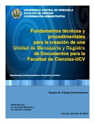
Always keep temporary data labels with specimenson spreading boards or associated with them in some wayto insure that there is no confusion or loss of data whenthey are removed from the boards.Spreading is a highly individualistic skill, subject towide variation. Nearly everyone, with practice, evolves hisor her own technique, so that two workers may appear tofollow different procedures <strong>and</strong> yet produce equally goodresults. There is no single st<strong>and</strong>ardized technique withrespect to the fine points of spreading.References: Lewis 1965; Tagestad 1974, 1977.3.6 - Riker MountsIt is sometimes desirable to prepare specimens forexhibition in such a way that they may be h<strong>and</strong>led freelyfor close examination without risk of damage. Rikermounts have long been used for this purpose. They may bepurchased from entomological supply houses, but similarcases may easily be constructed. The Riker mount (fig. 28)is simply a flat cardboard box about 3 cm deep, filled withcotton, <strong>and</strong> having a pane of glass or plastic set into thecover. Unpinned specimens are placed upsidedown on theglass of the cover, spread into position with some cottonheld in place by small weights, <strong>and</strong> allowed to drythoroughly (about 2 weeks). Then the weights are removed,enough cotton is added to hold the specimensfirmly in place, a little fumigant is added to kill any pestsor their eggs that might have been laid in the box, <strong>and</strong> thebottom part of the box is put in place. When the box isclosed, it should be sealed completely to prevent access to<strong>Collecting</strong> <strong>and</strong> <strong>Preserving</strong> Insects <strong>and</strong> Mitespests. Plant material may also be dried in place with thespecimens.Riker mounts are practical only for relatively largeinsects, such as butterflies, larger moths, beetles, <strong>and</strong>dragonflies, that are suitable for such display. AlthoughRiker-mounted specimens are useful for classroominstruction <strong>and</strong> general display, they are not used forstorage of insects in a scientific collection, where specimensmust be available for examination from all anglesunder magnification. Riker mounts should be inspectedperiodically for pests <strong>and</strong> kept out of sunlight, which willcause fading of colors <strong>and</strong> general deterioration.It is sometimes desirable to put pinned specimensinto Riker mounts. To do so, remove the pin or cut it offflush with the surface of the insect (see Holbrook 1927).3.7 - Inflation of LarvaeA common practice in the 19th <strong>and</strong> early 20thcenturies was to preserve larvae, mainly caterpillars, byinflation. That practice has largely been ab<strong>and</strong>oned infavor of alcoholic preservation or freeze-drying. Theselatter methods permit more thorough examination of allparts of the specimens, even internal organs, which mustbe removed before inflation. Some of the colors of largerlarvae are better preserved in inflated specimens than inalcohol, but color photography has made preservation ofthe larval colors less essential. However, the technique isstill potentially useful <strong>and</strong>, if well done, is not to bediscouraged. For instructions on how to inflate larvae, thefollowing references may be consulted.References: Banks 1909 (pp.69-70); Hammond1960; Martin 1977.3.8 - Artificial DryingThe most widely used method of artificial dryingFig. 28. A Riker mount.Fig. 29. A critical point dryer <strong>and</strong> its accompanyingCO 2supply.38
now in use at most museums <strong>and</strong> other institutions iscritical point drying (fig. 29). In critical point drying(CPD), specimens are immersed in absolute ethanol <strong>and</strong> aspecial machine is used to exchange the alcohol withliquid carbon dioxide under pressure. The liquid CO 2isthen warmed <strong>and</strong> passes through the "critical point" <strong>and</strong> isbled off. The effect of this process is to remove all waterfrom soft tissues <strong>and</strong> in effect "freeze" them in position. Inthis way, soft bodied specimens can be dried without thedistortion that normally results when soft tissues are airdryed. CPD machines are still fairly expensive <strong>and</strong>generally beyond the range of individuals although theyare very common at larger institutions.A more "low-tech" method of drying soft-bodiedinsects <strong>and</strong> other arthropods in a very lifelike manner <strong>and</strong>with no loss of color is by freeze-drying. While the cost ofspecialized freeze-drying equipment is high, it is possibleto freeze dry specimens in an ordinary freezer if donecarefully.Techniques <strong>and</strong> Toolscolors, one of the features sought in artificial drying (Berte1979).Another method of drying involves the use ofhexamethyldisilazane (HMDS) (Brown 1993; Nation1983; van Noort 1995). Using this method specimens aresoaked in absolute alcohol until all water has beenremoved. They are then moved into a small amount ofHMDS for a few minutes, then into a second bath ofHMDS (larger specimens may require a third transfer),which replaces the alcohol in the specimen with HMDS.Finally, the HMDS is allowed to evaporate. This methodhas proven quite effective in preventing distortion ofspecimens, but HMDS can be toxic <strong>and</strong> must be used withadequate ventilation, preferably within a fume hood. Avariation of this method uses acetone in place of HMDS.References: Berte 1979; Dodge & Seago 1954;Fisher & Jursic 1958; Gordh & Hall 1979; Harris 1964;Hower 1979; Woodring & Blum 1963.Briefly, the procedure consists of killing the insectby first freezing it in a natural position <strong>and</strong> then dehydratingit under vacuum in a desiccator jar kept inside a freezerat -4° to -7° C. With a vacuum of 0.1 micrometer at -7°, amedium sized caterpillar will lose about 90 percent of itsmoisture <strong>and</strong> about 75 percent of its weight in 48 hours. Itsfrozen condition prevents distortion while drying. Thetime required to complete drying is variable, at least a fewdays with small specimens <strong>and</strong> more than a week withlarger ones. When dry, they can be brought up to roomtemperature <strong>and</strong> pressure, <strong>and</strong> permanently stored in acollection. Like all well-dried insect specimens, they arerather brittle <strong>and</strong> must be h<strong>and</strong>led carefully. Freeze-dryingyields excellent specimens of plant galls formed byinsects.An inexpensive method of freeze-drying (Fisher &Jursic 1958) requires about 100 days to dry a mediumsizedlarva. The use of acetone is recommended beforedrying pinned specimens for better preservation of theirFig. 30. Typical materials used in slide making.3.9 - EmbeddingPreservation of various kinds of biological specimensin polymerized transparent plastics was popular inthe 1940’s <strong>and</strong> 1950’s <strong>and</strong> is still of some interest. Theprocess is rather complicated <strong>and</strong> laborious, but if carefullydone it will yield useful preparations, especially forexhibits <strong>and</strong> teaching. Directions for embedding insectspecimens may be found in the references below<strong>and</strong> indirections furnished by suppliers of the materials.References: Fallis & Smith 1964; Fleming et al.1940; Hocking 1953.3.10 - Mounting Specimens for MicroscopicExaminationThe small size of mites, thrips, whiteflies, aphids,scale insects, fleas, parasitic wasps, <strong>and</strong> many otherinsects, as well as the necessity of clearly seeing minutedetails of larger insects, requires examination under acompound microscope at high magnification. Suchspecimens <strong>and</strong> parts of specimens must therefore bespecially prepared <strong>and</strong> placed temporarily or permanentlyon microscope slides. If large <strong>and</strong> thick or complexstructures that must be examined from several angles makeslide mounting inadvisable, they may be examined in aliquid <strong>and</strong> preserved in microvials. Whichever course isadopted, their preliminary treatment is the same.The techniques <strong>and</strong> materials (fig. 30) used inpreparing specimens for high-power microscopic examinationvary considerably according to the kind of insect ormite as well as the researcher’s preferences. The informationgiven here will provide the reader with a basic39