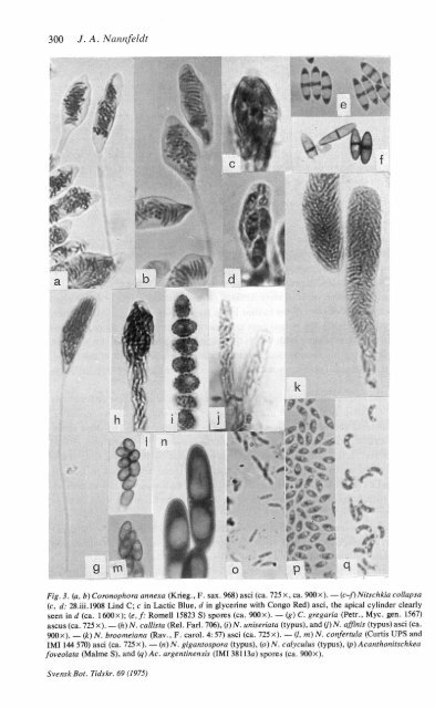
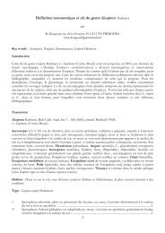
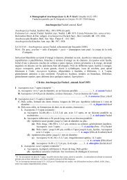
300 J. A. Nannfeldt4\*n»e1 ,iNlgM^ ?w *4 * W J * '?E H F vFig. 3. (a, b) Coronophora annexa (Krieg., F. sax. 968) asci (ca. 725x,ca. 900x). —(c-/)Nitschkia collapsa(c, d: 28.iii.1908 L<strong>in</strong>d C; c <strong>in</strong> Lactic Blue, d <strong>in</strong> glycer<strong>in</strong>e with Congo Red) asci, <strong>the</strong> apical cyl<strong>in</strong>der clearlyseen <strong>in</strong> d (ca. 1600x); (e, f: Romell 15823 S) spores (ca. 900x). — (g) C. gregaria (Petr.. Myc. gen. 1567)ascus (ca. 725x). — (h)N. callista (Rel. Farl. 706), (i)N. uniseriata (typus), and (j)N. aff<strong>in</strong>is (typus)asci (ca.900x). — (k) N. broomeiana (Rav., F. carol. 4: 57) asci (ca. 725x). — (I, m) N. confertula (Curtis UPS andIMI 144 570) asci (ca. 725 x). — («) N. gigantospora (typus), (o) N. calyculus (typus), (p) Acanthonitschkeafoveolata (Malme S), and (q)Ac. argent<strong>in</strong>ensis (IMI 38113a) spores (ca. 900x).Svensk Bot. Tidskr. 69
<strong>Studies</strong> <strong>in</strong> <strong>the</strong> <strong>Coronophorales</strong> 4-8 301Generally speak<strong>in</strong>g, <strong>the</strong> asci are stipitate, ra<strong>the</strong>r th<strong>in</strong>-walled and obviously unitunicate.As a rule <strong>the</strong> wall deliquesces ra<strong>the</strong>r early. The apical apparatus has lostits gun-function and is abortive.In Coronophora <strong>the</strong> shape of <strong>the</strong> ascus (Figs. 3 a, b, g) is most characteristic(comp. e.g. Parguey-Leduc 1966: 26-30) and repeatedly illustrated. The wall of <strong>the</strong>mature ascus is relatively firm and slightly thickened <strong>in</strong> <strong>the</strong> apical part, which iscyl<strong>in</strong>drical to conical and obtusely rounded. Downwards, often delimited by a slightconstriction of <strong>the</strong> lumen (a "bourrelet sous-apical"), follows <strong>the</strong> ma<strong>in</strong> body of <strong>the</strong>ascus as a bulg<strong>in</strong>g sack, downwards ± abruptly constricted <strong>in</strong>to a very long, almostfiliform stipe. In a subadult stage <strong>the</strong> "bourrelet" is much stronger, and so <strong>the</strong>subapical chamber of <strong>the</strong> spore-stuffed lumen looks like a stalked head "crown<strong>in</strong>g"<strong>the</strong> body of <strong>the</strong> ascus as depicted by <strong>the</strong> author of <strong>the</strong> genus (Fuckel 1870 tab. vi, fig.16) and commemorated <strong>in</strong> <strong>the</strong> generic name.In <strong>the</strong> Nitschkieae <strong>the</strong> asci are more varied. Those of N. collapsa are picturedwith an apical wall-thicken<strong>in</strong>g and a cyl<strong>in</strong>drical apical apparatus by Boudier (1910api. 574) and L<strong>in</strong>d (1913 tab. iii, fig. 31). Chenantais (1918:71-72, fig. 5b) depicts twoyoung asci and describes <strong>the</strong>m as follows: "A l'état jeune, ils sont surmontés d'uncyl<strong>in</strong>dre hyal<strong>in</strong> pourvu au centre d'une dépression en entonnoir fermée par unanneau réfr<strong>in</strong>gent qui se présente en coupe optique sous l'apparence de deuxguttules, analogues à celles des Diapor<strong>the</strong>, Melanconis, Laestadia etc.". Fitzpatrick(1923) notes an apical wall-thicken<strong>in</strong>g <strong>in</strong> <strong>the</strong> same species as well as <strong>in</strong> TV. cupularisand N. grevillii. Arx & Millier (1964:815=1973:92) describe and depict it <strong>in</strong> <strong>the</strong>last-mentioned species.These structures are only fa<strong>in</strong>tly seen <strong>in</strong> lactophenol (and <strong>in</strong> Lactic Blue) but wellshown <strong>in</strong> water, which certa<strong>in</strong>ly was <strong>the</strong> medium used by <strong>the</strong> early authors.Although <strong>the</strong> structures <strong>the</strong>mselves do not take Congo Red, this sta<strong>in</strong> makes <strong>the</strong>mstand out more clearly. Janus Green as a sta<strong>in</strong> has also sometimes proved to beuseful as well as glycer<strong>in</strong>e as a medium. In N. collapsa <strong>the</strong> apical thicken<strong>in</strong>g is justdiscernible <strong>in</strong> Lactic Blue (Fig. 3 c), but an ascus <strong>in</strong> <strong>the</strong> right stage shows <strong>in</strong> glycer<strong>in</strong>eand Congo Red an apical perforated cyl<strong>in</strong>der, up to 2 pm high and about as broad(Fig. 3d). In N. cupularis a dist<strong>in</strong>ct wall-thicken<strong>in</strong>g is seen with Congo Red. Theapex is often truncate and may even show a deep hemispherical <strong>in</strong>vag<strong>in</strong>ation.It seems likely that, at least dur<strong>in</strong>g some (short) stage, <strong>the</strong> ascus wall of most (all?)species is thickened apically and has a "bourrelet sous-apical". Thus e.g., <strong>in</strong> N.floridana <strong>the</strong> tip may show a slight thicken<strong>in</strong>g and be truncate and even shallowlyconcave. In N. brevisp<strong>in</strong>a, N. tetraspora, Ac. foveolata, and Ac. tristis asci havebeen seen with <strong>the</strong> tips constricted <strong>in</strong>to short, truncate cyl<strong>in</strong>ders with slightlythickened wall, and <strong>in</strong> <strong>the</strong> first also an apical apparatus <strong>in</strong> <strong>the</strong> shape of a low,perforated cyl<strong>in</strong>der tak<strong>in</strong>g Janus Green. In N. parasitans a dist<strong>in</strong>ct wall-thicken<strong>in</strong>ghas been observed and, occasionally, also an abortive r<strong>in</strong>g has glimpsed as two<strong>in</strong>dist<strong>in</strong>ct po<strong>in</strong>ts (comp. Chadefaud 1960: 587 fig. 444: 2 as N. cupularis).20-753873 Svensk Bot. Tidskr. 69 (19