Management of Melanotic Neuroectodermal Tumor of Infancy
Management of Melanotic Neuroectodermal Tumor of Infancy
Management of Melanotic Neuroectodermal Tumor of Infancy

You also want an ePaper? Increase the reach of your titles
YUMPU automatically turns print PDFs into web optimized ePapers that Google loves.
<strong>Management</strong> <strong>of</strong> <strong>Melanotic</strong> <strong>Neuroectodermal</strong><strong>Tumor</strong> <strong>of</strong> <strong>Infancy</strong>Márcia Gaiger De Oliveira, DDS, Lester D.R. Thompson, MD,Anna Cecília Moraes Chaves, DDS, Pantelis Varvaki Rados, DDS,Isabel da Silva Lauxen, Biol, and Manoel Sant’Ana Filho, DDS<strong>Melanotic</strong> neuroectodermal tumor <strong>of</strong> infancy is a rare congenital neoplasm involvingthe head and neck in young patients. The clinical assessment, histologic diagnosis,and management is reviewed, with an emphasis on different treatment alternativesin two new case reports.Ann Diagn Pathol 8: 207-212, 2004. © 2004 Elsevier Inc. All rights reserved.Index Words: <strong>Melanotic</strong> neuroectodermal tumor <strong>of</strong> infancy, clinical, pathology,prognosis, differential diagnosisFrom the School <strong>of</strong> Dentistry, Oral Pathology, Universidade Federal doRio Grande do Sul, Porto Alegre, Brazil; and Southern CaliforniaPermanente Medical Group, Woodland Hills, CA.Address reprint requests to Lester D. R. Thompson, MD, SouthernCalifornia Permanente Medical Group, Department <strong>of</strong> Pathology, 5601De Soto Ave, Woodland Hills, CA 91367.© 2004 Elsevier Inc. All rights reserved.1092-9134/04/0804-0003$30.00/0doi:10.1053/j.anndiagpath.2004.04.003MELANOTIC neuroectodermal tumor <strong>of</strong> infancy(MNTI) is a rare, usually benign neoplasm<strong>of</strong> neural crest origin composed <strong>of</strong> relativelyprimitive pigment-producing cells. MNTI usuallyarises in infants within the first 6 months <strong>of</strong> life.Because <strong>of</strong> the uncommon occurrence <strong>of</strong> this neoplasm,past terms used to describe the tumor include“pigmented epulis,” “retinal anlage tumor,”“congenital melanocarcinoma,” “melanotic epithelialodontoma,” “melanotic ameloblastoma,” and“melanotic progonoma,” to name just a few. Itusually arises in the head and neck region andpredominantly affects the maxilla, 1-3 althoughother sites, such as brain, epididymis, mediastinum,femur, and ovary have also been reported. 2,4-10The tumor is usually nonulcerated and presentsas a s<strong>of</strong>t tissue swelling, frequently affecting bone.Although the tumor cells produce melanin, pigmentationmay not be clinically evident. 2,4,9,10MNTI is a benign tumor, but can be locally aggressive,growing rapidly and resulting in tooth displacementas tumor cells invade bone. 1,11-13 Withplain radiographs, MNTI appear as intrabony expansiveareas <strong>of</strong> radiolucency, usually with poorlydemarcated margins, probably as a result <strong>of</strong> rapidtumor growth and a tendency to be locally invasive.Extensive tumor calcification may be identified. 5Teeth are usually displaced and appear within theradiolucent area <strong>of</strong> the tumor. 8,9 Computed tomography(CT) scans provide important informationregarding the extent <strong>of</strong> the lesion, thereby assistingin the development <strong>of</strong> a surgical plan. 9We report the clinical and histopathologic features<strong>of</strong> two cases <strong>of</strong> MNTI treated surgically, withand without incisional biopsy.Case 1Case ReportsA small mass was noted within the maxilla <strong>of</strong> amale patient at birth, with associated feeding problemsdeveloping during the ensuing 2 months.During this time the mass started growing rapidly,resulting in a referral at 2 months to the School <strong>of</strong>Dentistry, Universidade Federal do Rio Grande doSul (Porto Alegre, Brazil). No other physical, clinical,or laboratory abnormalities were identified,and there were no congenital anomalies. Clinicalexamination revealed a fluctuant blue mass coveredby an intact mucosa with expansion <strong>of</strong> the leftmaxillary alveolar ridge (Fig 1). Radiographic examinationrevealed a poorly defined osteolytic radiolucencyassociated with a left upper deciduouscentral incisor that was displaced anteriorly. CTshowed a radiolucent lesion with expansion <strong>of</strong> theAnnals <strong>of</strong> Diagnostic Pathology, Vol 8, No 4 (August), 2004: pp 207-212207
208 Gaiger de Oliveira et alFigure 1. Expansion <strong>of</strong> the left maxillaryalveolar ridge by a slightly “bluepurple”mass with intact overlying mucosa.surrounding bone in the area <strong>of</strong> the deciduouscentral incisor (Fig 2).An incisional biopsy performed under local anesthesiawas inconclusive, leading the surgeon toobtain diagnostic material under general anesthesia.Upon receiving a diagnosis <strong>of</strong> a MNTI, a secondFigure 2. Computed tomographyscan showing a radiolucent lesionwith expansion <strong>of</strong> the surroundingbone in the area <strong>of</strong> the deciduouscentral incisor. The macroscopicmass (inset) is well circumscribedand heavily pigmented.
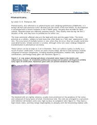
<strong>Melanotic</strong> <strong>Neuroectodermal</strong> <strong>Tumor</strong> <strong>of</strong> <strong>Infancy</strong>209Figure 3. An intermediate power illustratingthe biphasic tumor cell population,with large and small cells.Crush artifact can sometimes obscurethe underlying tumor.operation was required to achieve complete surgicalextirpation. Postoperatively, a lumbar puncturewas negative for malignant cells, and there was noevidence <strong>of</strong> metastatic or disseminated disease. Thepatient is alive and well 7.5 years after the originalsurgery without disease and no recurrent tumor.Case 2A 3-month-old girl was referred for investigation<strong>of</strong> a swelling <strong>of</strong> the left maxillary alveolus. Thelesion was first noted at birth, but started growingrapidly 8 weeks before the visit, causing difficultieswith feeding. Clinical examination revealed an expansion<strong>of</strong> the left maxillary alveolar ridge by adarkly pigmented sessile mass with intact overlyingmucosa. There were no other clinical or physicalfindings <strong>of</strong> significance. CT scan showed a radiolucentlesion expanding the surrounding bone, andconfirmed the displacement <strong>of</strong> the left and rightupper deciduous central incisor and the left upperdeciduous lateral incisor.Surgical excision was performed under generalanesthesia, with curettage <strong>of</strong> the bone. The tumorwas well demarcated. The crown <strong>of</strong> a deciduoustooth was attached to the enucleated specimen.The patient had an uneventful postoperativecourse and is alive and well without evidence <strong>of</strong>disease or recurrence 4 years after the initial presentation.Pathologic FindingsMacroscopically, both intact lesions had a darkblue tinctorial surface, with sectioning through thehard mass revealing a mottled white-grey and darkblue cut surface (Fig 2, inset). The tumors were 4cm in greatest dimension.Microscopically, both tumors were remarkablysimilar, displaying a densely sclerotic fibrous connectivetissue stroma separating a biphasic neoplasticproliferation. A fibrous capsule was seen in bothcases, but the tumors were considered circumscribedrather than encapsulated. One populationwas composed <strong>of</strong> centrally located small, darklystained cells with hyperchromatic nuclei and scantcytoplasm. This population was usually surroundedby a second group <strong>of</strong> larger epithelioid cells thatcontained nuclei with vesicular nuclear chromatin(less chromatic than the smaller cells). These nucleiwere surrounded by more abundant cytoplasmcontaining melanin pigment, distributed in a heavygranular arrangement (Figs 3 and 4). The neoplasticcells were occasionally arranged in alveolar nests(Fig 4), small nests, and solid sheets. Necrosis,hemorrhage, nuclear atypia, and mitotic figureswere absent.DiscussionSeveral theories have been proposed to explainthe pathogenesis <strong>of</strong> this neoplasm which recapitu-
210 Gaiger de Oliveira et alFigure 4. High power with fibrousconnective tissue stroma separatinga nest <strong>of</strong> large, heavily melanin-pigmentedcells separated from smallcells with high nuclear to cytoplasmicratios and hyperchromatic nuclei.lates the early stages <strong>of</strong> retinal development (retinalanlage tumor). A congenital dysembryogeneticneoplasm arising from neural crest cells is theposited theory best supported by embryologic, ultrastructural,biochemical, immunohistochemical,electron microscopic, and molecular genetic studies.9,14,15 Support for this proposed neuroectodermalorigin is given with secretion <strong>of</strong> vanilmandelicacid or other catecholamines by the neoplasticcells, a finding characteristic <strong>of</strong> other tumors <strong>of</strong>neural crest origin, such as pheochromocytoma,neuroblastoma, and ganglioneuroblastoma. 8 Aftertumor excision, vanilmandelic acid levels will usuallyreturn to normal. 2,10,11,16 Further support forthe neuroectodermal derivation is the expression<strong>of</strong> melanotransferrin (a melanoma-specific peptidethat may play a role in iron metabolism) inMNTI. 15<strong>Tumor</strong>s are circumscribed but not encapsulated.There is a biphasic tumor cell population arrangedin a background fibrous connective tissue stroma.The small, darkly staining cells comprise the majority<strong>of</strong> the cells and have a “neural” quality withscant, fibrillar cytoplasm surrounding round nucleiwith coarse and heavy nuclear chromatin deposition.Usually identified in a central location, these“neuroblastic-like” cells are surrounded by a secondpopulation <strong>of</strong> larger epithelioid cells. Thesecells have significantly greater amounts <strong>of</strong> opaquecytoplasm filled with melanin pigment granulessurrounding nuclei that contain a more vesicularnuclear chromatin. The melanin pigment can be sodense as to obscure the nucleus. By definition,mitotic figures are inconspicuous and these tumorslack necrosis and hemorrhage.Immunohistochemical studies have shown thatthe large epithelioid cells are variably positive forvimentin, cytokeratin, epithelial membrane antigen,neuron-specific enolase, glial fibrillary acidicprotein, synaptophysin, Leu 7, and HMB45. Thesmaller, hyperchromatic cells are positive for neuron-specificenolase, glial fibrillary acidic protein,and synaptophysin. 1,2,6,7,9,17 S-100 protein is usuallynonreactive, although rare cases may be focallyimmunoreactive. Whereas the neoplastic cells showpolyphenotypic expression with neural, melanocytic,and epithelial markers, no photoreceptor(retinol-binding protein) or myogenic differentiationis noted. Furthermore, alpha-fetoprotein andneur<strong>of</strong>ilament are also nonreactive, 17 helping toseparate other primitive neuroectodermal tumorsfrom MNTI.By ultrastructural examination, the biphasic nature<strong>of</strong> the tumor cells is also confirmed, withdense-core, membrane-bound neurosecretory granules,neur<strong>of</strong>ilaments, and cytoplasmic processesidentified in the small cell population and modifiedtight junctions, melanosomes at various stages
<strong>Melanotic</strong> <strong>Neuroectodermal</strong> <strong>Tumor</strong> <strong>of</strong> <strong>Infancy</strong>211<strong>of</strong> development, and a single cilium identified inthe epithelioid population. 6,17,18The differential diagnosis <strong>of</strong> MNTI is quitebroad, but must be separated from other pediatric“small round cell” neoplasms such as neuroblastoma,Ewing’s sarcoma, peripheral neuroepithelioma,rhabdomyosarcoma, peripheral primitiveneuroectodermal tumor, desmoplastic small roundcell tumor, malignant melanoma, and lymphoma.Histopathologically, the biphasic neoplastic populationand polyphenotypic immunohistochemicalexpression is quite distinctive and unique frommost <strong>of</strong> the other pediatric “small blue round cell”neoplasms. 2,19 MNTI may share a common histologicand immunophenotypic expression with cellularblue nevus, melanoma, neuroblastoma, andrhabdomyosarcoma, but MNTI does not expressdiffuse reactivity with S-100 protein, lacks othermarkers <strong>of</strong> neuroendocrine differentiation, andlacks myo-D1, myoglobin, myogenin, and musclespecificactin reactivity. Melanoma is distinctly rarein pediatric patients and even more so if the “mucosal”sites <strong>of</strong> development <strong>of</strong> MNTI are considered.Cellular blue nevus characteristically has aspindle cell population, the cytoplasm <strong>of</strong> which isfilled with pigment, while MNTI does not containspindle cells. Teratoma, especially immature types,may show isolated foci <strong>of</strong> MNTI, but these foci are<strong>of</strong> no prognostic significance. Curiously, eventhough there is a histologic similarity with neuroblastomaand other pediatric neoplasms, there isno genetic basis at present to link them. 7,20A limited review <strong>of</strong> the English literature (1990to 2003) confirms that most MNTI occur in themaxilla and oral cavity in pediatric patients, with90% occurring in infants 1 year <strong>of</strong> age. 2,20-23There is no gender predilection with patients usuallypresenting with a unilateral, nonulceratedmass, present since birth, frequently associatedwith recent rapid enlargement. Symptoms are usuallypresent for an average duration <strong>of</strong> about 2months. A radiolucent expansion <strong>of</strong> the bone withtooth displacement is noted on plain radiographs,while vascular enhancement will be appreciated onpostcontrast images. The extent <strong>of</strong> the lesion is bestdelineated with a CT.Given the typical clinical features <strong>of</strong> these tumorsin the oral cavity, we believe that complete surgicalexcision with negative margins can be performedwithout a prior biopsy. This approach avoids unnecessaryanesthetic risk and reduces manipulation<strong>of</strong> the lesion, because it has been reported that thetumor seems to grow faster at previous biopsysites. 8,12 Although a conservative approach, consisting<strong>of</strong> local excision and curettage, 7,21 has beenadopted for the management <strong>of</strong> MNTI, the extent<strong>of</strong> the surgical excision has been debated. 2,3,9,17Different authors have reported large blunt dissection,19 en bloc excision <strong>of</strong> the tumor with reconstruction<strong>of</strong> the defect with autogenous costochondralgraft, 13 the use <strong>of</strong> chemotherapy alone, 22 or inassociation with radiation therapy, or radiationtherapy alone. 8,18,22 However, the successful management<strong>of</strong> the two patients reported herein suggeststhat aggressive or radical surgery may not benecessary. Given the pigmentation <strong>of</strong> the lesion, itis usually possible to visualize the tumor limits andenucleate it as a whole (see Fig 2, inset). This wouldavoid tumor fragmentation that may lead to recurrence.Unfortunately, the biological behavior <strong>of</strong> thisneoplasm cannot be predicted by gross or histologiccharacteristics, requiring close and carefulclinical follow-up. 2,10,17,23 Rates <strong>of</strong> local recurrenceup to 45% after conservative excision have beenreported. 2,12,13,17 Recurrent tumors seem to growmore aggressively, tend to have indistinct borders,and may show osteoid formation, suggesting thatthe surgical procedures may trigger reactive boneformation. 11,24 Some authors have demonstrated asignificant presurgical reduction <strong>of</strong> the “neuroblastic-like”component with chemotherapy. 22 Whenmetastasis develops (up to 7% <strong>of</strong> cases), 2,6,10,17,18,21it is the “neuroblastic-like” component that is regardedas the aggressive part <strong>of</strong> the neoplasm.These malignant MNTI develop widespread metastasisand cause death within a few months. Consequently,these tumors are histologic mimics <strong>of</strong> neuroblastomarather than MNTI. 7,9 In this setting,surgery with adjuvant radiation and/or chemotherapyis the usual treatment. The usefulness <strong>of</strong> DNAploidy by flow cytometry in predicting tumor behavioris controversial, although aneuploidy is associatedwith tumor recurrence. 17Conclusion<strong>Melanotic</strong> neuroectodermal tumor <strong>of</strong> infancy is awell-characterized biphasic neoplastic lesion thatoccurs in infancy and may be treated conservativelyby surgical excision without an incisional biopsy,thus avoiding further manipulation <strong>of</strong> the lesion.
212 Gaiger de Oliveira et alThe distinguishing features <strong>of</strong> a biphasic tumor cellpopulation with melan pigment allow for separationfrom other pediatric neoplasms, thereby avoidingunnecessary therapy. Close clinical follow-up issuggested for the first few years after presentationto identify recurrence or the rare development <strong>of</strong>metastatic disease.References1. Kaya S, Ünal OF, Saraĉ S, et al: <strong>Melanotic</strong> neuroectodermaltumor <strong>of</strong> infancy: Report <strong>of</strong> two cases and review <strong>of</strong> literature.Int J Pediatr Otorhinolaryngol 2000;52:169-1722. Kapadia SB, Frisman DM, Hitchcock CL, et al: <strong>Melanotic</strong>neuroectodermal tumor <strong>of</strong> infancy. Clinicopathological, immunohistochemical,and flow cytometric study. Am J Surg Pathol1993;17:566-5733. Mast BA, Kapadia SB, Yunis E, et al: Subtotal maxillectomyfor melanotic neuroectodermal tumor <strong>of</strong> infancy. PlastReconstr Surg 1999;103:1961-19634. Regezzi JA, Scuibba JJ: Oral Pathology. Clinical-PathologicCorrelations. Philadelphia, PA, Saunders, 1989, p 1635. George JC, Edwards MK, Jakacki RI, et al: <strong>Melanotic</strong>neuroectodermal tumor <strong>of</strong> infancy. AJNR Am J Neuroradiol1995;16:1273-12756. Nelson ZL, Newman L, Loukota RA, et al: <strong>Melanotic</strong>neuroectodermal tumour <strong>of</strong> infancy: An immunohistochemicaland ultrastructural study. Br J Oral Maxill<strong>of</strong>ac Surg 1995;33:375-3807. Sharma MC, Mahapatra AK, Sudha K, et al: <strong>Melanotic</strong>neuroectodermal tumour <strong>of</strong> infancy: Immunohistochemicaland histogenetic consideration. J Assoc Physicians India 1996;44:278-2808. Kim YG, Oh JH, Lee SC, et al: <strong>Melanotic</strong> neuroectodermaltumor <strong>of</strong> infancy. J Oral Maxill<strong>of</strong>ac Surg 1996;54:517-5209. Bouckaert MM, Raubenheimer EJ: Gigantiform melanoticneuroectodermal tumor <strong>of</strong> infancy. Oral Surg Oral MedOral Pathol Oral Radiol Endod 1998;86:569-57210. Johnson RE, Scheithauer BW, Dahlin DC: <strong>Melanotic</strong> neuroectodermaltumor <strong>of</strong> infancy. A review <strong>of</strong> seven cases. Cancer1983;52:661-66611. Howell RE, Cohen MM Jr: Pathological case <strong>of</strong> themonth. <strong>Melanotic</strong> neuroectodermal tumor <strong>of</strong> infancy. ArchPediatr Adolesc Med 1996;150:1103-110412. Hoshina Y, Hamamoto Y, Suzuki I, et al: <strong>Melanotic</strong> neuroectodermaltumor <strong>of</strong> infancy in the mandible: report <strong>of</strong> a case.Oral Surg Oral Med Oral Pathol Oral Radiol Endod 2000;89:594-59913. Eckardt A, Swennen G, Teltzrow T: <strong>Melanotic</strong> neuroectodermaltumor <strong>of</strong> infancy involving the mandible: 7-Year follow-upafter hemimandibulectomy and costochondral graft reconstruction.J Crani<strong>of</strong>ac Surg 2001;12:349-35414. Gotcher JE, Jaffrey BJ, Hudson JW, et al: Recurrent melanoticneuroectodermal tumor <strong>of</strong> infancy: report <strong>of</strong> case andtumor heterotransplantation studies. J Oral Surg 1980;38:702-70615. Nitta T, Endo T, Tsunoda A, et al: <strong>Melanotic</strong> neuroectodermaltumor <strong>of</strong> infancy: A molecular approach to diagnosis.Case report. J Neurosurg 1995;83:145-14816. Neville BW, Damm DD, Allen CM, et al: Oral and maxill<strong>of</strong>acialpathology. 1st ed. Philadelphia, PA, Saunders, 1995, pp385-38617. Pettinato G, Manivel JC, d’Amore ES, et al: <strong>Melanotic</strong>neuroectodermal tumor <strong>of</strong> infancy. A reexamination <strong>of</strong> a histogeneticproblem based on immunohistochemical, flow cytometric,and ultrastructural study <strong>of</strong> 10 cases. Am J Surg Pathol1991;15:233-24518. Cutler LS, Chaudhry AP, Topazian R: <strong>Melanotic</strong> neuroectodermaltumor <strong>of</strong> infancy: An ultrastructural study, literaturereview, and reevaluation. Cancer 1981;48:257-27019. Puchalski R, Shah UK, Carpentieri D, et al: <strong>Melanotic</strong>neuroectodermal tumor <strong>of</strong> infancy (MNTI) <strong>of</strong> the hard palate:presentation and management. Int J Pediatr Otorhinolaryngol2000;53:163-16820. Khoddami M, Squire J, Zielenska M, et al: <strong>Melanotic</strong>neuroectodermal tumor <strong>of</strong> infancy: A molecular genetic study.Pediatr Dev Pathol 1998;1:295-29921. el Saggan A, Bang G, Ol<strong>of</strong>sson J: <strong>Melanotic</strong> neuroectodermaltumour <strong>of</strong> infancy arising in the maxilla. J Laryngol Otol1998;112:61-6422. Mello RJ, Vidal AK, Fittipaldi HM Jr, et al: <strong>Melanotic</strong>neuroectodermal tumor <strong>of</strong> infancy: Clinicopathologic study <strong>of</strong> acase, with emphasis on the chemotherapeutic effects. Int J SurgPathol 2000;8:247-25123. Barrett AW, Morgan M, Ramsay AD, et al: A clinicopathologicand immunohistochemical analysis <strong>of</strong> melanotic neuroectodermaltumor <strong>of</strong> infancy. Oral Surg Oral Med Oral PatholOral Radiol Endod 2002;93:688-69824. Nagase M, Ueda K, Fukushima M, et al: Recurrent melanoticneuroectodermal tumour <strong>of</strong> infancy. Case report andsurvey <strong>of</strong> 16 cases. J Maxill<strong>of</strong>ac Surg 1983;11:131-136