Final Report - SEDENTEXCT
Final Report - SEDENTEXCT
Final Report - SEDENTEXCT

You also want an ePaper? Increase the reach of your titles
YUMPU automatically turns print PDFs into web optimized ePapers that Google loves.
imaging technique for its intended purpose and to ensure its safe clinical use. It is usually performedby using a test “phantom” in conjunction with software routines that help in the interpretation of theresults. The phantom, constructed of materials of known characteristics, essentially acts as a“standard patient”, allowing repeated x-ray exposures to be performed and image quality measured.Preliminary tests before the start of this project on a dental CBCT unit showed that using a phantomdesigned for QA of medical CT equipment resulted in images with worse resolution than the medicalCT scan. Furthermore, discrimination between objects with different density was not alwayssuccessful. It was speculated that this was due to the fact that dental CBCT units are optimized forimaging of hard tissues, i.e. bone. This is also related to the relatively low dose delivered comparedwith medical CT. Therefore, the development of a specifically designed phantom, with a size anddensities resembling those of dental interest was necessary, including special software tools for theinterpretation of the results and the evaluation of image quality.There are a multitude of diagnostic applications of radiology in dentistry. The existing literature onCBCT was considerably diluted by numerous non-systematic reviews and single case reports. Suchopinion-led papers, invariably enthusiastic in their support for using CBCT, added little to ourknowledge of diagnostic usefulness. Radiology for orthodontics is a key clinical application forCBCT. Most orthodontic treatments are performed in the first or second decades of life, whenradiation-related risk is highest. Concerns over the use of x-rays in orthodontic treatments washighlighted in the European Guidelines on Radiation Protection in Dental Radiology (EuropeanCommission, 2004), emphasising the need for careful clinical assessment prior to selecting theneed for x-ray imaging. Many patients do not require complex imaging for orthodontic treatmentplanning and some no imaging at all. This European Commission document included indications forX-ray of patients for orthodontic treatment but did not consider CBCT as, at that time, there wasextremely limited literature available on CBCT use in orthodontics. Preliminary review of theliterature by the applicants shows that this literature was dominated by anecdotal reports and‘reviews’. Similar criticisms could be addressed at publications suggesting the usefulness of CBCTfor a range of dental applications, including dental caries (decay), bone health around teeth, jawjoint problems and various minor surgical procedures including wisdom tooth assessment. Some ofthese studies also failed to demonstrate good research design.Before accepting a new method for clinical diagnosis in dentistry, evidence should be available thatthe new method does more good than harm. This is particularly important with X-ray basedmethods. It should also be checked whether the costs for the new method are reasonable.Economic evaluation attempts to weigh effects and costs of alternative methods with the goal thatavailable resources are used to achieve maximum benefits for patients in terms of health and qualityof life. In emerging technologies, this is particularly important to avoid inappropriate and excessiveuse. Before the start of the project, dental CBCT was an emerging technology and no studies hadbeen performed that reported how examination with CBCT benefits the patient and how much anexamination actually costs compared with traditional radiological examinations, for different dentalproblems.There was very little education and training of general dentists about CBCT before the projectstarted. What was available was provided by manufacturers, who have a vested interest inportraying their equipment, and its capabilities, in the best light. CBCT was not covered as part ofthe undergraduate dental curriculum. CBCT equipment was reducing in price, resulting in thepossibility of ordinary “high street” dentists purchasing this equipment for their practices. Evenwhere the equipment was not immediately available to a dentist in his practice, referral to aspecialist centre required him/her to interpret the CBCT image and report the findings to a patient.Clearly, this situation was not favourable to good patient care.
2.2 Project objectivesWork package 1: Justification and Guideline DevelopmentThe overall aim of this work package was to develop guidelines on referral criteria for CBCT, qualityassurance and optimisation of CBCT use. The specific objectives were:• to perform a systematic review of CBCT based on ‘dose and risk’, ‘diagnostic accuracy’ and‘quality assurance’• to develop provisional guidelines• to incorporate knowledge gained from other parts of the <strong>SEDENTEXCT</strong> project• to develop definitive referral criteria and guidelines on quality assurance and optimisationWork package 2: DosimetryThe overall aim of this work package is to determine the level of (1) patient dose in dental CBCT,paying special attention to paediatric dosimetry, and (2) personnel dose in dental CBCT. Thesegoals corresponded to the following sub-objectives:• The development of a standardised technical dose index to characterise dose distribution inCBCTs• The estimation of the effective dose in anatomical phantoms• In vivo skin dose measurements• The development of mathematical models for dental CBCT dosimetry• Measurements of the scatter dose around scanners and to explore the consequences forradiation protection of personnel and helpersWork package 3: OptimisationThe overall aim of Work package 3 was to produce tools (phantom and software) and a protocol toassist in periodic quality assurance (QA) testing. The specific objectives were:• to develop, design and test a phantom for QA tests on dental CBCT equipment• to develop software tools for the evaluation of image quality and for routine QA testing• to form and implement a routine QA protocol, for periodic QA tests in daily clinical practice• to form an Image Quality testing protocol and determine its implementation on CBCT units.Work package 4: Diagnostic accuracyThe overall aim of Work package 4 was to answer the questions:• What additional information does CBCT exams provide compared with clinical and twodimensionalradiological methods?• What is the accuracy of such CBCT information?• Would any of the additional information on three dimensions change the treatment in anessential way?To answer these questions, various in vitro and clinical studies, often observer-based, wereperformed, the objectives being:
1. To determine the segmentation, linear and diagnostic accuracy of CBCT, using various scannersin vitro2. To determine diagnostic accuracy for CBCT for specific clinical applications: implant placement,impacted teeth (canines and 3 rd molars) and maxillary sinus grafting procedures in relation to dentalimplant planning.Work package 5: Cost effectivenessThe goal of Work package 5 was to analyse the cost-effectiveness of CBCT in different clinicalsituations, health care contexts and countries. The specific objectives were:• To analyse how much examinations with CBCT cost both the health care provider and thepatient for different clinical problems in dentistry, compared with the costs of conventionalradiological methods• To analyse any differences in costs of CBCT between the centres participating in the study• To analyse how access to CBCT radiographs influence the decisions of radiologists and theclinicians who are treating the patientWork package 6: Training and valorisationThe overall aim of Work package 6 was to develop a website providing information resources andtraining materials on CBCT, for use by dental professionals, medical physicists and othersinterested in CBCT, e.g. students, equipment manufacturers and the general public. The specificobjectives were:• to perform a needs analysis amongst the professional community to inform the design of thewebsite and its content• to provide a robust and cost-effective means of delivery of on-line training and informationdissemination• to provide an open repository of knowledge and experience on CBCT, including the Guidelinesdeveloped by Work package 1• to ensure continued support and maintenance of the resources developed in the Work packagebeyond the lifetime of the project
3. Main Scientific and Technical Results / Foregrounds3.1 Work package 1: Justification and Guideline Development3.1.1 Initial workThe first step in this Work package (WP1) was to assemble a multidisciplinary group of experts andto form a “Guideline Development Panel” (referred to as the “Panel” hereafter). The project partnersincluded individuals with a wide range of backgrounds, including dentists, dental specialistsincluding dental radiologists, medical physicists and scientists with a special interest in CBCT. Anexpert in evidence-based dentistry, with experience of clinical guideline development, was alsoavailable. The Panel membership was agreed at the first project meeting. Through a consensusprocess, the scope of the guidelines was discussed and agreed. The following key topic areas wereinitially identified:• Diagnostic accuracy studies• Radiation dose and Risk• Optimisation of radiation dose for patients and staff• Quality standards/assurance• Cost/Benefit Analysis• CBCT useAccording to the project plan, the guideline development process was to be built upon systematicreview of the scientific literature, incorporating any available national or specialist guidelines.Systematic review is focused on a research question that tries to identify, appraise, select andsynthesize all high quality research evidence relevant to that question. By following this approach,the influence of bias and opinion can be minimized. As a first step, a search strategy was developed(Table 1). A “search strategy” is a way of interrogating computerized databases to extract relevantinformation from the mass of scientific publications which form the broad scientific literature.Table 1: Search strategy developed for Medline (OVID)Search terms1 cone beam computed tomography.mp.2 volumetric radiography.mp.3 volumetric tomography.mp.4 digital volumetric tomography.mp.5 digital volume tomography.mp.6 Cone-beam.mp. or exp Cone-Beam Computed Tomography/7 (volume ct or volumetric ct).mp.8 (volume computed tomography or volumetric computed tomography).mp.9 (cbct or qcbct).mp.10 or/1-911 (dental or dentistry).mp.12 exp dentistry/13 (intra-oral or intraoral).mp. [title, original title, abstract, name of substance word,subject heading word]14 oral surgery.mp. or exp surgery, oral/15 endodontics$.mp. or exp endodontics/16 orthodontics$.mp. or exp orthodontics/17 (periodontic$ or periodontology).mp. or exp periodontics/
18 exp dental caries/19 maxillofacial.mp.20 or/11-1921 10 and 20The following databases were searched: 13• MEDLINE (OVID) (1950 onwards)• EMBASE (OVID) (1980 onwards)• Web of Science• Scopus• UK Clinical Research Network• Clinical Trials.gov• Register of Controlled Trials (www.controlled-trials.com)• NICE guidelines (www.nice.org.uk)• FDI World Dental Federation Guidelines (www.fdiworldental.org).3.1.2 Basic Principles of the use of Dental CBCTEarly in 2008, it became apparent that there was an urgent need to provide some basic guidance tousers of dental CBCT because of concerns over inappropriate use. These concerns were voiced bythe European Academy of DentoMaxilloFacial Radiology (EADMFR), an organisation whoseobjective is to promote, advance and improve clinical practice, education and/or researchspecifically related to the specialty of dental and maxillofacial radiology within Europe. EADMFR hasa membership exceeding 300 individuals whose special interest is imaging of the dental andmaxillofacial region. It is multi-disciplinary, including dental radiologists, medical physicists,radiographers and scientists. It includes both academics (teachers and researchers) and clinicians.In view of the mutual aims of EADMFR and <strong>SEDENTEXCT</strong>, a decision was taken to collaborate inthe development of a set of “Basic Principles” for the use of dental CBCT, based upon existingstandards. These standards include fundamental international principles, EU Directives andprevious Guidelines.A set of 20 “Basic Principles” on the use of dental CBCT was established using a consensusprocess (Horner et al, 2009). Consensus is an organised method to achieve agreement of themajority with mitigation of minority views, avoiding a “top/down” approach. Draft statements weredeveloped by a small team of collaborators but then discussed and adapted in a large EADMFRmeeting held in Budapest in 2008. The final draft questions were then presented to the EADMFRmembers for scoring using an online questionnaire, to which members were directed by email.Consensus was achieved and the Principles (Table 2) were published, describing the minimumrequirements for using CBCT.Table 2: The “Basic Principles” on the use of Cone Beam CT, established by consensus ofmembers of the European Academy of Dental and Maxillofacial Radiology (Horner et al, 2009).1 CBCT examinations must not be carried out unless a history and clinical examination havebeen performed2 CBCT examinations must be justified for each patient to demonstrate that the benefitsoutweigh the risks3 CBCT examinations should potentially add new information to aid the patient’s management4 CBCT should not be repeated ‘routinely’ on a patient without a new risk/benefit assessmenthaving been performed5 When accepting referrals from other dentists for CBCT examinations, the referring dentist mustsupply sufficient clinical information (results of a history and examination) to allow the CBCTPractitioner to perform the Justification process6 CBCT should only be used when the question for which imaging is required cannot be
answered adequately by conventional (traditional) radiography7 CBCT images must undergo a thorough clinical evaluation (‘radiological report’) of the entireimage dataset8 Where it is likely that evaluation of soft tissues will be required as part of the patient’sradiological assessment, the appropriate imaging should be conventional medical CT or MR,rather than CBCT9 CBCT equipment should offer a choice of volume sizes and examinations must use thesmallest that is compatible with the clinical situation if this provides less radiation dose to thepatient10 Where CBCT equipment offers a choice of resolution, the resolution compatible with adequatediagnosis and the lowest achievable dose should be used11 A quality assurance programme must be established and implemented for each CBCT facility,including equipment, techniques and quality control procedures12 Aids to accurate positioning (light beam markers) must always be used13 All new installations of CBCT equipment should undergo a critical examination and detailedacceptance tests before use to ensure that radiation protection for staff, members of the publicand patient are optimal14 CBCT equipment should undergo regular routine tests to ensure that radiation protection, forboth practice/facility users and patients, has not significantly deteriorated15 For staff protection from CBCT equipment, the guidelines detailed in Section 6 of the EuropeanCommission document ‘Radiation Protection 136. European Guidelines on RadiationProtection in Dental Radiology’ should be followed16 All those involved with CBCT must have received adequate theoretical and practical training forthe purpose of radiological practices and relevant competence in radiation protection17 Continuing education and training after qualification are required, particularly when new CBCTequipment or techniques are adopted18 Dentists responsible for CBCT facilities who have not previously received ‘adequate theoreticaland practical training’ should undergo a period of additional theoretical and practical trainingthat has been validated by an academic institution (University or equivalent). Where nationalspecialist qualifications in DMFR exist, the design and delivery of CBCT training programmesshould involve a DMF Radiologist19 For dento-alveolar CBCT images of the teeth, their supporting structures, the mandible and themaxilla up to the floor of the nose (eg 8cm x 8cm or smaller fields of view), clinical evaluation(‘radiological report’) should be made by a specially trained DMF Radiologist or, where this isimpracticable, an adequately trained general dental practitioner20 For non-dento-alveolar small fields of view (e.g. temporal bone) and all craniofacial CBCTimages (fields of view extending beyond the teeth, their supporting structures, the mandible,including the TMJ, and the maxilla up to the floor of the nose), clinical evaluation (‘radiologicalreport’) should be made by a specially trained DMF Radiologist or by a Clinical Radiologist(Medical Radiologist)3.1.3 Provisional Guidelines on CBCTFollowing the project work plan, the intention was to produce Provisional Guidelines at an earlystage in the project and an updated and comprehensive set of “Definitive” guidelines close to theend of the project.Using the search strategy described in Section 3.1.1, relevant literature was identified anddistributed to Panel members for appraisal and grading, using standard proformas for collection ofdata. The results from the assessment of all identified articles were tabulated to produce ‘EvidenceTables’. A meeting of members of the GDP was held to discuss the Evidence Tables and toformulate and grade provisional recommendations. When producing the provisionalrecommendations, members of the GDP were asked to consider:• Volume of evidence• Applicability of the findings to clinical practice• Generalisibility of the results presented to the guideline’s target population
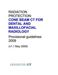
• Consistency of the results (highlight any major inconsistencies)• Clinical impact (e.g resource implications, balance of risk/benefit)Each provisional recommendation was linked, where applicable, to the relevant research evidence.It was graded according to an adaptation of the SIGN grading system (Table 3).Table 3: Grading systems used for levels of evidence [adapted from Scottish IntercollegiateGuidelines Network (SIGN), 2008].GradeABCAt least one meta analysis, systematic review, or RCT rated as 1++, and directly applicable tothe target population; or a systematic review of RCTs or a body of evidence consisting principallyof studies rated as 1+, directly applicable to the target population, and demonstrating overallconsistency of resultsA body of evidence including studies rated as 2++, directly applicable to the target population,and demonstrating overall consistency of results; or extrapolated evidence from studies rated as1++ or 1+A body of evidence including studies rated as 2+, directly applicable to the target population anddemonstrating overall consistency of results; or extrapolated evidence from studies rated as 2++D Evidence level 3 or 4; or extrapolated evidence from studies rated as 2+GPGood Practice (based on clinical expertise of the guideline group)Two additional gradings were used:• A grade of “ED” was applied where a statement was directly derived from The Council of theEuropean Union Directive 96/29/Euratom of 13 May 1996 (laying down basic safetystandards for the protection of the health of workers and the general public against thedangers arising from ionizing radiation) or Council Directive 97/43/Euratom of 30 June 1997(on health protection of individuals against the dangers of ionizing radiation in relation tomedical exposure).• A grade of “BP” was applied where a statement was identical to, or directly derived from, a“Basic Principle” of use of dental CBCT (3.1.2).The Panel developed 53 recommendation statements, of which 34 were related to clinical uses ofCBCT (referral criteria). The evidence grades were generally low, reflecting the limited evidenceavailable on which to base recommendations. The guideline development process was completedby the production of “Radiation Protection: Cone Beam CT for Dental and Maxillofacial Radiology.Provisional Guidelines” (v1.1) in May 2009 (Figure 1). This was published on the project website(www.sedentexct.eu) and also distributed widely to international and national professionalorganisations and societies. Specialist online newsgroups and newsletters were also used asmeans of dissemination. A press release was also used as a way of increasing publicity.
Figure 1: Cover page of “Radiation Protection: Cone Beam CT for Dental and MaxillofacialRadiology. Provisional Guidelines”. Produced by the <strong>SEDENTEXCT</strong> project in May 2009.3.1.4 Definitive GuidelinesFollowing the completion of the Provisional Guidelines, the work plan was aimed at development ofa “definitive” set of Guidelines which would incorporate the rapidly accumulating literature on CBCTand, specifically, the output from the other Work packages in the <strong>SEDENTEXCT</strong> project.The methodology used was broadly the same as that used for development of the ProvisionalGuidelines, with on-going systematic review, critical appraisal and recommendation developmentwith evidence grading. In addition to the identified literature, the Panel identified national guidelinedocuments on CBCT which had been produced since 2009. Seven such national documents wereidentified (from Belgium, Denmark, France, Germany, Norway and two from the UK). It was notedthat most of these included reference to, and inspiration from, the “Basic Principles” and ProvisionalGuideline documents produced by the <strong>SEDENTEXCT</strong> project.The Panel met in November 2010 and in March 2011 to consider aspects of the revision of theProvisional Guideline document. Evidence Tables were considered, along with copies of the originalpapers if required, and the provisional recommendations from 2009 reviewed and revised asnecessary. When producing the Definitive Guidelines, members of the Panel were asked toconsider:• Volume of evidence• Applicability of the findings to clinical practice• Generalisibility of the results presented to the guideline's target population• Consistency of the results (highlighting any major inconsistencies)• Clinical impact (e.g resource implications, balance of risk/benefit)Each guideline statement was linked, where applicable, to the relevant research evidence. It wasgraded according to an adaptation of the SIGN grading system (Table 3). To aid in the developmentof clinical referral criteria, GDPs were asked to consider two questions:
• Is CBCT indicated as a standard method for clinical use for this application?• Is CBCT indicated for selected clinical use for this application?A set of 68 recommendations were developed by the Panel, of which 43 were referral criteria. Thedocument includes recommendations for future research and development and a comprehensiveQuality Control Manual for CBCT systems (see 3.3, below). Examples of recommendations areshown in Table 4.Draft Definitive Guidelines (v1.0) were produced which underwent internal peer review within theproject and assessment by independent external reviewers to produce the Definitive Guidelinesv1.1. These were considered by further external reviewers and by members of the EADMFR. Thelatter completed an on-line consensus survey process to consider the guidelines rated 'bestpractice'. Following further revision, the final <strong>SEDENTEXCT</strong> Definitive Guidelines (v2.0) werereleased on 24 May 2011 (Figure 2).Table 4: Example Guideline statements taken from “Radiation Protection: Cone Beam CT for Dentaland Maxillofacial Radiology. Evidence-based Guidelines”. Produced by the <strong>SEDENTEXCT</strong> projectin May 2011.Guideline statementLimited volume, high resolution CBCT may be indicated in selected cases ofinfra-bony defects and furcation lesions, where clinical and conventionalradiographic examinations do not provide the information needed formanagement.Kilovoltage and mAs should be adjustable on CBCT equipment and must beoptimised during use according to the clinical purpose of the examination,ideally by setting protocols with the input of a medical physics expert.As a minimum target, no greater than 5% of CBCT examinations should beclassified as “unacceptable”. The aim should be to reduce the proportion ofunacceptable examinations by 50% in each successive audit cycle.GradeCBGPFigure 2: Cover page of “Radiation Protection: Cone Beam CT for Dental and MaxillofacialRadiology. Evidence-based Guidelines”. Produced by the <strong>SEDENTEXCT</strong> project in May 2011.
The content of the Guidelines was divided into the following:• Radiation dose and risk• Basic Principles of CBCT use• Justification and referral criteria• CBCT equipment factors in the reduction of radiation risk to patients• Quality standards and quality assurance• Staff protection• Economic evaluation• TrainingTo this were added four appendices:Appendix 1 Summary of recommendationsAppendix 2 Recommendations for research and developmentAppendix 3 Glossary and abbreviationsAppendix 4 Quality Control Manual for dental CBCT systemsIn summary, the guidelines highlight the fact that the radiation dose and risk from dental CBCT aregenerally higher than conventional radiography undertaken by the dentist, but lower than for CT.Clear guidance on optimising doses for patients were included. CBCT machines should offer avariety of settings, and examinations should be undertaken using those settings which arecompatible with the clinical situation whilst providing the lowest achievable dose.It was shown that CBCT has been used for a wide variety of clinical situations within dentistry, suchas identifying the position of unerupted teeth, assessment of cleft lip and palate, diagnosis of caries,the effects of gum disease and trauma. Research evidence suggests that CBCT is only indicatedfor certain situations, particularly those where CT is the current imaging method of choice or whenthe question for which imaging is required cannot be answered adequately by lower doseconventional (traditional) radiography.The Guidelines highlighted that it is essential that a qualified expert is consulted over the installationand use of CBCT to ensure that staff dose is as low as reasonably achievable and that all relevantnational requirements are met. A quality assurance programme should be followed to ensureconsistently adequate diagnostic information, while radiation doses are controlled to be as low asreasonably achievable.A written record of this programme should be maintained by staff to ensure adherence to theprogramme and to raise its importance among staff. In addition, assessment of the clinical imagesand other clinical audit should be undertaken on a regular basis to confirm that the equipment isbeing used correctly to produce clinically useful images.Further information on the Quality Control manual is given in Section 3.3 below.No set of guidelines is permanent. In the context of a rapidly growing new technology like dentalCBCT, the need for review and development is even more important. This is particularly needed forreferral criteria. The first formal statement in the Guideline document was, therefore, to recommendthat the Guidelines are reviewed after a period no longer than five years after its publication.3.2 Work package 2: DosimetryRadiation “dose” is a measure of the energy imparted to the patient (or other person exposed, suchas a worker) when exposed to X-rays or other form of ionising radiation. An accurate understandingof the doses involved in dental CBCT is of fundamental importance in planning radiation protection
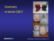
3.2.2 Measurement of the dose distribution in anatomical phantoms and subsequent calculation ofeffective doseAn adult phantom was used for these measurements, as well as two (10 year old and adolescent)paediatric phantoms. For the adult phantom, the effective dose for different CBCT devices showed a20-fold range (19-368 µSv). The largest contributions to the effective dose were from the remaindertissues (37%), salivary glands (24%), and thyroid gland (21%). For all organs, there was a widerange of measured values apparent, due to differences in exposure factors and in diameter, heightand positioning of the FOV relative to the radiosensitive organs.For the 10 year old and adolescent phantom, average effective doses were 116 μSv and 79 μSvrespectively which are comparable to adult doses. Similar to the adult phantom, a wide range ineffective dose was observed. There was a fourfold increase in the thyroid dose of the 10 year oldcompared with the adolescent because of its smaller size. The remainder tissues, salivary andthyroid glands contributed the most to the effective dose for a 10 year old while for an adolescent,the remainder tissues and the salivary glands contributed the most.The results show that a distinction is needed between small-, medium-, and large-field CBCTscanners and protocols, as they are applied to different groups of patients, because the dosereceived is strongly related to FOV size (Figure 3). Furthermore, the dose should always beconsidered relative to the image quality, seeing that image quality requirements also differ forpatient groups. The results from the current study indicate that the optimisation of dose should beperformed by an appropriate selection of exposure parameters and FOV size, depending on thediagnostic requirements. Furthermore, it was concluded that it is imperative that dental CBCTexaminations on children should be fully justified over conventional X-ray imaging and that doseoptimisation by FOV size restriction is particularly important in young children.Figure 3: Average effective dose for CBCT devices, divided into groups based on field of view size.Standard deviations are shown for each group. “FOV” means “Field of View”, i.e. the total volume ofthe patient which is imaged. The trend for higher doses with larger FOVs is evident.3.2.3 In vivo dose measurementsIn addition to phantom dose measurements, skin dose measurements were undertaken for differentscanner types. The skin dose measurements were done in adults and children, using differentclinical indications.A total of 248 patients were included in this study, encompassing six CBCT devices and a largenumber of exposure protocols, based on the clinical indication. A wide range of skin dose resultswas seen, due to patient factors (size and constitution) and scanning factors (FOV size and position,beam quality, amount of exposure).
These results aid in the establishment of diagnostic reference levels for dental CBCT, and providefurther evidence that dose limitation is crucial for child patients, and that the amount of exposureshould not be fixed but based on patient size.3.2.4 Development of mathematical models for dental CBCT dosimetryPerforming dosimetry in patients is labour-intensive and costly. Computer programmes areavailable, however, which “model” the distribution of radiation as it passes through material. It ispossible to use this to model what happens to the X-rays passing through a patient during aparticular X-ray examination and to calculate dose without involving real patients. One computermethod which is widely used in radiology (and for other comparable situations in science) is theMonte Carlo simulation method.A large number of CBCT devices and phantoms were modelled on a validated Monte Carloframework, and conversion factors were determined to obtain the effective dose from thesesimulations. Furthermore, the relationship between these simulated effective doses and themeasured dose indices was investigated.The conversion factors (mSv/mAs) from mAs to effective dose for an adult and child computationalphantom were calculated for a range of dental CBCT machines and clinical examinations. The errorin the computational models was quantified in two stages and it was found to be less than 17%. Theconversion factors increase as the irradiated volume increases due to the higher amount ofscattered radiation. In addition, the conversion factors increase at higher tube voltages for the samefiltration.The conversion factors for small FOVs were calculated for different examination protocols, forexample, mandibular and maxillary wisdom teeth. The conversion factors verify the general trendthat was found with the phantom dose measurements, that the closer the isocentre (i.e. the centreof rotation of X-ray tube and detector, also the central point of the FOV) is to the salivary glands andthyroid, the higher are the dose and the conversion factors.The relationship between the conversion factors and the two dose indices was investigated for thetwo computational phantoms. A linear relationship between the logarithms of the dose indices andthe logarithms of the conversion factors was found with dose index 2 giving a better fit than doseindex 1 for both phantoms. The fitted equations could be used to derive the conversion factors fromthe dose indices.It should be noted that the relationships between the dose index and the conversion factors areempirical and further work should be done using the Monte Carlo simulations and additional doseindex measurements to investigate the physical principles behind the relationships. Further workshould be done on a range of machines to investigate whether these relationships are machinespecific.3.2.5 Measurements of scatter dose and radiation protection of personnel and helpersWhen X-rays pass through material, some pass through unhindered, some are absorbed completelywhile others are scattered. Scattered radiation can expose other parts of the body of the patient toradiation but, of special interest here, can expose other people in the immediate environment, suchas operators and patient helpers. It is important to know about scatter for CBCT so that evidencebasedguidelines on staff protection can be used.Scatter dose measurements were performed on ten different models of CBCT devices. Themeasurements were collected using two techniques: one “active” where a scattering material was
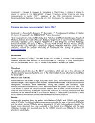
placed in the CBCT and a radiation detector positioned at various locations in the room to measureduring the exposure; the other “passive” where small dosimeters were attached to walls around theCBCT for a period of 3 to 12 weeks while normal, clinical and non-clinical, exposures were carried.The active measurements, which used the maximum exposure parameters, found that scatter doseper scan at a distance of 1 m from the isocentre of the CBCT was in the range 4.1 – 46.8 µSv(mean: 11.3 µSv, median: 7.4 µSv). The passive measurements, carried out on four models ofCBCT, ranged from 2.0 µSv to 8.1 µSv per scan at 1 m.Information was also gathered on the average number of patients seen in different CBCT facilitiesand the national requirements or guidelines on the design of such facilities with respect to radiationprotection. Example calculations of the shielding requirements, combining all the data, were offeredand recommendations based on the measurements were proposed. These results were used inWork package 1 guideline development.3.3 Work package 3: Dose optimisationOptimisation is a fundamental aspect of radiation protection. It requires that radiation doses shouldbe kept as low as reasonably achievable. While dose limits apply to workers and the general public,there are none for patients, so limiting doses to this group is particularly important on a day-to-daybasis. In practice, optimisation involves several aspects, but the focus of this Work package was todevelop a quality assurance programme for dental CBCT, including the design and production ofquality control phantom(s) for commercialisation. A quality control phantom is a test objectcontaining various inserts which can be X-rayed routinely (in place of the patient). The images whichare obtained can be measured as a means of checking if equipment is of a satisfactory standardand is performing consistently. It also allows clinicians to test the effect on image quality of reducingexposure factors and achieve lower doses in clinical work without “experimenting” on real patients.3.3.1.Phantom designThis research involved an iterative process of design and production of prototype phantoms andtesting, culminating in the production of a definitive phantom (Figure 4). This consists of a cylindricalphantom housing made of clear perspex and a number of test inserts (Figure 5) for evaluatingdifferent physical properties of the CBCT technique. Each column within the cylinder includes aseparate threaded cap which allows the user to fill and empty a single column of inserts withoutdisturbing the other columns. A 20mm deep section at the bottom of the phantom is included whichis used for homogeneity measurements (testing of the uniformity of the image). A threaded hole atthe bottom of the phantom means it can be securely attached to a support (e.g. a tripod or table).The phantom and inserts were scanned on a wide range of CBCT devices and have been refined inthree rounds of development, with feedback from validation informing the next round. The designchanges that were implemented into the definitive phantom were found appropriate for both thebody and the inserts for the different image quality tests.
Figure 4: The phantom body with seven columns for test insert accommodation, the white engravedlines (black arrow) for accurate positioning / alignment and the homogeneity 20mm deep section(white arrow) with the threaded hole at the bottom.Figure 5: The lettered threaded caps and the orientation rails3.3.2: Software design, implementation and validationFor the semi-automatic evaluation of phantom images, a specific software program was developed.The general idea behind the software is that the user imports the datasets of the scanned phantominto the software, performs the analysis of certain image quality parameters and enters these resultsinto a Quality Control (QC) report. These image quality parameters are either assessed throughvisual analysis or user-interactive measurements.There are two different parts to the software: (1) the graphic user interface (GUI) which allows theuser to import and visualise the datasets of the scanned phantom and allows for the visual analysisof certain image quality properties, and (2) different executables (i.e. a set of instructions in aspecific computer language) that allow the user to extract certain regions of interest from the datasetfor automated measurement of all other image quality parameters. The two parts are merged intoone package which allows for a full assessment of all phantom-related Quality Control (QC)parameters.
First part – user interface and insert selectionThe user is able to open datasets by selecting ‘File, Open set of images’ in the software, thenbrowsing to the folder containing the dataset and selecting any slice in that folder. Subsequently,reformatting in other planes is achieved using the stack of axial slices (Figure 3). The interface ofthe software contains four windows. On the left side, three small windows show the three planeslices. The main window can display one of these small windows in full size. After importing adataset, by default, the axial (horizontal) slices are shown in the main window. The user can switchto the other slices by selecting one of the other small windows on the left side. By scrolling throughthe slices, there is a possibility for: (1) visual analysis of certain image quality parameters, (2) linearmeasurements to assess geometric accuracy, (3) the selection of regions of interest for automatedimage quality analysis. For this third step, an ‘insert selection tool’ is implemented, which enablesthe free selection of certain parts of the phantom for automated analysis.Second part – region extraction and automated analysisTo start using the selection tool, the user has to click and drag to create a selection box while usingthe axial view, after which the borders of the selection can be adjusted in every direction and usingall three slice windows (Figure 6). After the appropriate selection of a region of interest, themeasurement of a certain image quality parameter is performed and the results are displayed in apop-up window (Figure 7).Validation of the associated software by five consortium partners was successful. A clear protocolfor all measurements was established. It was found that the final software is easy to work with, thatthe working speed has been significantly increased, and that the measurements can be sufficientlyreproducible for QC purposes. Namely, the software is able to open datasets from all availableCBCT devices, it allows for the measurement of all image quality parameters that are relevant forquality control and there is a sufficient agreement between repeated measurements, either bydifferent observers or by the same observer.Figure 6: Software with main (right) and side (left) windows, the insert selection tool and toolbar.
Figure 7: Pop-up window with image analysis results.3.3.3 Quality Assurance Programme for CBCT.A Quality Assurance (QA) procedure protocol was formed comprising two parts. The first is ageneric part about the implementation of a QA programme in CBCT that was formed based onprevious knowledge on QA programmes with special consideration on the particularities of theCBCT technology. The second part, the Quality Control Manual, in general lays out the necessarytesting to ensure that all parameters during the examination procedure are in accordance with thestandard operating protocol, thus resulting in images with diagnostic value, without exposing thepatient to unnecessary risk.The programme of equipment tests for dental cone beam CT considers the following aspects:− Performance of the X-ray tube and generator− Patient dose− Quantitative assessment of image quality− Display screen performanceThis protocol outlines those physical tests and measurements that are considered to be part of astandard quality control programme for a dental CBCT unit. It does not cover quality assurance ofthe clinical image (dealt with in the Guidelines developed in Work package 1).A range of tests are appropriate for dental CBCT looking at different aspects of the equipment andimage display. Some of the tests are straightforward and can be readily performed by the clinicalstaff using the CBCT equipment. Other tests are more complex and the input of a medical physicistis required. Therefore, the expertise required for each test is indicated in the protocol.Routine quality control tests primarily involve comparison of results with those determined duringcommissioning. Significant variation, as indicated by pre-determined action levels, should beinvestigated, either with the help of a medical physics expert (MPE) or the equipment serviceengineer.
Not all possible methods of assessment are considered essential. It is important to perform enoughtests to confirm that the equipment is operating as intended. More complex tests do add extrainformation that is helpful in the optimisation process and they are detailed here for completeness.However, whether the more detailed tests are undertaken will depend on the availability of expertsupport and the necessary resources.The generic part is followed by a specific part on how to use the <strong>SEDENTEXCT</strong> tools (the phantomand the software) for running the respective image quality tests. More specifically, in the specificpart of the QA procedure protocol, there is detailed instructions on the phantom handling andpositioning, test inserts selection, inserts placing in the phantom and using the software for specificimage quality tests, based on the knowledge acquired during the project.The tests described in the routine QA protocol including the periodic QA tests are summarised inTable 5.Table 5: Quality Control tests as part of the Quality Assurance programme for Dental Cone BeamComputed Tomography (CBCT) Systems.X-ray tubeandgeneratorQuantitativeimageQualityDisplayspecificTest Priority Level ofexpertise*SuggestedfrequencyAction levels**Output repeatability Essential MPE 12 monthly Mean ±10%Output reproducibility Essential MPE 12 monthly Baseline ±10%Filtration Essential MPE When new, ifoutput changesor tube headdismantled< 2.5mm aluminium(of which 1.5mmshould bepermanent)Tube potential Essential MPE 12 monthly > ±5% of intendedkVField size andalignmentEssential MPE 12 monthly >10% expected fieldsizeLeakage Essential MPE When new and ifdamagesuspected> 1000µGy hr-1 atmaximum tuberating.Image density values Recommended In house/MPE Monthly >10% from baselineUniformity andartifactsEssential In house Monthly Visible artefacts onthe image or >±10%of the meanNoise Recommended In house/MPE 12 monthly > ±10% frombaselineLimiting resolution Essential In house/MPE 12 monthly > ±20% frombaselineContrast detail Recommended In house/MPE 12 monthly Dependent onmethod used.Geometrical accuracy Essential In house/MPE 12 monthly within ±2mm and ±2ºGeneral condition Essential In house Monthly Failure to resolvedifferent contrasts intest pattern/ notconsistent betweenmonitorsMonitor resolution Recommended In house Monthly Not consistent withbaseline imagePatient dose Patient dose index Recommended MPE 12 monthly Outside ±15% ofmanufacturer’sspecificationPatient dose audit Essential In house/MPE At least 3 yearly > national orinternationalreference level3.4 Work package 4: Diagnostic accuracyIt is obvious that a diagnostic technique, such as CBCT, should be “accurate”. In other words, that itcan correctly differentiate health from disease and that associated software can perform correct
measurements of length, volume and angles. In addition, there are other aspects related todiagnostic accuracy, including the impact of the diagnostic method on treatment planned and on theconfidence of clinicians. For CBCT, there are a vast range of clinical uses, even within a relativelysmall area like dentistry. In this Work package, “key” applications of CBCT were chosen to study indepth. In studies of diagnostic accuracy, there is a need for a “gold standard” against which tehperformance of the diagnostic technique can be compared. This gold standard can be direct visionat surgery or histopathology, but in this work micro-CT (µCT) was sometimes used. Micro-CT is anextremely detailed radiological method used on small, laboratory, samples.3.4.1 Determination of the linear, segmentation and diagnostic accuracy of CBCT, using variousscanners in vitro.Linear accuracy was assessed by measuring distances on CBCT images, obtained with differentimaging parameters. Our results showed that, for the device used, the number of frames (= scantime) can be reduced without implications for linear measurements. Measuring the distancebetween two points does not require the smallest voxel size. Although there were no significantdifferences in the observers’ assessments according to kV and mA changes, it should be stated thateach device needs to be tested separately, which was covered in WP3, where a quality controlprogramme was developed.Segmentation involves splitting the image data (usually by thresholding the picture elements by greyscale value) so that only relevant parts are retained. Segmentation accuracy was analysed both forsegmenting the skull surface, useful for example in orthodontic measurements, and for segmentingthe trabecular bone structure, useful in assessing bone lesions. It goes without saying that theresults of this study need to be evaluated keeping in mind the radiation dose associated which eachdevice and each setting (WP2).Surface models generated from CBCT images deviated from 5 to 29% when compared to µCT.Intra-device comparison of deviation from the gold standard (µCT), showed a preference-setting foreach device. This could be inspiring for further research on preference-settings per device perapplication. In trabecular bone evaluation, the parameter of overlap was found to be the most robustparameter to compare devices and assess trabecular bone structure. Overlap calculates the amountof voxels that coincide with voxels on a gold standard volume (i.e. µCT). This parameter was leastinfluenced by the choice of the threshold value of bone. The average voxel deviation was 31%, witha range from 22 to 42%. Based on the overlap of CBCT images with µCT images, a ranking couldbe made between the scanners, that had great similarity with the intuitive classification of anobserver.Diagnostic accuracy was assessed by creating lesions in bone and tooth specimens, after whichseveral observers assessed the presence of such lesions. The results of the in vitro tooth lesionstudy suggest that the CBCT technique could be a reliable diagnostic tool for detecting canineimpaction and associated lateral incisor root resorption. Lesions as small as 200µm could be easilydiagnosed. From a previous study it was found that additional conventional panoramic radiographsare redundant, thus additional X-ray exposure of the patient can be avoided. In addition, theradiation dose of CBCT is significantly lower compared with conventional CT, and the typicaloverlap of dental structures on panoramic radiography is not observed.When assessing bone lesions, a number of defects remained undiagnosed in the CBCT images.This number differed depending on the CBCT scanners and on the size of the lesions. Identificationof lesions sized 175µm was possible for the best performing CBCT. Lesions were most difficult toidentify in the cortico-trabecular area (area between dense outer bone and the spongy inner bone).This is due to the inherent irregular pattern of the area. Presumably computer-aided analysis oflesions in this area would yield better results.
Another study on diagnostic accuracy of bone lesions was done on animal material: pig jaws. Theresults showed that the sensitivity for detecting bone lesions with various CBCT scanners rangedfrom 72 to 80%. Specificity ranged from 60 to 77%. It is clear that differences exist between differentCBCTs in the detection of apical lesions. Although the result of this in vitro study based on ananimal model cannot be considered a standard reference when simulating clinical situations, ithighlighted the importance of combining radiological and clinical findings in the diagnosis of apicallesions.3.4.2 Determination of diagnostic accuracy for CBCT for specific clinical applications: implantplacement, impacted teeth (canines and mandibular 3 rd molars) and sinus grafting procedures.Implant placementThe difference in planning implants based on two dimensional and CBCT images appeared mostclearly as a difference in the length of the implant and in the confidence of observers to performsurgery with the information available. It is important to use the available bone space in an optimalway, and the choices about the implants to be placed can be made with more conviction when the‘critical boundaries’ can be assessed in all planes. Any surgical event could be better predicted with3D than with two-dimensional images. However, more in depth research on this is required to drawfirm conclusions that can be generalised. Potential complications can be foreseen by CBCT, butdepend on the surgeon and followed surgical procedure as well.It needs saying that the benefit of 3D imaging is related to volume rendering, making it possible tofully integrate data prior to surgery: anatomical, pathological, biomechanical and esthetical aspects.In implant therapy, not only the surgical approach, yet an integrated approach is indeed warranted,in contrast to e.g. wisdom tooth removal.Impacted teeth: third molarsThird molars (wisdom teeth) in the lower jaw are commonly impacted and removed by dentists(Figure 8). In a minority of cases, the tooth is closely related to the large nerve which lies within thejaw. The risk of sensory damage caused by inferior alveolar nerve exposure during the surgicalremoval of impacted mandibular third molars can be limited with an accurate preoperative predictionof nerve exposure. Based on our results, we could not state that CBCT is better than panoramicradiography in predicting nerve exposure for average cases of impacted third mandibular molars:evidence of a close relation on two-dimensional images as well as too simple cases were excludedfor ethical reasons. Difficult cases do require 3D imaging, to avoid permanent nerve injuriesfollowing third molar removal. The value of a third dimension for pre-operative planning of impactedmandibular third molars has been stressed by numerous authors. More precise information whenthere is a close relationship between the impacted third molar and the inferior alveolar nerve, maysimplify the surgical procedure and make it considerably safer. Additionally, the patient can receivemore adequate information about the procedure and the associated risk.
Figure 8: an impacted wisdom tooth on CBCTImpacted teeth: caninesCBCT was more accurate than two-dimensional conventional radiography in differentiating theposition of an impacted canine and its impaction grade. The detection of ectopic canine relationsand root resorption was different in CBCT and panoramic images. Mild resorption of the root did notinfluence the type of treatment chosen. More advanced resorptions however, may influence thetherapeutic decision as in these cases it might be necessary to perform an extraction or anintervention on the root level. Based on the currently collected data, no statistically significantdifference could be found between the treatment plan recommended after the evaluation of CBCTcompared to assessment of conventional two-dimensional images. CBCT evaluation of impactedcanines prior to surgery could reduce the number of extractions due to a better assessment of theirposition and their relation to the adjacent teeth, and through a greater degree of confidence aboutthe therapy plan. To be able to draw stronger conclusions on this hypothesis, a study with a largerand more homogenous patient group would be required. The confidence of the examiners in theirtherapy plan based on the CBCT examination was significantly higher compared to the confidencerelated to two-dimensional radiographic images. None of the radiographic techniques used (twodimensionalradiography or CBCT) could accurately predict complications.In conclusion, based on the results of this study, CBCT can recommended in the following clinicalsituations:• To define the surgical access route: A vestibular or oral crown position can more accuratelybe defined on CBCT images.• To guide the direction of orthodontic traction. If the radiological appearance on the 2D imageshows a direct relationship with the roots of adjacent teeth. This may interfere with the pathof orthodontic treatment of the impacted canine.• To determine if root resorption is present. If there are clinical and/or radiological signssuggestive for root resorption of adjacent teeth, and if these resorption would require aspecific treatment (resorption degree II or III).• To differentiate the pain due to mechanic traction or due to a iatrogenic resorption.• To choose optimal treatment in case of doubt: when the treating dentist cannot decidebetween canine extraction or orthodontic treatment of the canine.Sinus grafting proceduresWhen placing implants in the posterior parts of the upper jaw, the air sinuses may be involved dueto their close inter-relationship. In such cases, a dentist may graft bone below the soft tissue liningof the sinus to “lift” the floor, giving more room to place implants. A CBCT examination may change
the treatment plan for a sinus lift procedure: sinus morphology, better visible on CBCT, is importantin the prevention of postoperative complications and implant loss. CBCT allows the estimation of thenecessary bone graft volume using specialized software. This in its turn increases the accuracy ofharvesting a bone graft in line with the estimated useful volume. A good planning of this harvestingreduces morbidity, traumatic surgery and the duration of the surgery. In our study, CBCT increasedthe confidence in the treatment planning compared to panoramic imaging. Further studies areneeded to follow up the postoperative complications for sinus lift planned only with conventionalradiological methods compared with those that were planned using cross-sectional imaging.3.5 Work package 5: Cost effectiveness3.5.1 Analysis of the costs of CBCT examinations in terms of radiation dosage and in monetarytermsAll patients involved in this study had common dental problems such as retained teeth, or had lostsome of their natural teeth and were going to get implants installed in the jaw bone for area(s)where teeth had been extracted. For these groups of patients dental X-ray examinations are alwaysperformed before treatment and conventional radiological methods are used. Conventionalradiological methods are (1) an overview of teeth and jaw bone – a panoramic view and (2) detailedintraoral images of teeth and jaw bone. To be able to compare the conventional methods with CBCTall patients in our study were examined with conventional methods as well as with CBCT, afterethical approval and informed consent by the patient or, for children, their parent(s).To do a cost-analysis it is important to identify, measure and value all resources used in performingthe examinations. For this purpose, we proposed a model for cost-analysis and developed differentprotocols that were applied during examinations with CBCT and during examinations that wereperformed with conventional radiological methods. The protocols deal with direct costs (such as howmuch the equipment costs and how much the labour costs for people involved in the examinations).Indirect costs are also identified, measured and valued and involve costs for the patient to come tothe clinic, both out-of-pocket costs and the cost for time used and being away from work.When comparing costs of conventional methods and CBCT it appeared that CBCT is more costly,which was mainly due to the higher direct costs, in particular the capital costs that comprised 43%for the new method and 17% for the conventional method of the total cost per examination. Also thecost for maintenance, accommodation and labour was higher for CBCT.In conclusion, CBCT is more costly than conventional radiological methods used for patients withcommon clinical situations in dentistry. Furthermore, the model that we presented and used mayassist other researchers in doing cost evaluations of methods in dentistry.3.5.2 Comparison of the costs of CBCT between countriesWe anticipated that costs of using a radiological method in dentistry differ in countries havingdifferent health care systems. We used the model describe above to calculate and compare costsfor CBCT examinations in four countries – Belgium, Lithuania, Romania and Sweden. CBCTexaminations were performed on patients with three different clinical conditions and costs werecalculated by identifying different resources used, measurement of the amount of the resourcesused and a monetary valuation of the quantity of resources used.The estimates of direct costs and indirect costs of CBCT examinations varied across the health caresystems, being highest in Sweden (Malmö) and lowest in Belgium (Leuven), irrespective of theclinical condition examined. The variation in direct costs was mainly due to different capital costs forthe CBCT-equipment arising from differences in purchase prices of the equipments. The average
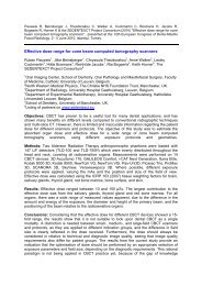
indirect costs per patient were highest in Sweden, second highest for Lithuania (Vilnius), thirdhighest for Romania (Cluj) and lowest for Belgium. Overall, where the examination fees werecharged, these comprised the major part of the indirect costs.In conclusion, a cost evaluation of a dental radiographic method cannot be generalised from onehealth care system to another but must take into account the specific circumstances. The model forcost analysis provides an important input for economic evaluations in comparing costs andconsequences of diagnostic methods in different health care systems, and for planning of servicedelivery in both public and private sectors.3.5.3 Analysis of additional diagnostic information (diagnostic accuracy and diagnostic thinkingefficacy)In this last part of the study we investigated if radiographs from CBCT examinations change whatthe radiologist writes in her/his report or what the clinician decides about treatment. Protocols forassessment of radiographs were developed as well as protocols for decisions regarding diagnosisand treatment plan. Both radiologists and clinicians registered their level of confidence for every“clue” they considered.Regarding the radiologists´ decisions there were no difference if they were assessing radiographsobtained with conventional methods as compared when having access to CBCT images. Theywere, however, in general more confident in their decision about the different “clues” when havingaccess to CBCT images as compared with images from the conventional radiological methods. Thetime it took to assess radiographs were significantly longer for assessing CBCT images.Clinicians changed their treatment decision in between 25% and 50% of their decisions dependingon which clinical situation they were dealing with, when having access to CBCT images ascompared to having access to images from the conventional methods. The change in treatmentdecision was, however, not always due to the information they got from the radiographic report,even if they were more confident in their decision when having access to CBCT images.3.6: Work package 6: Training and valorisationThere were two aspects to this work package. The first was to produce a website for the project(including secure intranet and a depository for documents and discussion tools for the projectpartners) and the second, main, aspect was the development of a means of delivery of informationand training on dental CBCT to stakeholders (professional groups, scientists and the public).As a first step at the start of the project, a Work package 6 implementation group (WP6 Group) wasformed from amongst the project partners. A decision had already been reached to use a webbasedapproach to addressing the needs of CBCT users.3.6.1 Training needs analysisThe WP6 group began by developing a questionnaire to identify the training needs of potentialwebsite users. Transposed into an online format, it was piloted amongst Consortium members of<strong>SEDENTEXCT</strong> prior to general release. Three important stakeholder groups were consulted on thecontent of the website: the membership of the EADMFR, the European Federation of Organizationsfor Medical Physics (EFOMP) and manufacturers. An internet questionnaire to 339 EADMFRmembers resulted in 282 valid email addresses and 139 replies. When the internet questionnairewas sent to members of the European Federation of Organizations for Medical Physics, 28physicists replied. Four manufacturers also replied to the questionnaire. In addition, a focus group
of general dental practitioners was held to determine their needs. We used this information todevelop a set of training priorities to determine the most urgent requirements for each stakeholdergroup. Both dentists and radiologists had similar priorities. Table 6 summarises the contentrequirements identified as a result of the Needs Analysis, as well as content included through othermeans.Table 6: Desirable website content derived from the results of the survey with EADMFR (principallyradiologists) and EFOMP (principally physicists).SourceProjectDescriptionof WorkSurveys(ranked)DevelopmentteamContent requirements• Guidelines document (WP1)• Work generated by other Work Packages• Information about Radiation doses and risks of CBCT• A Quality Assurance Programme for users of CBCT• Information about Quality Control Test Tools for CBCT• A strategy for Radiation Protection of patients• A strategy for Radiation Protection of staff• Evidence-based Referral/ Selection Criteria for CBCT• Descriptions of the Dental clinical uses of CBCT• Anatomical CBCT reference image library• Dose Maps for CBCT• A detailed technological description of how CBCT works• A regularly updated database of links to scientific publications on CBCT• An objective technical comparison of different CBCT machines• Information about Radiation doses and risks of CBCT• A Quality Assurance Programme for users of CBCT• Information about Quality Control Test Tools for CBCT• A strategy for Radiation Protection of patients• Information for patients undergoing CBCT scans3.6.2 Website developmentThe implementation of the site was divided into two distinct phases; the prototype site and the predefinitivesite. A decision was taken to use an off-the-shelf Drupal Content Management System(CMS) application. This offered the appropriate functionality to allow Wiki development, text-basedinformation, PowerPoint lectures, diagnostic forums/ discussion boards, appropriate accessibilitystandards, a secure Intranet and levels of user access. The Prototype web site was designed andbuilt, based on the design model with iterative rapid prototyping of each major design element withpartners and major stakeholder representatives. This was an exercise involving minimal content, butthe technical issues were explored e.g. the look and “feel” of the website, prior to developingmaterials to populate it.The pre-definitive website development was concerned with building the content through multiauthorcontributions to for the Wiki and through developing the training programme.The needs analysis results were used to develop a ten module training package which would beachievable and which reflected accurately the requirements of stakeholders (Table 7). The WP6group developed Learning Outcomes for each module using a brainstorming approach on theproject intranet, detailing Knowledge and Understanding, Skills and Abilities, and Judgement andStance.The delivery of the training modules was considered by the WP6 group and a three-componentapproach was considered the most appropriate:
• Powerpoint lectures with voiceover• “Additional materials”• Use of the Wiki“Additional materials” were made specific to each module. This included activities of a practicalnature to allow the learner to engage in active learning. For example, the additional activitiesinvolved the learner in (1) clicking on the correct word and moving it to the correct box and (2)labelling diagrams. For some modules the “additional activity” required the reading of specificdocuments (e.g. Guidelines). The display of 3D DICOM models through a web interface was alsodeveloped.Table 7: The training modules..Title1 How does CBCT work - Part 12 How does CBCT work - Part 23 Principles of Radiation dose and risk4 Radiation dose and risk in CBCT5 Justification - principles6 Justification - referral criteria7 Dose optimisation - patients and staff8 Dose optimisation - quality assurance9 Anatomy on CBCT images10 Interpretation of pathology on CBCT imagesThe project partners were each allocated responsibility for the production of assessment materialson the same basis as the training materials. It was decided that a form of multiple choice questions(MCQs) would be used in the assessment section using a series of true or false questionstatements where at least one statement is always true. This section was designed to allow users tosave their answers in order to complete tests at a later date and to review answers after completinga test. The assessments were developed using HTML/PHP with a MySQL database recording theanswers given.All of the questions were based on the information found in the training and additional materials.Users are given a percentage mark at the end of the assessment with 70% being the condition for apass. If this is reached a certificate can be downloaded with the user’s details and those of the unithaving been passed. If this mark is not reach the user can attempt to retake the assessment. Inmost of Europe there is no well-established system for recording or validating ContinuousProfessional Development (CPD). The modules and certification in the <strong>SEDENTEXCT</strong> trainingprogramme, however, do meet the requirements of the UK General Dental Council for accreditationof CPD. In the UK, dentists are required to achieve a target number of validated CPD points over a5-year cycle for re-accreditation purposes. The <strong>SEDENTEXCT</strong> training programme would contributeto such requirements.3.6.3: Definitive websiteVisitors to the website at www.sedentexct.eu are met by a home page with links to other pages(Figure 9). The general design has been consistent, although “banners” highlighting new materialhave been added and removed as appropriate during the project lifetime. One of the key home pagelinks is to the Guidelines developed through WP1 (see Section 3.1). Also highlighted is a link to
“Information for patients”, a one page series of questions and answers designed to support patientunderstanding of dental CBCT (Figure 10).Figure 9: <strong>SEDENTEXCT</strong> home page.Figure 10: Part of the “Information for patients” web page.At the bottom of the home page are six picture links:• CBCT Info• Forums• Guidelines• The project• CBCT Publications• CBCT Training“CBCT Info” is the link into the wiki. This element consists of many interlinked information pages,constructed by registered users, with internal and external links. The “Forums” permit users to postitems of interest and permit discussion (Figure 11). “Guidelines” is an additional link to the<strong>SEDENTEXCT</strong> Guideline document, but also includes national guidelines identified during theproject. “The project” link leads to descriptions of the <strong>SEDENTEXCT</strong> project and its Work packages.
“CBCT publications” provides abstracts of scientific publications on dental CBCT obtained from theWP1 literature searches. The final picture link is to the CBCT Training programme (see below).Figure 11: a page from the Discussion forum.CBCT Training ProgrammeThe training programme is accessed from either the picture link or a banner on the home page(Figure 12). These link to a main training contents page (Figure 13) and from here the individualModules and elements (training materials, exercises and assessments) can be accessed.Figure 12: Opening page for the CBCT training programme.A common element in each module is a Powerpoint presentation with voiceover. This opens in aseparate window (Figure 14) and can be viewed as often as the user wishes.
Figure 13: A module page from the training programme.Figure 14: A Powerpoint presentation for one of the modules.Future work and development.At the conclusion of the <strong>SEDENTEXCT</strong> project, a functional web-based source of information andtraining for stakeholders on dental CBCT has been produced. The website content has been copiedonto the EADMFR website (www.eadmfr.info). EADMFR is the natural custodian of the training andeducational material beyond the project lifetime. The material will inevitably require updating andfurther development, reflecting the continued evolution of dental CBCT and accumulating scientificknowledge. The website and training programme developed, however, forms a valuable foundationfor the future.
4. Potential Impact4.1 Impact and main exploitation resultsThis section describes the expected impact of the project, including the socio-economic impact andwider societal applications. It also describes the main exploitation results to date and the outputs ofthe project that are expected to be sustainable after the project, by commercial or non-commercialexploitation.The key stakeholder groups addressed through the <strong>SEDENTEXCT</strong> project are:• Policy makers, e.g. national and international radiation protection agencies and professionalbodies in dentistry and medical physics• Researchers in dentistry, medical physics and health economics• Practising dentists and medical physicists• Manufacturers of cone beam CT equipment• The general public<strong>SEDENTEXCT</strong> is expected to have a major influence on the development of national andinternational guidelines for use of CBCT produced by radiation protection agencies and asEuropean Guidelines. As CBCT examinations are associated with higher radiation dose than mostconventional radiological methods in dentistry, it is of utmost importance to draw attention toimportant radiation safety aspects related to the use of CBCT technology. One important way ofreducing radiation dose to patients is to use proper selection criteria to use CBCT only when itbenefits the patient. The increased knowledge on advantages and disadvantages of the use ofCBCT that is a result of our work will increase patient safety as there will be more relevant referralson CBCT examinations and use of CBCT. The work may also inform social security regulations onallowable treatments for reimbursement.The results of this study will ensure that clinicians have a clearer understanding of when to useCBCT as well as the importance of dose limitation when undertaking cone beam computedradiography. Consideration has also been given to the ethical use of CBCT as well as developingstrategies to reduce inappropriate practice and also inconsistency in clinical care. In addition, thestudy found that research was urgently needed in the field of diagnostic accuracy.The Basic Principles were published in 2009 (Horner et al.) and provide a framework of standardsachieved by consensus for the clinical use of cone beam CT. The Basic Principles have alreadyinfluenced the Norwegian, Belgian, French and UK Guideline documents. The evidence-based<strong>SEDENTEXCT</strong> Guidelines are available at http://www.sedentexct.eu/content/guidelines-cbctdental-and-maxillofacial-radiology.Both the Basic Principles and Guidelines are expected to bereproduced in other documents, papers etc. by scientists, clinicians and regulatory authorities,including national and international guideline and standards documents. The <strong>SEDENTEXCT</strong>Guidelines also direct research into diagnostic accuracy studies where evidence is missing orlacking. The <strong>SEDENTEXCT</strong> project has remained in contact with the Article 31 Expert group (laterWP-MED) throughout the project, with a view to acceptance of the <strong>SEDENTEXCT</strong> Guidelines asEuropean guidelines on CBCT, and WP-MED will consider this formally at their meeting in October2011.The <strong>SEDENTEXCT</strong> results on the clinical applications of CBCT are the subject of severalpapers in preparation and provide information about the diagnostic efficacy of CBCT indentomaxillofacial applications, thus contributing to the development of national and internationalguidelines.
The Quality Assurance (QA) Manual forms part of the <strong>SEDENTEXCT</strong> Guidelines and is alsoavailable independently. The QA Manual provides the basic quality assurance principles andmethods for optimised performance of dental CBCT systems and optimised radiation doses forpatient and staff. Dentists and radiologists will benefit from following the QA protocol, ensuring thattheir CBCT equipment operates efficiently in terms of output image quality. In developing the QAManual, the project's conclusions on personnel protection from measurements of the scatter dosewere considered. It is expected that the QA Manual will be reproduced in other documents, suchas information sheets accompanying newly purchased CBCT devices and phantoms. The QAManual already accompanies the <strong>SEDENTEXCT</strong> phantom. The quality control programme isadditionally included in the information pages of the <strong>SEDENTEXCT</strong> and EADMFR websites.The <strong>SEDENTEXCT</strong> quality control (QC) phantom has a patent pending and commercial saleshave begun through LTO and appointed regional distributors. The phantom is a tool for measuringimage quality and dose for dental CBCT systems. Apart from the routine use of the QA protocoland phantom for long-term assessment of CBCT performance, the phantom can also be used as atool for CBCT optimisation. The phantom is applicable on all CBCT devices that are currently on themarket. Furthermore, it can be applied to any new or upgraded devices that will be released in thecoming years, providing an initial assessment of imaging performance. By using the QC phantomand evaluating technical image quality parameters in relation with radiation dose, it can be ensuredthat new and upgraded CBCT devices are optimized for dental imaging. Potentially every CBCTsystem in clinical or research use throughout the EU and beyond could use the phantoms as acommon standard for measurement of image quality and dose. Furthermore, a customizeddosimetry phantom was produced and is being marketed, enabling the measurement of two doseindices which can be used for device intercomparison, dose optimization or quality control.The <strong>SEDENTEXCT</strong> phantom software facilitates interpretation of the results obtained fromscanning the QC phantom, aiding the speed of use of the <strong>SEDENTEXCT</strong> phantoms. LTO planeither to sell the software or bundle it with the <strong>SEDENTEXCT</strong> phantoms, and will continue todevelop and refine the software. An Intellectual Property Rights (IPR) agreement will shortly befinalised allowing both co-developers of the software (LTO and the Katholieke Universiteit Leuven)to continue to develop the software.The availability of the quality control (QC) phantom, software and QA Manual is expected to have agreat impact on several stakeholder groups. Medical physicists may use the same phantom andsoftware for advanced imaging performance tests on CBCT units. The research community mayuse the phantom and the software for further studies on imaging characteristics. The QC phantomcan be used as a research tool for various applications. Algorithmic improvement of CBCT imagequality can be investigated using raw data of the phantom. Improvements of image reconstruction interms of advanced image properties, (e.g. noise, spatial and contrast resolution, metal artefacts)can be evaluated this way. <strong>Final</strong>ly, the CBCT unit manufacturers may use the phantom and thesoftware for testing prototypes units, their new equipment before delivery and any new featuresadded to their units.Researchers and manufacturers can use the image analysis results obtained from the range ofCBCT devices and protocols used in the <strong>SEDENTEXCT</strong> project to continue research anddevelopment on image quality optimisation. Using the <strong>SEDENTEXCT</strong> phantom, newmeasurements can be obtained from any new device or protocol, and results can be directlycompared to previously obtained measurements. Thus, <strong>SEDENTEXCT</strong> provides standardisedmeasurement of image quality on CBCT, definition of different optimisation strategies, andquantification of the improvement of image quality through exposure adjustment or reconstruction.The dose index definitions and conversion factors are the subject of papers in preparation andprovide a relatively quick and easy way of estimating the effective radiation dose received by typicaladult and paediatric patients for different CBCT devices and exposure protocols, compared with
using direct measurements. The risk to a typical patient could be estimated and optimisationtechniques could be developed. A thorough investigation of the dose distribution has led to thedefinition and validation of different specific dose indices. Additional information was obtainedthrough in vivo skin measurements and Monte Carlo simulations, and conversion factors weredefined to relate a technical dose index to the effective dose. It is expected that the dose indicesdefined in WP2 will be incorporated into practice and lead to a standardisation of the measurementof CBCT exposures. Furthermore, the results will lead to an optimisation of patient and personneldoses by adhering to the principles defined in the <strong>SEDENTEXCT</strong> Guidelines.A clear understanding of the dose ranges obtained with CBCT imaging, the factors that influencethis dose, and the possibilities for reducing it in practice can lead to a more optimised use of CBCTin terms of dose reduction. The project has provided a large amount of dosimetric data, greatlyincreasing the knowledge of radiation dose in CBCT as well as addressing a few major gaps in theliterature. The effect of different exposure parameters on patient dose was assessed, and can berelated to image quality parameters. The dose measurement results increase the generalknowledge on the dose range in CBCT imaging, and enable clear and evidence-based definitions ofdose optimisation strategies. The results are expected to have a significant direct or indirect impacton the research community, clinical users of CBCT and CBCT manufacturers. The research hasbeen incorporated into the <strong>SEDENTEXCT</strong> Guidelines, providing different dose limitation strategies.Also, the dosimetric work fed into the QA Manual, as part of the work encompassed radiation dose.<strong>SEDENTEXCT</strong> provides a valuable contribution to diagnostic accuracy studies in the researchcommunity. There has been much discussion at national and international scientific meetings onthe research subject of diagnostic accuracy of CBCT. <strong>SEDENTEXCT</strong> has provided some importantfindings that might be important for similar research. At the very beginning of the process, we cameacross a boundary: to perform diagnostic accuracy studies, in the strict definition thereof, is notobvious with the type of data and applications with which we worked. Indeed, diagnostic accuracyrequires a gold standard. In an in vivo context, it is not always feasible to work with this principle.That is why, early in the process, we decided to focus on the comparison between 2D and 3Dimages as well as the surgical outcome. We feel it might be worth developing a scientifically soundapproach for studies on the justification of the use of new radiological technology, a guide withresearch standards for scientists to follow when performing research in this area. As such, it wouldbecome also easier to compare previous research. This is quite laborious to date, due to the ratherunsystematic approach of different authors.Another general comment concerns the follow-up of patients. Within the framework of the currentproject, a long-term follow-up was not feasible. However, it is exactly this long-term follow-up thatmight strongly differentiate the 2D from the 3D approach. This goes most of all for the canine study,where the approach might show its implications only after the entire therapy process has beencompleted.The diagnostic accuracy work on surface segmentation and will assist in CBCT-based surgicalplanning. Its main applicability will be the planning of implant placement. On the basis of theoutcome of present surface segmentation studies, companies specialising in CBCT-based surgicalplanning software and/or CBCT manufacturers may integrate guidelines and protocols for theircustomers on how to optimise exposure parameters for a specific CBCT scan of either an upper orlower jaw. The present surface segmentation study might be elaborated to provide data based onlarger datasets.The work on diagnostic accuracy of trabecular bone segmentation will help provide objectivemeasures of bone quality. Its main applicability will be the planning of implant placement,assessment of periodontal tissues and follow-up of bone lesions such as osteonecrosis, bonetumours, etc. Companies specialising in CBCT-based surgical planning software and/or CBCTmanufacturers may integrate guidelines and protocols for their customers on how to interpret bonequality from a specific CBCT scanning. Dentists could apply this technique on CBCT to obtain more
insight in the local bone structure and thus predict if a potential host bed is of good quality, ishealing well and could potentially allow a successful implant placement. The present method fortrabecular segmentation might be elaborated to provide data based on larger datasets.In May 2011, a consensus meeting on the guidelines on preoperative imaging for implant placementtook place in Warsaw. This meeting was attended by <strong>SEDENTEXCT</strong> delegates, the EuropeanAssociation for Osseointegration (EAO), the European Academy of Dentomaxillofacial Radiology(EADMFR) and the Computer Aided Implantology Academy (CAIA).The health economics methodology is the subject of papers in preparation and can be used tocompare the economic costs of CBCT in different dental health care contexts and the factorscontributing to those costs, leading to an improved return on investment in health care resourcesand increased efficiency in health care resource utilisation. In recent decades there has beenincreasing recognition of the need to consider cost in diagnostic methods in medicine and dentistry.Cost-effectiveness analysis is an objective systematic technique of comparing alternative healthcare strategies on both costs and effects simultaneously. The research on costs and effects in<strong>SEDENTEXCT</strong> is among the first studies within the field of diagnostic imaging in dentistry and willhave impact on practice guidelines on the use of CBCT. Furthermore, there will be an impact on thedesign of future studies on diagnostic methods and support for researchers within the field.<strong>Final</strong>ly, the <strong>SEDENTEXCT</strong> website www.sedentexct.eu, including the training materials, providesa valuable information resource and training for a wide range of people interested in CBCT. The<strong>SEDENTEXCT</strong> website is designed to bring information about CBCT in dentistry to dentists, medicalphysicists, radiologists and other concerned healthcare professionals, CBCT equipmentmanufacturers, undergraduate and postgraduate students, as well as the general public. Aprominent feature of the website home page is a link to ‘information for patients’ which consists of a‘Frequently Asked Questions’ answering a wide range of queries patients may have beforeundergoing a scan themselves. The hope is that this will allay any fears members of the public mayhave regarding such a procedure, by laying out the details of the procedure for them, prior to theirappointment.At a general level the site provides detailed information on CBCT in dentistry to any interested party,including the general public. The CBCT Information section of the site provides general informationon the subject for the layperson. The <strong>SEDENTEXCT</strong> newsletter also includes readily accessibleinformation on the subject, and the project itself. For professionals with an interest in CBCT indentistry, the website provides a deeper layer of information, beyond the general information whichis likely to be accessed by members of the public. By following links from these general pages it ispossible for dentists to learn more about the physics behind the operation of CBCT machines, thereasons for artefacts in images and how to minimise them and a wealth of other information which isunlikely to have been covered during their formal training in dentistry. Likewise physicists can studythe ways in which CBCT is useful in dentistry and the issues which are faced by dentists, such asminimising field of view and patient protection. The website also acts as a vehicle for the delivery ofthe <strong>SEDENTEXCT</strong> Guidelines document. This document is freely available as a download from thesite.Of even greater interest to the healthcare professionals will be the training section of the website.This training programme is divided into ten modules which cover a wide range of topics relevant tousers of CBCT devices in a modern dental setting. Introductory modules set the scene and ensurethat a basic level of understating is reached before the later modules tackle issues such as patientprotection, diagnostic efficacy, and interpretation of normal dental anatomy and dental pathology.The resources will have an impact on education at different levels, undergraduate as well as postgraduateand in continuing professional development courses, which consequently will benefitpatients. The training materials are already suitable for accreditation of Continuous ProfessionalDevelopment (CPD) for dentists in the UK. However, there is so much variation in how CPD is
accredited across different European countries that further work would be needed after the projectto make the training materials more widely accredited. By transferring the website's informationresources to the European Academy of DentoMaxilloFacial Radiology, the project team has ensuredthat these resources can be maintained and further developed after the end of the project.4.2 Main dissemination activitiesThe <strong>SEDENTEXCT</strong> dissemination activities were guided by the Communication Action Plan, whichwas reviewed as the project developed. This stated that '<strong>SEDENTEXCT</strong> aims to communicate withthose inside and outside the research, medical physics and dental communities'. Our principaltypes of dissemination activity were:<strong>SEDENTEXCT</strong> newsletter<strong>SEDENTEXCT</strong> WorkshopJournal papersPresentations at conferencesInfluencing national andinternational guidelines for use ofCBCTCommercial disseminationactivities with regard to sales ofthe <strong>SEDENTEXCT</strong> phantoms<strong>SEDENTEXCT</strong> websiteMembers of professional associations, radiation protectionagencies, manufacturers, researchersMembers of professional associations, radiation protectionagencies, manufacturers, researchersResearchers, members of professional associations,manufacturersResearchers, members of professional associations,manufacturersPolicy makersPractising radiologists, medical physicists and researchersAll stakeholdersAt an early stage in the project, a mailing list was developed, comprising principally of contacts atprofessional associations, radiation protection agencies, CBCT manufacturers, universities,researchers, and individuals who requested to be included on the mailing list. This list includeddissemination to the European Academy of DentoMaxilloFacial Radiology (EADMFR), the EuropeanFederation of Organisations for Medical Physics, and the ORADLIST mail base. The estimatedaudience for the newsletter is up to 75,000 people, subject to the professional organisationscontacted circulating the newsletter to their members.<strong>SEDENTEXCT</strong> newsletterThe project produced twice-yearly newsletters, which were disseminated via the mailing list andremain available on the <strong>SEDENTEXCT</strong> website. The newsletters included an editorial fromProfessor Keith Horner, information about progress in the project, significant results, papers andpresentations, and biographies of <strong>SEDENTEXCT</strong> young researchers and other project personnel.<strong>SEDENTEXCT</strong> workshopAn end-of-project workshop entitled "The State of the Art" <strong>SEDENTEXCT</strong> Workshop on dental ConeBeam CT' took place on 31 March 2011 in Leeds, UK. This was a public workshop organised by,and taking place as part of, a conference of the British Society of Dental and MaxillofacialRadiology. The workshop described the main outputs of the project to an enthusiastic audience ofover 100 people from the main stakeholder groups. CBCT manufacturers were well represented, asthey were interested in the comparisons of CBCT machines undertaken within the project. Severalmanufacturers ran marketing stands in association with the workshop, and <strong>SEDENTEXCT</strong>'s
commercial partner Leeds Test Objects took the opportunity to market the <strong>SEDENTEXCT</strong>phantoms.Journal papersThere is always a long delay between submission of papers to journals and their eventualpublication. Three papers were published during the lifetime of the project, including the 'BasicPrinciples' paper which provided the framework agreed by consensus on which the <strong>SEDENTEXCT</strong>Guidelines would be based. A further three papers have been accepted for publication, and 24papers are in development or have been submitted, covering all the major research outputs of theproject. The papers have been published in, or are targeted at, major journals in dentomaxillofacialradiology and medical physics.Conference presentationsThe major peer-reviewed conferences specialising in dentomaxillofacial radiology are the EuropeanCongress of Dentomaxillofacial Radiology (ECDMFR) and its worldwide equivalent, the InternationalCongress of Dentomaxillofacial Radiology (ICDMFR), each on a two yearly cycle such that one orother takes place each year. These conferences provided major annual dissemination opportunitiesfor the project.ECDMFR (Budapest, 2008) provided an opportunity for a workshop discussion with experts, chairedby one of the <strong>SEDENTEXCT</strong> partners, which formed part of the groundwork for development of theBasic Principles. <strong>SEDENTEXCT</strong> members gave peer-reviewed conference presentations of workundertaken within the project, giving nine presentations at ICDMFR (Amsterdam, 2009) and sevenpresentations at EADMFR (Istanbul, 2010). One of the Amsterdam papers (Pauwels et al, 2009)was a finalist in the IADMFR Research award. ICDMFR 2011 took place in Hiroshima, Japan,which limited attendance to presentation of four papers (two funded by <strong>SEDENTEXCT</strong> with theagreement of the EC Project Officer), though by this stage of the project, partners were focusing onjournal papers. One of the <strong>SEDENTEXCT</strong>-funded papers at Hiroshima was accepted as a keynotepresentation (Rohlin et al, 2011) and a non-funded paper achieved second prize in the IADMFRresearch award (Vandenberghe et al, 2011).Partners presented <strong>SEDENTEXCT</strong> work at a number of other peer-reviewed conferences, includingthe World Conference on Medical Physics and Biomedical Engineering (WCMPBE), conferences ofthe Institute of Physics and Engineering in Medicine (IPEM) and the European Society of Head andNeck Radiology (ESHNR).In addition to the presentations specifically disseminating aspects of <strong>SEDENTEXCT</strong> research,partners undertook general dissemination of the project, as a slide or two within presentations atother conferences and events. While not funded by <strong>SEDENTEXCT</strong>, these general disseminationactivities have been important in raising wider awareness of the project. General dissemination hastaken place at research conferences, within training courses for professionals and universitystudents, and to professional associations.Influencing national and international guidelines for use of CBCTThroughout the project, <strong>SEDENTEXCT</strong> members have sought opportunities to influence nationaland international guidelines for use of CBCT. Early in the project, the Basic Principles documentwas passed to the Euratom Article 31 Expert group, a group of experts in radiation and public healthwho are attached to the European Commission in an advisory role(http://ec.europa.eu/energy/nuclear/radiation_protection/article_31_en.htm). The Chair of the groupresponded by distributing the document to members and offering to aid in distribution to memberstates. Later in the project, the WP-MED group advising the European Commission on radiationprotection issues was asked to comment on the draft of the <strong>SEDENTEXCT</strong> Guidelines version 2.0,
with a view to possible publication of the Guidelines in its official "Radiation Protection" publicationseries. The WP-MED group will discuss publication of the Guidelines at their meeting in October2011.Individual <strong>SEDENTEXCT</strong> members have also been directly involved in influencing policy through,for example, membership of the UK Health Protection Agency's Working Group on Dental ConeBeam CT, which led to the development of UK national guidelines, and through informal contactswith people developing guidelines in other European countries.<strong>Final</strong>ly, the <strong>SEDENTEXCT</strong> Guidelines (versions 1.0 and 2.0) have been disseminated to policymakers and others via the <strong>SEDENTEXCT</strong> mailing list.Commercial dissemination activitiesThe <strong>SEDENTEXCT</strong> partner Leeds Test Objects (LTO) produced a marketing plan for the<strong>SEDENTEXCT</strong> phantoms and is marketing them through commercial distributors. While much ofthe marketing is undertaken by the distributors, LTO has raised awareness of the phantoms in anumber of ways. The phantoms are marketed on LTO's website, and LTO exhibited the phantomsat the European Congress of Radiology (Vienna, 2011). LTO have also produced an informationsheet (flyer) describing the phantoms, and have used a mailshot to raise awareness with CBCTdevice manufacturers. In addition, the marketing of the phantom has benefited from awarenessraisingthrough inclusion in the <strong>SEDENTEXCT</strong> newsletter, which has a very wide distribution.<strong>SEDENTEXCT</strong> and EADMFR websiteThe <strong>SEDENTEXCT</strong> website www.sedentexct.eu has provided information about the project andCBCT from an early stage in the project, including an information sheet for patients. The websiteappears on the first page of the Google search for 'cone beam CT' (22 July 2011). During theproject, the website was enhanced to include training materials on CBCT, aimed at novices inCBCT. The training materials have been carefully written to be accessible to anyone with a generalscientific background, so are suitable for continuing professional development, university educationand members of the general public interested in CBCT.At the end of the project, the <strong>SEDENTEXCT</strong> website information resources were transferred to thewebsite of the European Academy of DentoMaxilloFacial Radiology (EADMFR). The<strong>SEDENTEXCT</strong> resources are available directly from the EADMFR home page www.eadmfr.info.Other dissemination activitiesDuring the course of the project, <strong>SEDENTEXCT</strong> has collaborated with a number of other projectsrunning in partner institutions in Leuven, Belgium and Cluj-Napoca, Romania. It has alsocollaborated with projects in national organisations, such as the Greek Atomic Energy Commission.In February 2009, <strong>SEDENTEXCT</strong> issued a joint press release with EADMFR concerning the BasicPrinciples, and a further press release concerning the Definitive Guidelines will be issued inSeptember 2011.<strong>Final</strong>ly, <strong>SEDENTEXCT</strong> was mentioned in an article in the New York Times ("Radiation worries forchildren in dentists' chairs"; 22 November 2010), within an article on safety of CBCT for paediatricuse.
5. Contact DetailsWebsite:Contact:www.sedentexct.euProfessor Keith HornerUniversity of ManchesterSchool of DentistryCoupland ThreeHigher Cambridge StreetManchester M13 9PLUKTel: +44 (0) 161 275 6640E-mail: keith.horner@manchester.ac.ukPartnersThe University of Manchester (UK) (Project Coordinator)The National and Kapodistrian University of Athens (Greece)"Iuliu Hatieganu" University of Medicine and Pharmacy in Cluj-Napoca (Romania)Leeds Test Objects Ltd. (UK)Katholieke Universiteit Leuven (Belgium)Malmö University (Sweden)Vilnius University (Lithuania)ReferencesHorner K, Islam M, Flygare L, Tsiklakis K, Whaites E (2009) “Basic Principles for Use of DentalCone Beam CT: Consensus Guidelines of the European Academy of Dental and MaxillofacialRadiology”, Dentomaxillofacial Radiology 38, 187-195.Pauwels R, Stamatakis CH, Manousaridis G, Walker A, Michielsen K, Bosmans H, Jacobs R,Horner K, Tsiklakis K, The <strong>SEDENTEXCT</strong> Project Consortium (2009), “Image analysis of a qualityassurance phantom for cone beam CT” presented at the 17th International Congress ofDentomaxillofacial Radiology 28 June - 02 July, 2009 Amsterdam, The Netherlands.Rohlin M, Christell H, Birch B, Horner K, Lindh C, the <strong>SEDENTEXCT</strong> Project Consortium (2011)"Cost evaluation of CBCT imaging in oral health care with examples of <strong>SEDENTEXCT</strong>" presented atthe 18th International Congress of Dentomaxillofacial Radiology 25-29 May 2011, Hiroshima, Japan.Vandenberghe B, Luchsinger S, Hostens J, D'hoore E, Jacobs R, the <strong>SEDENTEXCT</strong> ProjectConsortium (2011) "The influence of exposure parameters on jaw bone model accuracy" presentedat the 18th International Congress of Dentomaxillofacial Radiology 25-29 May 2011, Hiroshima,Japan.
TEMPLATE A1: LIST OF SCIENTIFIC (PEER REVIEWED) PUBLICATIONS, STARTING WITH THE MOST IMPORTANT ONESNO.Title1. Basic Principles for Use of DentalCone Beam CT: ConsensusGuidelines of the EuropeanAcademy of Dental andMaxillofacial Radiology2. Effective dose range for dentalcone beam computedtomography scanners.3. Paediatric organ and effectivedoses in Dental Cone BeamComputed TomographyMain authorTitle of theperiodical or theseriesK. Horner DentomaxillofacialRadiologyR. Pauwels European Journalof RadiologyC.TheodorakouWC2009, IFMBEProceedingsNumber, dateor frequencyPublisher38 The BritishInstitute ofRadiologyPlace ofpublicationYear ofpublicationMonthly Elsevier USA 31/12/2010ePub ahead ofprintRelevantpagesPermanent identifiers 1(if available)UK 16/02/2009 187-195 doi: 10.1259/dmfr/74941012 No5doi:10.1016/j.ejrad.2010.11.02825/3 SpringerLink 7/09/2009 305-308 doi: 10.1007/978-3-642-03902-7_86Is/Will openaccess 2provided tothispublication?NoNo1 A permanent identifier should be a persistent link to the published version full text if open access or abstract if article is pay per view) or to the final manuscript accepted for publication (link toarticle in repository).2Open Access is defined as free of charge access for anyone via Internet. Please answer "yes" if the open access to the publication is already established and also if the embargo period for openaccess is not yet over but you intend to establish open access afterwards.1
TEMPLATE A2: LIST OF DISSEMINATION ACTIVITIESNO.Type ofactivities 1Main leader Title Date Place Type of audience 2Size ofaudienceCountriesaddressed1 Workshop UNIMAN "The State of the Art" <strong>SEDENTEXCT</strong> Workshop on DentalCone Beam CT2 Workshop UNIMAN “Dental radiology – the forgotten problem” (K. Horner, JointEC and International Atomic Energy Authority Workshop onJustification of Diagnostic Imaging Exposures)31 March 2011 Leeds, UK Scientific community,civil society, industry,policy makers2-4 September2009Brussels,Belgium3 Workshop NKUA, UNIMAN “Cone Beam CT Debate / Discussion” (ECDMFR) 28 June 2008 Budapest,Hungary4 Workshop UNIMAN Interactive session on CBCT justification (K. Horner,ICDMFR)5 Conference UNIMAN "Risk from dental CBCT and risk reduction-<strong>SEDENTEXCT</strong>"(C. Theodorakou, 2 nd NOTE AnnualMeeting)6 Conference VU "<strong>SEDENTEXCT</strong>: a 7th Framework Programme EuratomProject to Increase Safety and Efficacy of Cone BeamComputed Tomography’"(D. Ivanauskaite, 3 rd BalticScientific Conference in Dentistry)7 Conference UNIMAN "Risk from dental cone beam computed tomography and riskreduction-<strong>SEDENTEXCT</strong> project" (C. Theodorakou, UK RC09)28 June – 2 July200915-18September20086-8 November2008Amsterdam,Netherlands112 UK, EuropePolicy makers 100 InternationalScientific community,industry, civil society,policy makersScientific community,civil society, industry,policy makers250 Europe350 InternationalGalway, Eire Scientific community 100 EuropeVilnius,Lithuania8-10 June 2009 Manchester,UKScientific community,industryScientific community,civil society, industry350 Lithuania,Latvia,Estonia,otherEuropeancountries2,000 UK1A drop down list allows choosing the dissemination activity: publications, conferences, workshops, web, press releases, flyers, articles published in the popular press, videos, mediabriefings, presentations, exhibitions, thesis, interviews, films, TV clips, posters, Other.2 A drop down list allows choosing the type of public: Scientific Community (higher education, Research), Industry, Civil Society, Policy makers, Medias ('multiple choices' is possible.
NO.Type ofactivities 1Main leader Title Date Place Type of audience 2Size ofaudienceCountriesaddressed8 Conference CLUJ “The accuracy of CBCT in the assessment of artificiallyinduced periapical bone lesions for deciduous andpermanent teeth” - O 09, p 29 (M Baciut, ICDMFR)28 June – 2 July2009Amsterdam,NetherlandsScientific community,civil society, industry,policy makers350 International9 Conference MAHOD “Economic evaluation in oral health” (H. Christell, ICDMFR) 28 June – 2 July2009Amsterdam,NetherlandsScientific community,civil society, industry,policy makers350 International10 Conference UNIMAN “Entrance skin dose measurements in dental CBCT” (L.Cockmartin, ICDMFR)28 June – 2 July2009Amsterdam,NetherlandsScientific community,civil society, industry,policy makers350 International11 Conference CLUJ “CBCT accuracy for detection and measurement of bonedefects – a comparative study with stereomicroscopy as agold standard” O-76, p 73. (M. Hedesiu, ICDMFR)28 June – 2 July2009Amsterdam,NetherlandsScientific community,civil society, industry,policy makers350 International12 Conference KUL “Radiographic detection of artificial bone lesions in an invitro mandible” (S. Martens, ICDMFR)13 Conference MAHOD Measuring absorbed dose in a humanoid phantomirradiated with a dental cone beam CT comparison toconventional dental CT (M Nilsson, ICDMFR)28 June – 2 July200928 June – 2 July2009Amsterdam,NetherlandsAmsterdam,NetherlandsScientific community,civil society, industry,policy makersScientific community,civil society, industry,policy makers350 International100 International14 Conference KUL “Image analysis of a quality assurance phantom for conebeam CT” (R. Pauwels, ICDMFR)28 June – 2 July2009Amsterdam,NetherlandsScientific community,civil society, industry,policy makers350 International15 Conference NKUA “The use of a specifically developed CBCT quality controlphantom for examining the correlation between CBCT pixelintensity values and medical CT numbers” (H. Stamatakis,ICDMFR)28 June – 2 July2009Amsterdam,NetherlandsScientific community,civil society, industry,policy makers350 International16 Conference UNIMAN “A survey of organ and effective doses for seven dentalcone beam computed tomography (CBCT) units” (C.Theodorakou, ICDMFR)28 June – 2 July2009Amsterdam,NetherlandsScientific community,civil society, industry,policy makers350 International17 Conference UNIMAN "Paediatric organ and effective doses in dental cone beamcomputed tomography" (C. Theodorakou, WCMPBE)7-12 September2009Munich,GermanyScientific community,civil society, industry3,000 International18 Conference CLUJ “CBCT assessment of well defined radiolucent jawbonelesion" P 014 (M. Hedesiu, ESHNR)1-3 October2009Verona, ItalyScientific community,civil society, industry200 International
NO.Type ofactivities 1Main leader Title Date Place Type of audience 2Size ofaudienceCountriesaddressed19 Conference UNIMAN "Radiation protection considerations for dental cone beamcomputed tomography equipment" (Anne Walker, IPEM)16 December2009Manchester,UKScientific community,civil society, industry100 UK20 Conference UNIMAN Survey of paediatric organ and effective doses in dentalCBCT" (C. Theodorakou, IPEM)16 December2009Manchester,UKScientific community,civil society, industry100 UK21 Conference NKUA “Development of a Quality Control phantom specificallydesigned for CBCT” (H. Stamatakis, ECDMFR)2-5 June 2010 Istanbul,TurkeyScientific community,civil society, industry,policy makers250 Europe22 Conference CLUJ "CBCT bone quantification for preoperative planning of oralimplant placement" (M. Hedesiu, ECDMFR)2-5 June 2010 Istanbul,TurkeyScientific community,civil society, industry,policy makers250 Europe23 Conference KUL "Effective dose range for cone beam computedtomography scanners" (R. Pauwels, ECDMFR)2-5 June 2010 Istanbul,TurkeyScientific community,civil society, industry,policy makers250 Europe24 Conference KUL "Density and morphology of jaw bone assessed in 2D and3D imaging methods" (O. Nackaerts, ECDMFR)2-5 June 2010 Istanbul,TurkeyScientific community,civil society, industry,policy makers250 Europe25 Conference UNIMAN "Paediatric organ and effective doses in dental cone beamcomputed tomography" (C. Theodorakou, ECDMFR)2-5 June 2010 Istanbul,TurkeyScientific community,civil society, industry,policy makers250 Europe26 Conference MAHOD "A model for cost-analysis of diagnostic methods in oralhealth care. An application comparing a new imagingtechnology with a conventional one for maxillary canines witheruption disturbances " (H. Christell, ECDMFR)2-5 June 2010 Istanbul,TurkeyScientific community,civil society, industry,policy makers250 Europe27 Conference MAHOD "Evidence of Cost-Analysis on Diagnostic Imaging Methodsin Oral Health Care is Insufficient. A Systematic Review"(M. Rohlin, ECDMFR)2-5 June 2010 Istanbul,TurkeyScientific community,civil society, industry,policy makers250 Europe28 Conference UNIMAN "A survey of scattered radiation in the vicinity of dentalcone beam computed tomography equipment" (L.Sweetman, Medical Physics and Engineering Conference)15 September2010Nottingham,UKScientific community,civil society, industry500 UK
NO.Type ofactivities 1Main leader Title Date Place Type of audience 2Size ofaudienceCountriesaddressed29 Conference KUL "Segmentation of trabecular jaw bone on CBCT and µCTdatasets" (O. Nackaerts, Skyscan user meeting).Proceedings book pages 254-256, ISBN 978908167810012-15 April2011Leuven,BelgiumScientific community,civil society, industry100 International30 Conference KUL "A comparison of six CBCT systems for the detection ofsimulated canine impaction-induced external rootresorption in maxillary lateral incisors" (A. Alquerban,American Association of Orthodontics)13-17 May 2011 Chicago, USA Scientific community,civil society, industry450 International31 Conference KUL "Dose distribution for CBCT and its implication for defininga dose index" (R. Pauwels, ICDMFR)25 – 29 May2011Hiroshima,JapanScientific community,civil society, industry,policy makers450 International32 Conference KUL "Pre-surgical assessment of impacted canines using 2Dand 3D imaging" (O. Nackaerts, ICDMFR)33 Conference MAHOD "Cost evaluation of CBCT imaging in oral health care withexamples of <strong>SEDENTEXCT</strong>" (M. Rohlin, keynote speaker,ICDMFR)34 Conference KUL "The influence of exposure parameters on jaw bone modelaccuracy" (B. Vandenberghe, ICDMFR). Second prize inIADMFR research award35 Conference KUL “Dosimetry and image quality of Cone Beam ComputedTomography in dentistry” (R. Pauwels, invited speaker,Brazilian Dentomaxillofacial Meeting)25 – 29 May201125 – 29 May201125 – 29 May201119-22 October2011Hiroshima,JapanHiroshima,JapanHiroshima,JapanSalvador,BrazilScientific community,civil society, industry,policy makersScientific community,civil society, industry,policy makersScientific community,civil society, industry,policy makersScientific community,civil society36 Publication UNIMAN <strong>SEDENTEXCT</strong> Newsletter No. 1 July 2008 N/A Civil society, scientificcommunity, industry,policy makers450 International450 International450 International500 International75,000 Europe37 Publication UNIMAN "Euratom Project on Dental Cone Beam CT" (in EuropeanMedical Physics News)Winter 2008 N/A Civil society, scientificcommunity, industry2,500 Europe38 Publication UNIMAN <strong>SEDENTEXCT</strong> Newsletter No. 2 January 2009 N/A Civil society, scientificcommunity, industry,policy makers39 Publication UNIMAN '<strong>SEDENTEXCT</strong>' in ‘Euratom FP7 Research and TrainingProjects’ volume 1, European Commission, 32-33.2009 N/A Scientific community,industry, policy makers75,000 Europe1,000 Europe
NO.Type ofactivities 1Main leader Title Date Place Type of audience 2Size ofaudienceCountriesaddressed40 Publication MAHOD Lecture on Provisional Guidelines to the 45th AnnualSwedish Dental Congress summarised in The Journal ofthe Swedish Dental Association, 2009;142009 N/A Scientific community,civil society, industry8,000 Sweden41 Publication UNIMAN <strong>SEDENTEXCT</strong> Newsletter No. 3 July 2009 N/A Scientific community,civil society, industry,policy makers75,000 Europe42. Publication CLUJ “Recomandari provizorii privind utilizarea tomografieivolumetrice dentare (Cone beam computed tomography –CBCT) in medicina dentara” (M. Hedesiu, CMDRnewsletter)43 Publication KUL Proceedings edited by Claude Hodez and Pierre Bravetti:L’imagerie par faisceau conique “cone beam” (SaurampsMédical, Paris, March 2010) including Imagerie dentomaxillaireet cone beam: un nouveau regard à Louvain byReinhilde Jacobs, pp143-15844 Publication MAHOD Lecture on Evidence for CBCT examinations on the 46 thAnnual Swedish Dental Congress l Congress summarisedin The Journal of the Swedish Dental Association, 201045 Publication CLUJ Short version of the Provisional Guidelines in “Buletinulinformativ al medicilor dentisti” nr. 3-4/2009, ISSN: 1844-220XDecember 2009 N/A Scientific community,civil society, industry,policy makers2010 N/A Scientific community,civil society, industry2010 N/A Scientific community,civil society, industryJanuary 2010 N/A Scientific community,civil society, industry1,000 Romania500 France,Belgium8,000 Sweden1,200 Romania46 Publication UNIMAN <strong>SEDENTEXCT</strong> Newsletter No. 4 January 2010 N/A Scientific community,civil society, industry,policy makers47 Publication UNIMAN <strong>SEDENTEXCT</strong> Newsletter No. 5 July 2010 N/A Scientific community,civil society, industry,policy makers48 Publication UNIMAN <strong>SEDENTEXCT</strong> provisional guidelines discussed in theeditorial of the American Journal of Orthodontics andDentofacial Orthopedics "Clinical guidelines and the use ofcone-beamcomputed tomography" (D. Turpin, AJODO 2010;138:1-2,doi:10.1016/j.ajodo.2010.05.007)July 2010 N/A Scientific community,civil society75,000 Europe75,000 Europe15,000 International
NO.Type ofactivities 1Main leader Title Date Place Type of audience 2Size ofaudienceCountriesaddressed49 Publication UNIMAN <strong>SEDENTEXCT</strong> Newsletter No. 6 December 2010 N/A Scientific community,civil society, industry,policy makers50 Publication UNIMAN EADMFR Newsletter Issue 7 Spring 2011 refers to<strong>SEDENTEXCT</strong> in its editorial and other articles, includingan article by Keith Horner "<strong>SEDENTEXCT</strong> <strong>Report</strong>"51 Publication UNIMAN "Estimation of paediatric organ and effective doses fromdental cone beam computed tomography usinganthropomorphic phantoms" (C. Theodorakou), acceptedfor publication in British Journal of Radiology52 Publication KUL " A comparison of six CBCT systems for the detection ofsimulated canine impaction-induced external rootresorption in maxillary lateral incisors" (A. Alqerban),accepted for publication in American Journal ofOrthodontics53 Publication KUL " Development and applicability of a quality controlphantom for dental cone beam CT" (R. Pauwels), acceptedfor publication in Journal of Applied Clinical MedicalPhysics54 Publication MAHOD “A framework for costing diagnostic methods in oral healthcare. An application comparing a new imaging technologywith the conventional approach for maxillary canines witheruption disturbances” (H. Christell et al. ) submitted toCDOE (Community Dentistry and Oral Epidemiology)55 Publication MAHOD “Variation in costs of CBCT-examinations among healthcare systems” (H. Christell et al) submitted to DMFRDentoMaxilloFacial Radiology)56 Publication KUL “Dose distribution for dental cone beam CT and itsimplication for defining a dose index” (R. Pauwels et al.)submitted to DentoMaxilloFacial Radiology (DMFR)57 Publication CLUJ "Is CBCT justified in oral implant treatment? Comparativestudy between 2D and 3D implant planning" (M. Hedesiu)submitted to Journal of Oral RehabilitationSpring 2011 N/A Scientific community,civil society2011 N/A Scientific community,civil society2011 N/A Scientific community,civil society2011 N/A Scientific community,civil society2011 N/A Scientific community,civil society2011 N/A Scientific community,civil society2011 N/A Scientific community,civil society2011 N/A Scientific community,civil society75,000 Europe350 International1,000 International1,000 International1,000 International1,000 International1,000 International1,000 International1,000 International
NO.Type ofactivities 1Main leader Title Date Place Type of audience 2Size ofaudienceCountriesaddressed58 Publication CLUJ "Comparison of different CBCT scans and protocols for thedetection of simulated apical bone lesions" (M. Hedesiu)submitted to DentoMaxilloFacial Radiology (DMFR)59 Publication UNIMAN “An analysis of training needs for continuing professionaldevelopment in cone beam CT” (H. Devlin et al), inpreparation for DMFR (DentoMaxilloFacial Radiology)60 Publication MAHOD “Economic evaluation in oral health care with emphasis ondiagnostic methods. A systematic literature review” (H.Christell et al), in preparation for CDOE (CommunityDentistry and Oral Epidemiology)61 Publication MAHOD “Observer performance and confidence in assessingradiographs from conventional methods compared withCBCT images for maxillary canines with eruptiondisturbances” (C. Lindh et al) in preparation for Journal ofOrthodontics2011 N/A Scientific community,civil society2011 N/A Scientific community,civil society2011 N/A Scientific community,civil society2011 N/A Scientific community,civil society1,000 International1,000 International1,000 International1,000 International62 Publication UNIMAN "A systematic review of the effectiveness of CBCT in theidentification of caries lesions" (V. Rushton et al) inpreparation for Journal of Dentistry63 Publication UNIMAN "A systematic review of the effectiveness of CBCT in theidentification of periodontal disease" (V. Rushton et al) inpreparation for Journal of Dentistry64 Publication UNIMAN “Effective dose conversion factors for dental cone beamcomputed tomography” (C. Theodorakou et al) inpreparation for Physics in Medicine and Biology65 Publication UNIMAN “A survey of scattered radiation in the vicinity of dentalcone beam tomography - requirements for shielding”, (L.Sweetman et al.) in preparation for Journal of RadiologicalProtection66 Publication NKUA “Modification of a Quality Control CT phantom for usingwith dental CBCT” (K. Tsiklakis et al) in preparation forBritish Journal of Radiology (BJR)2011 N/A Scientific community,civil society2011 N/A Scientific community,civil society2011 N/A Scientific community,civil society2011 N/A Scientific community,civil society2011 N/A Scientific community,civil society1,000 International1,000 International1,000 International1,000 International1,000 International
NO.Type ofactivities 1Main leader Title Date Place Type of audience 2Size ofaudienceCountriesaddressed67 Publication NKUA “The use of a specifically developed CBCT quality controlphantom for examining the correlation between CBCT pixelintensity values and medical CT numbers” (K. Tsiklakis etal), in preparation for DentoMaxillofacial Radiology (DMFR)68 Publication NKUA “Development of a quality assurance protocol for dentalCone Beam CT” (K. Tsiklakis et al) in preparation forDentoMaxillofacial Radiology (DMFR)69 Publication KUL “Dose optimization in CBCT through field of view limitationand half-scanning” (R. Pauwels et al.), in preparation forDentoMaxilloFacial Radiology (DMFR)70 Publication KUL “Validation of a standardized CBCT dose index andconversion to effective dose” (R. Pauwels et al.) inpreparation for European Journal of Radiology71 Publication KUL “In vivo skin dose for CBCT“ (R. Pauwels et al) inpreparation, journal not yet decided.72 Publication KUL “Quantification of metal artifacts on cone-beam computedtomography images” (R. Pauwels et al.), in preparation forClinical Oral Implants Research73 Publication KUL “Spatial and contrast resolution of CBCT devices: anobserver study” (R. Pauwels et al.), in preparation for OralSurgery, Oral Medicine, Oral Pathology, Oral Radiologyand Endodontology (OOOOE)74 Publication KUL “Contrast, noise and uniformity of CBCT” (R. Pauwels etal.), in preparation, journal not yet decided75 Publication KUL “Correlation between CBCT voxel values and CT numbers”(R. Pauwels et al) in preparation for Clinical Oral ImplantsResearch76 Publication KUL "Neurosensory disturbances following impacted wisdomtooth removal based on 2D and 3D imaging: a pilot study"in preparation for JOr77 Publication CLUJ "Pre-surgical assessment and postoperative follow-up ofsinus grafting procedures using cone-beam computed2011 N/A Scientific community,civil society2011 N/A Scientific community,civil society2011 N/A Scientific community,civil society2011 N/A Scientific community,civil society2011 N/A Scientific community,civil society2011 N/A Scientific community,civil society2011 N/A Scientific community,civil society2011 N/A Scientific community,civil society2011 N/A Scientific community,civil society2011 N/A Scientific community,civil society2011 N/A Scientific community,civil society1,000 International1,000 International1,000 International1,000 International1,000 International1,000 International1,000 International1,000 International1,000 International1,000 International1,000 International
NO.Type ofactivities 1Main leader Title Date Place Type of audience 2Size ofaudienceCountriesaddressedtomography (CBCT) compared with panoramicradiographs" (M. Baciut et al) in preparation for ClinicalOral Implant Journal78 Press release UNIMAN Joint press release with EADMFR concerning BasicPrinciplesFebruary 2009 N/A Civil society, scientificcommunity, industry,policy makers75,000 Europe79 Articlespublished inpopular pressUNIMAN"Radiation worries for children in dentists' chairs" (NewYork Times)22 November2010N/A Civil society 900,000 USA,international80 Articlespublished inpopular pressLTOUK distributor MediScientific advertises SedentexCT inRAD magazine.81 Exhibitions LTO Leeds Test Objects exhibits the SedentexCT phantoms atthe European Congress of Radiology 2011 (Vienna)82 Exhibitions LTO UK distributor MediScientific exhibits <strong>SEDENTEXCT</strong>phantom at BSDMFR Leeds, UK.February 2011 N/A Scientific community,civil society, industry,policy makers4-8 March 2011 Vienna,AustriaScientific community,civil society, industry,policy makers31 March 2011 Leeds, UK Scientific community,civil society, industry,policy makers18,000 UK20,000 Europe100 Europe83. Flyer LTO Leeds Test Objects’ February newsletter informsdistributors of release of SedentexCT phantoms.February 2011 N/A Industry 50 International84 Flyer LTO Leeds Test Objects send a mail shot to CBCT devicemanufacturers.January 2011 N/A Scientific community,civil society, industry,policy makers30 International85 Presentation UNIMAN "Update on <strong>SEDENTEXCT</strong> Project" (Anne Walker, TheChristie Hospital)26 February2008Manchester,UKScientific community,civil society, industry20 UK86 Presentation VU "Possibility for dosimetry in Lithuania" (J. Ziliukas,<strong>SEDENTEXCT</strong> Physics Group)9 June 2008 Leuven,BelgiumScientific community 10 Europe87 Presentation NKUA “Current position on CBCT licensing and training inEurope” (K. Tsiklakis, ECDMFR)25-28 June2008Budapest,HungaryScientific community,civil society, industry,policy makers250 Europe88 Presentation UNIMAN "The <strong>SEDENTEXCT</strong> project" (Anne Walker, to colleagueson research committee at North West Medical Physics)1 July 2008 Manchester,UKScientific community,civil society20 UK
NO.Type ofactivities 1Main leader Title Date Place Type of audience 2Size ofaudienceCountriesaddressed89 Presentation MAHOD "<strong>SEDENTEXCT</strong> – description of project"(C. Lindh, tocolleagues in Malmö Dental School)20 August 2008 Malmö,SwedenScientific community,civil society20 Sweden90 Presentation UNIMAN "CBCT Applications and Selection Criteria" (K. Horner,BSDMFR)26 September2008London, UKScientific community,civil society, industry150 UK91 Presentation NKUA "CBCT or Dental Volumetric Tomography" (K. Tsiklakis,28 th Panhellenic Dental Conference)23-26 October2008Thessaloniki,GreeceScientific community,civil society, industry300 Greece92 Presentation UNIMAN "The use of cone beam computed tomography" (V.Rushton, University of Warwick)28 October2008Coventry, UKScientific community,civil society10 UK93 Presentation NKUA "Imaging of Benign and Malignant tumors of the jaws" (K.Tsiklakis, AAOMR)29 October – 1November 2008Pittsburgh,USAScientific community,civil society, industry200 International94 Presentation NKUA "Round table on CBCT. Basic Principles and dosimetry" (K.Tsiklakis, 11 th Panhellenic Congress of the GreekOrthodontic Society)6-7 December2008Athens,GreeceScientific community,civil society, industry250 Greece95 Presentation MAHOD Presentations to different groups of clinicians in order torecruit patients more actively96 Presentation UNIMAN Dissemination of <strong>SEDENTEXCT</strong> Provisional Guidelines toImplantology MSc students, University of WarwickJan-Mar 2009September2009Malmö,SwedenScientific community,civil society20 SwedenCoventry, UK Civil society 10 UK97 Presentation KUL Looking to teeth & jaws in depth: CBCT in dentistry (RJacobs, JABRO)3-5 September2009Ouro Preto,BrazilScientific community,civil society, industry100 International98 Presentation KUL "The impact of CBCT on diagnosis and therapy in oralhealth care (R Jacobs, WCMPBE)7-12 September2009Munich,GermanyScientific community,civil society, industry200 International99 Presentation KUL "Patient risk factors/patient assessment (includingradiographic assessment) (R Jacobs, Implant guidelinesmeeting – Belgian Society of Periodontology)16 October2009Leuven,BelgiumScientific community,civil society, industry150 Belgium100 Presentation CLUJ CBCT preliminary recommendation for use in dentalradiology, oral presentation (M. Hedesiu, InternationalCongress of dentistry, NAOPCABIODENT and Symposiumof Oral and Maxillofacial Radiology)5-7 November2009Cluj-Napoca,RomaniaScientific community,civil society, industry50 International101 Presentation MAHOD "Evidence of new methods – in a European perspective"(C. Lindh, 45 th Annual Swedish Dental Congress)12-14November 2009Stockholm,SwedenScientific community,civil society, industry25 Sweden
NO.Type ofactivities 1Main leader Title Date Place Type of audience 2Size ofaudienceCountriesaddressed102 Presentation UNIMAN “Developments in Dental Radiology – a ClinicalPerspective” (K. Horner, IPEM)103 Presentation KUL "Imagerie dento-maxillaire et cone beam: un nouveauregard à Louvain" (R. Jacobs, conference: L’imagerie parfaisceau conique “cone beam” )104 Presentation UNIMAN “Imaging for implants”, lecture to MSc students in DentalImplantology, University of Manchester (K. Horner)105 Presentation UNIMAN “Imaging for implants”, lecture to MSc students in DentalImplantology, University of Central Lancashire (K. Horner)106 Presentation KUL "In the depth of teeth, is cone beam CT improvingendodontics or rather promoting implant surgery" (R.Jacobs, National Dental Congress and ARE 2010Romanian Association Endodontics)107 Presentation CLUJ Dissemination of Training Module 10, "Interpretation ofCBCT images" (M. Baciut, EACMFS Course)108 Presentation KUL "La dosimétrie sera-t-elle un frein au passage à la 3D?" (R.Jacobs, 56 th Conference of the Société Francophone deMédecine Buccale et de Chirurgie Buccale109 Presentation MAHOD "Cost-effectiveness of CBCT" disseminated to organisationof Swedish dentists in Germany16 December2009Manchester,UK13 March 2010 Nancy,France15 March 2010 Manchester,UK18 March 2010 Manchester,UK24-28 March201016-17 April201022-24 April2010Craiova,RomaniaConstanta,RomaniaNîmes,France7-9 June 2010 LaWantzenau,FranceScientific community,civil society, industryScientific community,civil society, industry75 UK200 France,BelgiumCivil society 20 UKCivil society 20 UKScientific community,civil society, industryScientific community,civil society, industryScientific community,civil society, industryScientific community,civil society, industry200 Romania250 Romania100 France,Belgium50 Germany,Sweden110 Presentation VU “CBCT in endodontology: assessment of periapical tissue,diagnosis of root fracture” (D. Ivanauskaite, Endodontology2010)26-27November 2010Vilnius,LithuaniaScientific community,civil society, industry250 Lithuania111 Presentation UNIMAN “Using CBCT” (Aspects of justification and referral criteria)at the Health Protection Agency training course fordentists.July 2011Chilton,Oxfordshire,UKScientific community,civil society30 UK112 Presentation MAHOD Specialist trainee course "The use of CBCT in differentclinical situations" (C. Lindh)September2011Malmö,SwedenScientific community,civil society27 Sweden113 Presentation UNIMAN “CBCT equipment and performance issues” at theEuropean Workshop to Introduce Radiation Protection 162.September2011Dublin, IrelandPolicy makers; scientificcommunity75 International
NO.Type ofactivities 1Main leader Title Date Place Type of audience 2Size ofaudienceCountriesaddressed114 Presentation MAHOD "The Sedentexct project and specifically Cost-effectivenessof CBCT" (C. Lindh, Swedish Dental Association)19 November2011Gothenburg,SwedenScientific community,civil society40 Sweden115 Presentation UNIMAN Co-chair and speaker at European Association ofOsseointegration training course for implant dentists onCBCT116 Presentation MAHOD Lecture on Evidence for CBCT examinations on the 46 thAnnual Swedish Dental Congress Nov, 2010117 Web UNIMAN Short note on UK Medical Physics and Engineering mailbase(Anne Walker)October 2012Copenhagen,Denmark19 Nov 2010 Gothenburg,SwedenScientific community,civil societyScientific community,civil society, industry,policy makersSummer 2008 N/A Scientific community,civil society, industry200 International50 Sweden500 UK118 Web UNIMAN <strong>SEDENTEXCT</strong> website – wiki information on CBCT andinformation for patients became available2009 N/A Scientific community,industry, civil society,policy makers9999International119 Web VU Collaboration in dose measurement with the RadiationProtection Centre of Lithuania120 Web VU Collaboration regarding WPs 2 & 5 with the Zalgirio Clinicof Hospital of Vilnius University2009 N/A Policy makers 20 Lithuania2009 N/A Scientific community,civil society, industry50 Lithuania121 Web UNIMAN Short note on UK Medical Physics and Engineering mailbase(Anne Walker)122 Web CLUJ The Provisional guidelines (English version) were postedon the site of Romanian Dentist Professional Association(CMDR)http://www.cmdr.ro/doc/Noutati/Ghid%20CBCT.pdf123 Web LTO Leeds Test Objects’ website product pages featureSedentexCT IQ and SedentexCT DI phantoms.124 Web LTO UK distributor MediScientific’s website product pagesfeature SedentexCT phantoms.Spring 2009 N/A Scientific community,industryDecember 2009 N/A Scientific community,industry, policy makersJanuary 2011 N/A Scientific community,civil society, industry,policy makersJanuary 2011 N/A Scientific community,civil society, industry,policy makers500 UK10,000 Romania425 International120 UK125 Web (email) UNIMAN The ‘Basic Principles’ guideline document was passed to theEuratom Article 31 Expert GroupNovember 2008 N/A Policy makers 15 Europe
NO.Type ofactivities 1Main leader Title Date Place Type of audience 2Size ofaudienceCountriesaddressed126 Web (email) UNIMAN Communications with Tomohiro Okano, Director, ShowaUniversity Dental Hospital Professor of Radiology, ShowaUniversity School of Dentistry, Japan, with a view totranslating the Basic Principles document into Japanese127 Web (email) UNIMAN Cooperation with DMFR providing input into Germannational guidelines on CBCT128 Web (email) UNIMAN Dissemination of Provisional Guidelines to national andinternational professional organisations in dentistry andmedical physics129 Web (email) UNIMAN Dissemination of Provisional Guidelines to practising anduniversity dentists and postgraduate students130 Web (email) UNIMAN Dissemination of Provisional Guidelines to members ofradiation protection organisations131 Web (email) CLUJ Dissemination of Provisional Guidelines to CBCTequipment dealers in Romania132 Web (email) NKUA Dissemination of WP2 dosimetry and scatter dose to theGreek Atomic Energy Commission (GAEC)133 Web (email) UNIMAN Simplant email to its UK mailbase "Important informationfor (CB)CT owners/users" recommending reading<strong>SEDENTEXCT</strong> Provisional Guidelines and HPA <strong>Report</strong> onCBCT.134 Web (email) VU Dissemination of <strong>SEDENTEXCT</strong> Provisional Guidelines tothe Riga Stradins University135 Web (email) UNIMAN Dissemination of <strong>SEDENTEXCT</strong> Workshop to national andinternational professional organisations in dentistry andmedical physics136 Web (email) UNIMAN Dissemination of <strong>SEDENTEXCT</strong> Workshop to practisingand university dentists and postgraduate students137 Web (email) UNIMAN Dissemination of <strong>SEDENTEXCT</strong> Workshop to members ofradiation protection organisations138 Web (email) UNIMAN Dissemination of <strong>SEDENTEXCT</strong> Workshop to CBCTequipment manufacturers and dealersNovember 2008 N/A Scientific community,policy makers10 Japan2009 N/A Policy makers 10 GermanyMay 2009 N/A Scientific community,civil society, industryMay 2009 N/A Scientific community,civil society65,000 International8,500 InternationalMay 2009 N/A Policy makers 500 InternationalMay 2009 Romania Industry 5 RomaniaMay 2009 N/A Policy makers 10 Greece15 February2010N/AScientific community,civil society, industry200 UK2011 Riga, Latvia Scientific community 100 Latvia27 January201127 January201127 January2011N/AN/AScientific community,civil society, industryScientific community,civil society65,000 International8,500 InternationalN/A Policy makers 500 International27 January N/A Industry 20 International
NO.Type ofactivities 1Main leader Title Date Place Type of audience 2Size ofaudienceCountriesaddressed2011139 Web (email) UNIMAN Communications with WP-MED of EC regarding publicationof Definitive Guidelines by Office for Official Publications ofECJune 2011 N/A Policy makers 15 Europe140 Web (email) UNIMAN Dissemination of Definitive Guidelines to national andinternational professional organisations in dentistry andmedical physics141 Web (email) UNIMAN Dissemination of Definitive Guidelines to practising anduniversity dentists and postgraduate students20 June 2011 N/A Scientific community,civil society, industry20 June 2011 N/A Scientific community,civil society65,000 International8,500 International142 Web (email) UNIMAN Dissemination of Definitive Guidelines to members ofradiation protection organisations143 Web (email) UNIMAN Dissemination of Definitive Guidelines to CBCT equipmentmanufacturers and dealers144 Workshop UNIMAN K. Horner and A. Walker were members of the UK HealthProtection Agency’s Working Group on Dental Cone BeamCT20 June 2011 N/A Policy makers 500 International20 June 2011 N/A Industry 20 InternationalSeptember2008 - 2009N/A Policy makers 20 UK145 Workshop KUL Collaboration with a project at KUL on a voxel model forCBCT simulation studies2008 -2011 Leuven,BelgiumScientific community 50 Belgium146 Workshop CLUJ Collaboration with project CNCSIS 146/2007 2008-2011 Cluj-Napoca,RomaniaScientific community 6 Romania147 Workshop CLUJ Collaboration with project ' Imaging Laboratory forMaxillofacial Surgery' (CNCSIS no. 155/2006)148 Workshop CLUJ Collaboration with project 'STEMCELLS' (CEEX 2005. PC-D01-PT11-39)2008-2011 Cluj-Napoca,Romania2008-2011 Cluj-Napoca,RomaniaScientific community 50 RomaniaScientific community 20 Romania149 Workshop CLUJ Collaboration with project 'CMDIAGNOSIS' (CEEX 2006) 2008-2011 Cluj-Napoca,RomaniaScientific community 10 Romania150 Workshop UNIMAN Discussion with executive members of EADMFR aboutcloser links between both organisations' websitesSeptember2010Spain Policy makers 15 Europe151 Press release UNIMAN Press release regarding Definitive Guidelines (planned) September2011N/AScientific community,civil society, industry,policy makers, media9999 International
Section BType ofExploitableForeground 1EXPLOITATION VIASTANDARDSEXPLOITATION VIASTANDARDS,EXPLOITATION VIA EUPOLICIESGENERALADVANCEMENT OFKNOWLEDGEEXPLOITATION VIASTANDARDSGENERALADVANCEMENT OFKNOWLEDGEDescriptionofexploitableforegroundBASICPRINCIPLESConfidentialClick onYES/NOForeseenembargodatedd/mm/yyyyExploitableproduct(s) ormeasure(s)NO NA CONSENSUSGUIDELINESGUIDELINES NO NA EVIDENCE-BASEDGUIDELINESSTUDYRESULTS ONCLINICALAPPLICATIONSOF CBCTNO NA STUDY RESULTS ANDMETHODOLOGICALCONSIDERATIONSSector(s) ofapplication 2MEDICALQ86.1.0, Q86.2.3MEDICALQ86.1.0, Q86.2.3MEDICAL, POLICYQ86.1.0, Q86.2.3QA MANUAL NO NA QA PROCEDURES 1.MEDICAL/DENTAL2. CBCTEQUIPMENTPHYSICISTINSPECTIONQ86.1.0, Q86.2.3IMAGEANALYSISRESULTS ONCBCTNO NA RESULTS ON IMAGEANALYSIS FORDIFFERENT IMAGEQUALITYALL USES OF CBCTM72.1.9Timetable,commercial orany other usePatents orother IPRexploitation(licences)2009 NONE PLANNED UNIMANOwner & OtherBeneficiary(s)involved[COPYRIGHT OF TEXT OFPUBLISHED PAPERTRANSFERRED TOPUBLISHERS: BRITISHINSTITUTE OFRADIOLOGY]2011 NONE PLANNED UNIMAN, NKUA, UMFCLUJ,KUL, MAHOD, VU.NOT YET SPECIFIED NONE PLANNED UNIMAN, NKUA, UMFCLUJ,KUL, MAHOD, VU.2011 NONE PLANNED UNIMANNOT YET SPECIFIED NONE PLANNED UNIMAN, NKUA, UMFCLUJ,KUL, MAHOD, VU.19A drop down list allows choosing the type of foreground: General advancement of knowledge, Commercial exploitation of R&D results, Exploitation of R&D results via standards,exploitation of results through EU policies, exploitation of results through (social) innovation.2 A drop down list allows choosing the type sector (NACE nomenclature) : http://ec.europa.eu/competition/mergers/cases/index/nace_all.html1
Type ofExploitableForeground 1GENERALADVANCEMENT OFKNOWLEDGEGENERALADVANCEMENT OFKNOWLEDGEGENERALADVANCEMENT OFKNOWLEDGEGENERALADVANCEMENT OFKNOWLEDGEGENERALADVANCEMENT OFKNOWLEDGEGENERALADVANCEMENT OFKNOWLEDGEDescriptionofexploitableforegroundDOSE INDEXDEFINITIONSANDCONVERSIONFACTORSDOSEMEASUREMENTRESULTSSURFACESEGMENTATIONTRABECULARBONESEGMENTATIONHEALTHECONOMICSMETHODOLOGYConfidentialClick onYES/NOForeseenembargodatedd/mm/yyyyExploitableproduct(s) ormeasure(s)PARAMETERSNO NA THREE POTENTIALDOSE INDICES ANDCONVERSIONFACTORS TOPATIENT DOSENO NA ALL DOSEMEASUREMENTS(DOSE INDEX,EFFECTIVE DOSE, INVIVO DOSE)NO NA SURFACESEGMENTATION:EXPOSUREPARAMETERS XCBCT DEVICES XJAW TYPENO NA TRABECULAR BONESEGMENTATION:CBCT DEVICES XJAW TYPE X BONEQUALITYYES 2013 METHODOLOGY FORASSESSINGEFFECTIVENESS OFDENTAL CBCTWEBSITE NO NOT YETKNOWNINFORMATIONRESOURCES ONCBCTSector(s) ofapplication 2ALL USES OF CBCTM72.1.9ALL USES OF CBCTM72.1.9CBCT-BASEDSURGICALPLANNING:IMPLANTOLOGY,ORTHODONTICSM72.1.9GENERALDENTISTRY,PERIODONTOLOGY,ORAL SURGERYTimetable,commercial orany other usePatents orother IPRexploitation(licences)Owner & OtherBeneficiary(s)involvedNOT YET SPECIFIED NONE PLANNED KUL, UNIMANNOT YET SPECIFIED NONE PLANNED KUL, UNIMAN, NKUA,MAHOD, VU.NOT YET SPECIFIED NONE PLANNED KULNOT YET SPECIFIED NONE PLANNED KULM72.1.9M72.1.9 NOT YET SPECIFIED NONE PLANNED MAHOD, UNIMANGENERALDENTISTRYP85.4, P85.52011 NONE PLANNED UNIMAN, NKUA, UMFCLUJ,LTO, KUL, MAHOD, VU.2
Basic PrinciplesPurpose:The 'Basic Principles' provide a consensus‐based framework for the development of basic standards for clinical use of dental CBCT, and can beused as a basis for developing operational guidelines on the use of dental CBCT.How the foreground might be exploited, when and by whom:The Basic Principles can be referenced in other documents, papers etc, by scientists, clinicians and regulatory authorities, with no time limit.IPR exploitable measures taken or intended:No IPR measures have been taken or are intended. The text of the paper published on the development of these guidelines (Horner K et al.Basic Principles for Use of Dental Cone Beam CT: Consensus Guidelines of the European Academy of Dental and Maxillofacial Radiology.Dentomaxillofacial Radiology 2009; 38: 187‐195) has been transferred to publisher.Further research necessary, if any:None.Potential / expected impact (quantify where possible):The Basic Principles can be incorporated into national and international guideline and standards documents. This has already been achieved forthe Norwegian, Belgian, French and UK Guideline documents.GuidelinesPurpose:The <strong>SEDENTEXCT</strong> evidence-based guidelines on the clinical use of CBCT in dentistry include referral criteria, quality assurance guidelines andoptimisation strategies, so provide strategic and operational guidance on best practice in the use of CBCT.How the foreground might be exploited, when and by whom:3
The Guidelines (in whole or in part) can be reproduced in other documents, papers etc, by scientists, clinicians and regulatory authorities overthe next five years.IPR exploitable measures taken or intended:No IPR measures have been taken or are intended.Further research necessary, if any:No further research is required at present. The guidelines will be revised after no longer than five years. The current intention is that this willbe led by the European Academy of Dental and Maxillofacial Radiology.Potential / expected impact (quantify where possible):The <strong>SEDENTEXCT</strong> Guidelines can be incorporated into national and international guideline and standard documents, which will lead toimprovements in the health of European and worldwide citizens undergoing CBCT examinations. The Guidelines are expected to be used inundergraduate, postgraduate and continuing education of dentists in Europe and beyond, to propagate best practice. The <strong>SEDENTEXCT</strong>Provisional Guidelines (2009) were referenced in national guidelines in Norway, Belgium and UK. It is likely that the Definitive Guidelines (2011)will be similarly influential. Agreement has been reached to allow translation into Portuguese by colleagues in Brazil.Study results on clinical applications of CBCTPurpose:The purpose of exploiting the results on clinical applications of CBCT is to provide reference works on diagnostic efficacy of CBCT indentomaxillofacial applications.How the foreground might be exploited, when and by whom:Results from the clinical studies contributed to the <strong>SEDENTEXCT</strong> Guidelines and thus will be helpful to clinicians consulting the Guidelines andpolicy makers developing related guidelines.IPR exploitable measures taken or intended:None4
Further research necessary, if any:Research in this field will continue to be necessary, as methodological and ethical challenges will remain present.Potential / expected impact (quantify where possible):Impact can be defined as the optimised use of CBCT for specific clinical applications.QA ManualPurpose:The QA Manual provides the user and the dental and medical physics community with the basic quality assurance principles and methods fordental CBCT systems.How the foreground might be exploited, when and by whom:The QA Manual already accompanies the LTO quality control phantom and has been included in the <strong>SEDENTEXCT</strong> Guidelines document.IPR exploitable measures taken or intended:Open accessFurther research necessary, if any:Further work is needed to collect and analyse quality assurance data from centres across Europe, to set baselines for acceptable performanceof CBCT devices and to optimise the QA protocols.Potential / expected impact (quantify where possible):The use of the QA Manual will lead to optimised performance of dental CBCT systems and optimised radiation doses for patients and staff.Content will influence decision on Suspension levels for CBCT being addressed currently (RP162) by the European Commission.5
Image analysis results on CBCTPurpose:The image analysis results from the <strong>SEDENTEXCT</strong> project increase the knowledge of the performance range of CBCT devices for differenttechnical image quality parameters.How the foreground might be exploited, when and by whom:Researchers and manufacturers can use the image analysis results obtained from the range of CBCT devices and protocols used in <strong>SEDENTEXCT</strong>to continue research and development on image quality optimisation.IPR exploitable measures taken or intended:No IPR measures have been taken or are intended.Further research necessary, if any:Using the <strong>SEDENTEXCT</strong> quality control phantom, new measurements can be obtained from any new device or protocol, and results can directlybe compared to previously obtained measurements.Potential / expected impact (quantify where possible):The work is expected to lead to standardized measurement of image quality on CBCT, definition of different optimisation strategies, andquantification of improvement of image quality through exposure adjustment or reconstruction.Dose index definitions and conversion factorsPurpose:The dose index definitions and conversion factors facilitate routine monitoring of the dose output from a CBCT device, to identify anydeviations. By using an index that can be related to patient risk, an estimate can be made of the effective dose received by typical adult andpaediatric patients.6
How the foreground might be exploited, when and by whom:Manufacturers and medical physicists can use a dose index measured in a standardized way, and use this index to estimate patient risk from acertain CBCT protocol. Manufacturers, medical physicists and dental professionals can use the conversion factors to estimate the effective doseto a typical adult or paediatric patient for their dental CBCT machine.IPR exploitable measures taken or intended:No IPR measures have been taken or are intended.Further research necessary, if any:Further research would be required to update the conversion factors and dose indices for new dental CBCT machines.Potential / expected impact (quantify where possible):It will become much more straightforward to compare CBCT exposure protocols when a standardised index is used. This facilitates doseoptimisation as the effect of certain dose reduction strategies can be quantified using routine measurements, rather than requiring tediouseffective dose measurements. The risk to a typical patient could be estimated and optimisation techniques could be developed.Dose measurement resultsPurpose:The dose measurement results increase general knowledge of the dose range in CBCT imaging, and enable clear and evidence‐based definitionsof dose optimisation strategies.How the foreground might be exploited, when and by whom:CBCT users, researchers and manufacturers can use the information obtained from these measurements. A clear understanding of the doseranges obtained with CBCT imaging, the factors that influence this dose, and how to reduce it in practice can lead to a more optimised use ofCBCT in terms of dose reduction.IPR exploitable measures taken or intended:No IPR measures have been taken or are intended.7
Further research necessary, if any:No further research is required.Potential / expected impact (quantify where possible):The work is expected to lead to a significant reduction in x‐ray doses from CBCT imaging.Surface segmentationPurpose:The purpose of surface segmentation is to provide models for CBCT‐based surgical planning. Its main applicability will be the planning ofimplant placement.How the foreground might be exploited, when and by whom:The present studies are the first to show and determine a critical exposure parameter setup for each CBCT device regarding segmentationaccuracy enabling to make guidelines and recommendations on machine and software settings. These findings are useful for both CBCTcompanies, third party software providers and dentists using these products. On the basis of the outcome of present surface segmentationstudies, companies specialising in CBCT‐based surgical planning software and/or CBCT manufacturers may integrate guidelines and protocolsfor their customers on how to set the optimal exposure parameters for a specific CBCT scan of either an upper or lower jaw. This will bepublished and thus made publicly available.IPR exploitable measures taken or intended:No IPR measures have been taken or are intended.Further research necessary, if any:The present surface segmentation study might be elaborated to provide data based on larger datasets. This would be research aiming atdefining imaging standards for specific applications and equipment.Potential / expected impact (quantify where possible):The impact can be defined as optimised use of CBCT for specific clinical applications.8
Trabecular bone segmentationPurpose:The purpose of trabecular bone segmentation is to provide objective measures of bone quality. Its main applicability will be the planning ofimplant placement, assessment of periodontal tissues and follow‐up of bone lesions such as osteonecrosis, bone tumours, etc.How the foreground might be exploited, when and by whom:The present study is the first on finding a possibility for quantifying bone quality on dental CBCT, as other measures developed for CT or 2Dmethods do not seem to readily apply. These findings are therefore useful for CBCT companies who can try and adapt their machines andsoftware to enable reliable bone quantification. It may also be useful for third party software providers making implant planning software,enabling them to integrated customized tools for CBCT bone quality assessment in their software. <strong>Final</strong>ly dentists may benefit from using thehardware and software, ensuring a better preoperative planning with a more predictable surgical outcome.On the basis of the outcome of present surface segmentation studies, companies specialising in CBCT‐based surgical planning software and/orCBCT manufacturers may integrate guidelines and protocols for their customers on how to interpret bone quality for a specific CBCT scanning.Dentists could apply this technique on CBCT to get more insight in the local bone structure and thus predict if a potential host bed is of goodquality, is healing well and would potentially allow a successful implant placement.IPR exploitable measures taken or intended:No IPR measures have been taken or are intended.Further research necessary, if any:The present method for trabecular segmentation might be elaborated to provide data based on larger datasets. This would be research aimingat defining imaging standards for specific applications and equipment.Potential / expected impact (quantify where possible):The impact can be defined as optimised use of CBCT for specific clinical applications.9
Health Economics MethodologyPurpose:A methodology for the economic evaluation of the introduction of new diagnostic radiology technologies in oral health care that includes theidentification, measurement and valuation of the impacts of technology on the costs to the health care system, patients and their families andsociety as well as on patient health and well being..How the foreground might be exploited, when and by whom:National and international professional organisations, health care providers (hospitals, dental Practices) in developing business plans for CBCTservices. Academic institutions involved in education and research on diagnostic methods and health economics. The methodology is availablenow.IPR exploitable measures taken or intended:None anticipated. The methodology developed in <strong>SEDENTEXCT</strong> will be published in academic/ professional journals and normal copyrighttransfer to publishers will be required.Further research necessary, if any:The methodology is complete, but research to extend the application of the methodology to Randomized Control Studies of the technologieswill be facilitated in the future.Potential / expected impact (quantify where possible):Improved clinical outcomes through more accurate diagnostic information; improved patient well being through more specific servicerecommendations; improved return on investment in health care resources by Service Providers and increased efficiency in health careresource utilisation.WebsitePurpose:10
The <strong>SEDENTEXCT</strong> website provides an unbiased, evidence‐based source of information about cone beam CT.How the foreground might be exploited, when and by whom:The website provides background information and training materials on CBCT, suitable for continuous professional development of dentists,dental radiologists and medical physicists, introductory training for undergraduates and postgraduates in these disciplines, and of generalinterest to researchers, CBCT manufacturers, policy makers and the general public. The <strong>SEDENTEXCT</strong> website has been available since the earlydays of the project and will remain available without further update for the foreseeable future.The website information resources and training materials have been transferred (June 2011) to the European Academy of DentoMaxilloFacialRadiology (EADMFR), assisting in the dissemination amongst dentists of best practice across Europe. The materials appear on the public part oftheir website, http://www.eadmfr.info and EADMFR will be responsible for future updates to the materials.IPR exploitable measures taken or intended:The <strong>SEDENTEXCT</strong> Consortium will formalise the IPR arrangement with EADMFR to provide a licence to use and update the materials, while IPRownership remains with the Consortium.Further research necessary, if any:The website requires on‐going update to reflect developments in research and changing policies.Potential / expected impact (quantify where possible):Better education and training of professional staff can be expected to lead to optimised x‐ray doses for patients and staff, and better patientoutcomes from optimised use of the CBCT devices.11