(GMR) Sensor Fabrication - Lamar University
(GMR) Sensor Fabrication - Lamar University
(GMR) Sensor Fabrication - Lamar University

You also want an ePaper? Increase the reach of your titles
YUMPU automatically turns print PDFs into web optimized ePapers that Google loves.
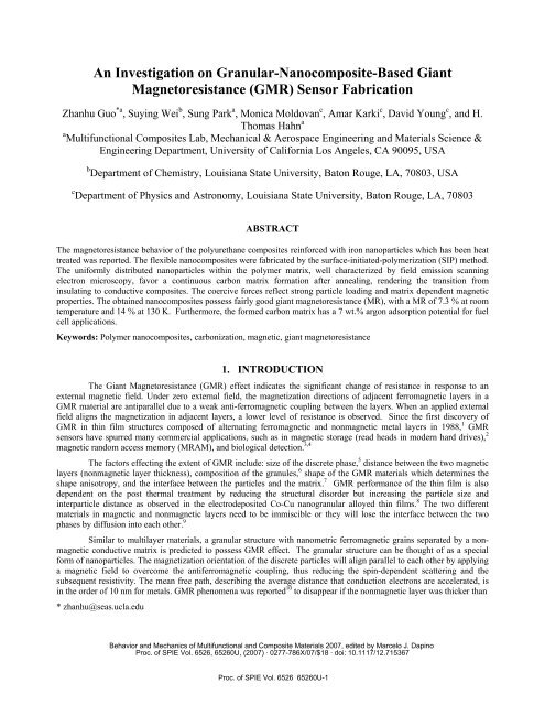
An Investigation on Granular-Nanocomposite-Based GiantMagnetoresistance (<strong>GMR</strong>) <strong>Sensor</strong> <strong>Fabrication</strong>Zhanhu Guo *a , Suying Wei b , Sung Park a , Monica Moldovan c , Amar Karki c , David Young c , and H.Thomas Hahn aa Multifunctional Composites Lab, Mechanical & Aerospace Engineering and Materials Science &Engineering Department, <strong>University</strong> of California Los Angeles, CA 90095, USAb Department of Chemistry, Louisiana State <strong>University</strong>, Baton Rouge, LA, 70803, USAc Department of Physics and Astronomy, Louisiana State <strong>University</strong>, Baton Rouge, LA, 70803ABSTRACTThe magnetoresistance behavior of the polyurethane composites reinforced with iron nanoparticles which has been heattreated was reported. The flexible nanocomposites were fabricated by the surface-initiated-polymerization (SIP) method.The uniformly distributed nanoparticles within the polymer matrix, well characterized by field emission scanningelectron microscopy, favor a continuous carbon matrix formation after annealing, rendering the transition frominsulating to conductive composites. The coercive forces reflect strong particle loading and matrix dependent magneticproperties. The obtained nanocomposites possess fairly good giant magnetoresistance (MR), with a MR of 7.3 % at roomtemperature and 14 % at 130 K. Furthermore, the formed carbon matrix has a 7 wt.% argon adsorption potential for fuelcell applications.Keywords: Polymer nanocomposites, carbonization, magnetic, giant magnetoresistance1. INTRODUCTIONThe Giant Magnetoresistance (<strong>GMR</strong>) effect indicates the significant change of resistance in response to anexternal magnetic field. Under zero external field, the magnetization directions of adjacent ferromagnetic layers in a<strong>GMR</strong> material are antiparallel due to a weak anti-ferromagnetic coupling between the layers. When an applied externalfield aligns the magnetization in adjacent layers, a lower level of resistance is observed. Since the first discovery of<strong>GMR</strong> in thin film structures composed of alternating ferromagnetic and nonmagnetic metal layers in 1988, 1 <strong>GMR</strong>sensors have spurred many commercial applications, such as in magnetic storage (read heads in modern hard drives), 2magnetic random access memory (MRAM), and biological detection. 3,4The factors effecting the extent of <strong>GMR</strong> include: size of the discrete phase, 5 distance between the two magneticlayers (nonmagnetic layer thickness), composition of the granules, 6 shape of the <strong>GMR</strong> materials which determines theshape anisotropy, and the interface between the particles and the matrix. 7 <strong>GMR</strong> performance of the thin film is alsodependent on the post thermal treatment by reducing the structural disorder but increasing the particle size andinterparticle distance as observed in the electrodeposited Co-Cu nanogranular alloyed thin films. 8 The two differentmaterials in magnetic and nonmagnetic layers need to be immiscible or they will lose the interface between the twophases by diffusion into each other. 9Similar to multilayer materials, a granular structure with nanometric ferromagnetic grains separated by a nonmagneticconductive matrix is predicted to possess <strong>GMR</strong> effect. The granular structure can be thought of as a specialform of nanoparticles. The magnetization orientation of the discrete particles will align parallel to each other by applyinga magnetic field to overcome the antiferromagnetic coupling, thus reducing the spin-dependent scattering and thesubsequent resistivity. The mean free path, describing the average distance that conduction electrons are accelerated, isin the order of 10 nm for metals. <strong>GMR</strong> phenomena was reported 10 to disappear if the nonmagnetic layer was thicker than* zhanhu@seas.ucla.eduBehavior and Mechanics of Multifunctional and Composite Materials 2007, edited by Marcelo J. DapinoProc. of SPIE Vol. 6526, 65260U, (2007) · 0277-786X/07/$18 · doi: 10.1117/12.715367Proc. of SPIE Vol. 6526 65260U-1
the electron mean-free path, which was consistent with the prediction of the RKKY theory (an abbreviation derived fromthe names: Ruderman, Kittel, Kasuya and Yosida). 11 Granular materials thus have the potential to be used for <strong>GMR</strong>sensing, similar to the multilayer counterparts normally used. However, the only reported granular <strong>GMR</strong> sensor is incopper-containing cobalt granules with a low <strong>GMR</strong> ratio. The poorer performance is attributed to a large granule sizeand a large interparticle distance resulting from annealing process during fabrication. Compared with the traditionalmetal matrix composites, polymer nanocomposites containing magnetic nanoparticles are more attractive because oftheir high flexibility, easy processing, and low cost. High-pressure prepared pellets of polystyrene composites filledwith magnetic nanoparticles after annealing are reported to have a room-temperature MR of 14 % through the tunnelingeffect.In this paper, <strong>GMR</strong> performance in the granular structured nanocomposites arising from the annealingpolyurethane nanocomposite filled with high-loading iron nanoparticles was observed to have about 7 % at roomtemperature and around 14 % at 130 K. Utilization of the high flexibility of the precursor nanocomposites, <strong>GMR</strong>materials for sensor measurement can be done immediately after the annealing process without any external treatment.The annealing temperature effect on the magnetic and magnetoresistance was also reported2. EXPERIMENTALThe processing starts with the fabrication of a polyurethane matrix composite reinforced with iron nanoparticles(NPs). The nanoparticles were reported to have an iron core/iron oxide shell structure and an approximate diameter of 20nm (provided by QuantumSphere Inc.). The surface-initiated-polymerization (SIP) method 12-14 allows a high loading, upto 65 wt%, of iron nanoparticles to be incorporated into the polyurethane matrix. In the SIP method, both the catalyst (aliquid containing aliphatic amine, parachlorobenzotrifluoride and methyl propyl ketone) and the accelerator(polyurethane STD-102, containing organo-titanate) are added into an iron-nanoparticle suspended tetrahydrofuransolution. The two-part monomers (diisocyanate and diol, CAAPCOAT FP-002-55X, CAAP Co., Inc.) are introducedinto the above solution to polymerize for 6 hours, and then poured into a mold for curing and solvent evaporation. Chart1 shows the chemical structures of the two part urethane monomers. All the operations are carried out withultrasonication.OCNRNdiisocyanateOCHOR OHChart 1. Chemical formulas of the two-part polyurethane monomers usedNanocomposites with two different particle loadings of 35 and 65 wt% were fabricated by the SIP method,respectively. They were heat treated at 250 o C for 2 hours in hydrogen gas balanced with ultra high purity argon (5%).In order to carbonize the matrix, the nanocomposite with 65 wt% particle loading was further heat treated at 450 oC for2 h in the same environment. Particle structures were characterized on a JEOL transmission electron microscope (TEM,JEOL TEM-2010) with an accelerating voltage of 200 KeV. TEM samples were prepared by dispersing the nanoparticlesinto ethanol and dropping some solution onto a holey carbon coated copper grid. The valence state in the Fenanoparticles was determined by X-ray photo-electron spectroscopy (XPS). Weight percentage of nanoparticles in thenanocomposites was determined by the thermogravimetric analysis (TGA, PerkinElmer) with an argon flow rate of 50cm 3 /min. The polyurethane, particle loading and heat treatment effect on the magnetic properties were investigated in a9-Tesla Physical Properties Measurement System (PPMS) by Quantum Design. The electric conductivity and magneticfield dependent resistance were carried out using a standard four-probe method.diol3. RESULTS AND DISCUSSIONFigure 1 shows the TEM bright field microstructures of the as-received nanoparticles. The obvious contrastwithin the particle in Figure 1(a) is due to the oxidation of the Fe nanoparticle surface. XPS studies show that the ironProc. of SPIE Vol. 6526 65260U-2
oxide is Fe 2 O 3 rather than other oxides (FeO and Fe 3 O 4 ) and the percentage of the iron and iron oxide is shown in table1. Therefore, elemental Fe in as-received Fe nanoparticles accounts for 57.05% while the oxidized Fe accounts for42.95%. The lattice distance of 0.204 nm (ring 1), 0.143 nm (ring 3), 0.116 nm (ring 4), 0.100 nm (ring 5) and 0.083 nm(ring 6) of the selected area electron diffraction (SAED) in the inset of Figure 1(a) can be assigned to (110), (200), (211),(220) and (222) planes of Fe (Standard XRD file: PDF#06-0696); and 0.167 nm (ring 2) arises from the (430) plane ofFe 2 O 3 (Standard XRD file: PDF#39-1346). The clear lattice fringes shown in Figure 1(b) indicate a high crystallinity ofthe NPs. The discontinuous fringes indicate the existence of a small number of defects within the NPs. The calculatedfringe spacing of 0.350 nm corresponds to the standard (211) plane of Fe 2 O 3 with a reported d-spacing of 0.3411 nm(ref: PDF#39-1346), indicating partial oxidation of the Fe NPs, consistent with SAED analysis.Table 1. XPS summary of the as-received nanoparticlespeak Position FWHM Raw height RSF Atomic Atomic Mass conc.BE (eV) (eV) (cps)mass conc. % %Fe 2p 708.077 1.978 4175.0 2.957 55.846 40.30 40.30Fe 2p 709.981 2.806 2756.0 2.957 55.846 26.63 26.63Fe 2p 721.314 1.476 1721.0 2.957 55.846 16.75 16.75Fe 2p 723.202 2.555 1676.1 2.957 55.846 16.32 16.321'VEFig. 1. (a) Bright field TEM micrographs and (b) HRTEM micrographs of as-received nanoparticles. The inset showsselected area electron diffraction (SAED) of the nanoparticles.Figure 2 shows the TEM bright field microstructures of the nanocomposite (65 wt%) after heat treatment at 250o C for 2 h and 450 o C for additional 2 h. There is almost no microstructural change in the first heat treatment stage (250o C), and the nanocomposite has little mass loss. However, a large shrinkage is observed in the second stage (450 o C),indicating decomposition of the polymer. The inner ring of the SAED patterns with a d-spacing of 0.34 nm in the inset ofFigure 2(a) clearly indicates the formation of graphite carbon. Clear lattice fringes shown in Figure 2(b) of highresolutionTEM indicate the formation of highly crystalline nanoparticles. The calculated lattice distance of 0.21 nmcorresponds to Fe naoparticles, and the surrounding lattice fringe spacing of 0.34 nm corresponds to the (002) plane ofProc. of SPIE Vol. 6526 65260U-3
graphite carbon. This indicates that Fe nanoparticles are embedded in a carbon matrix. No oxides observed remaining inthe NPs indicate that the high-temperature heat treatment favors the reduction of iron oxides.Fig. 2. (a) TEM and (b) HRTEM micrographs of the nanocomposite with a particle loading of 65 wt% after heattreatment at 450 o C. The inset shows corresponding SAED.The particle distribution within the polyurethane matrix before the heat treatment was characterized by ascanning electron microscope (SEM). The samples were prepared by embedding the flexible composite in a cured vinylestertab and polishing with 4000 grit sand paper. The inset of Figure 3 shows typical SEM images of the cross-sectionalarea of the nanocomposite with a particle loading of 65 wt%. The uniform particle distribution and no obvious particleagglomeration indicate that the SIP method yields a high-quality nanocomposite, as compared with a direct mixingmethod which results in a brittle nanocomposite.Figure 3 shows the room-temperature hysteresis loops of the as-received NPs and the nanocomposites. Thesaturation magnetization (M s , 97.6 emu/g, based on the total mass) of the as-received NPs is lower than that of the purebulk Fe (222 emu/g) 15 , which is as expected because of the presence of oxide shells. The lower coercive force(coercivity, H c ; 5 Oe) indicates superparamagnetic behavior of the as-received NPs. Little difference in M s is observedfor the NPs after they are embedded in the polymer matrix. The saturation magnetizations of the nanocomposites, 54emu/g and 31.6 emu/g for the particle loadings of 65 wt.% and 35 wt.%, respectively, correspond to 84 emu/g and 90.2emu/g for the nanoparticles. The slightly lower M s in the nanocomposites than in the as-received NPs may be attributedto the further oxidation of the NPs during the nanocomposite fabrication process and the particle-polymer surfaceinteraction effect. 16 The coercivities of the polyurethane nanocomposites are 685 and 900 Oe for 65 and 35 wt% loading,respectively, which are much larger than that of the as-received NP assembly. Such behavior, however, is typical ofmagnetic nanocomposites as explained later.The heat treatment at 250 o C does not show any significant changes in mass, volume, M s , or H c , indicating goodthermal stability of the nanocomposite. However, the heat treatment at 450 o C brings about many changes. First of all,it carbonizes the matrix and reduces the oxide shells. The mass loss and shrinkage in the matrix effectively increases theparticle loading for the composite. All these changes effectively increases M s while reducing H c .Proc. of SPIE Vol. 6526 65260U-4
In the 65 wt% nanocomposite, H c remains practically the same after heat treatment at 250 o C but decreases to165 Oe after the additional heat treatment at 450 o C. This trend is due to the interparticle dipolar interaction within thenanocomposite with a good dispersion of single-domain nanoparticles, consistent with particle-loading-dependentcoercivity in nanoparticle assembly. 17 Compared with the 35 wt% nanocomposite, the smaller coercivity in the 65 wt%nanocomposite arises from the decreased interparticle distance concomitant with a stronger dipolar interaction. Thefurther decrease in H c after the 450 o C heat treatment is for the same reason, i.e., the decreased interparticle distanceresulting from shrinkage. In addition, the presence of an oxide shell around the metallic core is reported to increase theblocking temperature of NPs through the exchange coupling interaction between the ferromagnetic metal core and theantiferromagnetic oxide shell. 18 Thus, the loss of the exchange coupling in the heat-treated nanocomposites due to thedisappearance of antiferromagnetic oxide shell also contributes to the smaller coercivity.Iagnetizaation (emu/total mass).Ic•) 1DI\. '\-100 I • I • • I •-20000-15000-10000 -5000 0 5000 10000 15000 20000Magnetic field (Oe)Fig. 3 Hysteresis loops of (a) as-received NPs; nanocomposites with a particle loading of (b) 35 wt% and (c) 65 wt%; andnanocomposite with 65 wt% particle loading with heat treatment at (d) 250 o C for 2 h and (e) 450 o C for additional 2 h.The inset shows the SEM image of nanocomposite with a 65 wt% particle loading.No electrical conductivity is detected in the polyurethane nanocomposites, even at 65 wt% loading, indicatingthe particle loading is still lower than the percolation threshold. The conductivity improves considerably after the heattreatment. Fig. 4 shows the temperature dependent resistance of the 65 wt% nanocomposite after heat treatment at 450o C. The resistance increases dramatically with decreasing temperature, characteristic of a non-metallic behavior.Equipment limitations precluded us from measuring the resistance at temperatures below 80 K. Contrary to the aspreparednanocomposites, those heat treated at 250 o C show somewhat improved electric conductivity. However, theresistance is still about ten times higher than that of the 450 o C heat treated specimen. Also, the lowest possiblemeasurement temperature decreases from 125 K to 80 K as the heating temperature is increased from 250 o C to 450 o C.In view of the high conductivity of iron, the high resistance observed in the 450 o C heat treated specimen is due to thepoor conductivity of the carbon matrix. The observed linear relationship between the logarithmic resistance and thesquare root of temperature T -1/2 shown in Fig. 4 indicates an interparticle tunneling/hopping conduction mechanism. 19Proc. of SPIE Vol. 6526 65260U-5
T (K)80 100 120 140 160 180 200 220 240 260 28010000Fig. 4. Resistance as a function of temperature after the nanocomposite with a 65 wt% particle loading after heat treatmentat 450 o C.0-1-2-3-4-5-8-9-10-11-12-13-1 £1Fig. 5. MR as a function of the applied field at room temperature and 130 K for the nanocomposite with a 65 wt% particleloadingProc. of SPIE Vol. 6526 65260U-6
Fig. 5 shows the MR as a function of the applied magnetic field H, where MR (%) is defined in the followingEquation (1):R(H ) − R(0)MR (%)= × 100(1)R(0)R(H) and R(0) is the resistance at zero and any applied field of H, respectively. The 250 o C heat treated nanocompositehas a MR of 7 % at 130 K whereas the 450 o C heat treated nanocomposite shows a MR of 7.3 % at room temperatureand 14 % at 130 K, all at a fairly high field of 90 kOe. Compared with multilayered <strong>GMR</strong> materials, a high magneticfield is required to saturate the MR, which is characteristic of the tunneling conduction mechanism. However, a 2 % MRobserved at 4 kOe still indicates that this <strong>GMR</strong> sensor could be used for biological targeting application. 20 An initialadsorption test shows that the heat-treated nanocomposite has a fairly high porosity adsorbing about 7 wt% of argon;indicating that this composite can be used for water distillation, 21 as well as for tail gas catalysis or hydrogen storage 22for fuel cell applications.4. CONCLUSIONIn conclusion, we have shown that a granular <strong>GMR</strong> nanocomposite can be synthesized using the surfaceinitiatedpolymerization (SIP) method to accommodate a high particle loading required and the subsequent heattreatment to induce carbonization of the matrix and reduce oxide shells in the nanoparticles. The final iron/carbonnanocomposites exhibit a room-temperature <strong>GMR</strong> of 7 % at 90 kOe, indicating a spin-dependent tunneling/hoppingconduction.5. ACKNOWLEDGEMENTThis work was partially supported by the Air Force Office of Scientific Research through AFOSR GrantFA9550-05-1-0138 with Dr. B. Les Lee as the Program Manager. DPY kindly acknowledges support from the NationalScience Foundation under Grant No. DMR 04-49022. Partial financial support and the generous donation of thenanoparticles from QuantumSphere Inc. are kindly acknowledged.REFERENCES1 M. N. Baibich, J. M. Broto, A. Fert, F. Nguyen Van Dau, F. Petroff, P. Eitenne, G. Creuzet, A. Friederich, andJ. Chazelas, Phys. Rev. Lett. 61, 2472-5 (1988).2 A. Moser, K. Takano, D. T. Margulies, M. Albrecht, Y. Sonobe, Y. Ikeda, S. H. Sun, and E. E. Fullerton, J.Phys. D-Appl. Phys. 35, R157-R167 (2002).3 R. L. Edelstein, C. R. Tamanaha, P. E. Sheehan, M. M. Miller, D. R. Baselt, L. J. Whitman, and R. J. Colton,Biosensors & Bioelectronics 14, 805-813 (2000).4 M. M. Miller, P. E. Sheehan, R. L. Edelstein, C. R. Tamanaha, L. Zhong, S. Bounnak, L. J. Whitman, and R. J.Colton, J. Magn. Magn. Mater. 225, 138-144 (2001).5 M. Y. Zhuravlev, H. O. Lutz, and A. V. Vedyayev, Physical Review B, 6317, 174409 (2001).6 A. E. Berkowitz, J. R. Mitchell, M. J. Carey, A. P. Young, D. Rao, A. Starr, S. Zhang, F. E. Spada, F. T. Parker,A. Hutten, and G. Thomas, J. Appl. Phys.,73, 5320-5325 (1993).7 T. Koide, H. Miyauchi, J. Okamoto, T. Shidara, A. Fujimori, H. Fukutani, K. Amemiya, H. Takeshita, S.Yuasa, T. Katayama, and Y. Suzuki, Phys. Rev. Lett. 87, 257201 (2001).8 G. R. Pattanaik, D. K. Pandya, and S. C. Kashyap, Thin Solid Films, 433, 247-251 (2003).9 A. Garcia Prieto, M. L. Fdez-Gubieda, C. Meneghini, A. Garcia-Arribas, and S. Mobilio, Phys. Rev. B: Conde.Matter Mater. Phys. 67, 224415 (2003).10 R. H. Yu, X. X. Zhang, J. Tejada, J. Zhu, and M. Knobel, J. Appl. Phys., 79, 1979-90 (1996).11 S. Tumanski, Thin film magnetoresistive sensors, Institute of Physics Pub, Bristol ; Philadelphia, (2001).12 R. C. Advincula, J.f Dispersion Sci. Technol., 24, 343-361 (2003).13 G. K. Jennings and E. L. Brantley, Adv. Mater., 16, 1983-1993 (2004).14 Z. Guo, S. Park, and H. T. Hahn, Nanocomposite fabrication through particle surface initiated polymerization,The 2006 AICHE Annual Meeting, San Francisco (2006).Proc. of SPIE Vol. 6526 65260U-7
15 B. D. Cullity, Introduction to Magnetic Materials; Addison-Wiley, New York (1972).16 D. Zhang, K. J. Klabunde, C. M. Sorensen, and G. C. Hadjipanayis, Phys. Rev. B: Conden. Matter Mater.Phys., 58, 14167-14170 (1998).17 D. Kechrakos and K. N. Trohidou, Phys. Rev. B, 58, 12169-12177 (1998).18 V. Skumryev, S. Stoyanov, Y. Zhang, G. Hadjipanayis, D. Givord, and J. Nogues, Nature, 423, 850-853 (2003).19 P. Sheng, Phys. Rev. Lett., 31, 44-47 (1973).20 D. L. Graham, H. A. Ferreira, and P. P. Freitas, Trends Biotechnology, 22, 455-462 (2004).21 P. K. Ghosh and L. Philip, J. Environ. Sci. Health, part B, 40, 425-441 (2005).22 J. W. Lee, H. S. Kim, J. Y. Lee, and J. K. Kang, Appl. Phys. Lett., 88, 143126 (2006).Proc. of SPIE Vol. 6526 65260U-8