CHALLENGES IN X-RAY OPTICS FOR MODERN X-RAY SOURCES
CHALLENGES IN X-RAY OPTICS FOR MODERN X-RAY SOURCES
CHALLENGES IN X-RAY OPTICS FOR MODERN X-RAY SOURCES

Create successful ePaper yourself
Turn your PDF publications into a flip-book with our unique Google optimized e-Paper software.
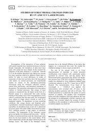
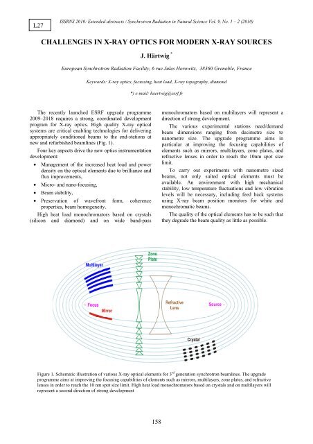
L27ISSRNS 2010: Extended abstracts / Synchrotron Radiation in Natural Science Vol. 9, No. 1 – 2 (2010)<strong>CHALLENGES</strong> <strong>IN</strong> X-<strong>RAY</strong> <strong>OPTICS</strong> <strong>FOR</strong> <strong>MODERN</strong> X-<strong>RAY</strong> <strong>SOURCES</strong>J. Härtwig *European Synchrotron Radiation Facility, 6 rue Jules Horowitz, 38360 Grenoble, FranceKeywords: X-ray optics, focussing, heat load, X-ray topography, diamond*) e-mail: haertwig@esrf.frThe recently launched ESRF upgrade programme2009–2018 requires a strong, coordinated developmentprogram for X-ray optics. High quality X-ray opticalsystems are critical enabling technologies for deliveringappropriately conditioned beams to the end-stations atnew and refurbished beamlines (Fig. 1).Four key aspects drive the new optics instrumentationdevelopment:• Management of the increased heat load and powerdensity on the optical elements due to brilliance andflux improvements,• Micro- and nano-focusing,• Beam stability,• Preservation of wavefront form, coherenceproperties, beam homogeneity.High heat load monochromators based on crystals(silicon and diamond) and on wide band-passmonochromators based on multilayers will represent adirection of strong development.The various experimental stations need/demandbeam dimensions ranging from decimetre size tonanometre size. The upgrade programme aims inparticular at improving the focusing capabilities ofelements such as mirrors, multilayers, zone plates, andrefractive lenses in order to reach the 10nm spot sizelimit.To carry out experiments with nanometre sizedbeams, not only suited optical elements must beavailable. An environment with high mechanicalstability, low temperature fluctuations and low vibrationlevels will be necessary, including feed back systemsusing X-ray beam position monitors for white andmonochromatic beams.The quality of the optical elements has to be such thatthey degrade the beam quality as little as possible.Figure 1. Schematic illustration of various X-ray optical elements for 3 rd generation synchrotron beamlines. The upgradeprogramme aims at improving the focusing capabilities of elements such as mirrors, multilayers, zone plates, and refractivelenses in order to reach the 10 nm spot size limit. High heat load monochromators based on crystals and on multilayers willrepresent a second direction of strong development158
ISSRNS 2010: Extended abstracts / Synchrotron Radiation in Natural Science Vol. 9, No. 1 – 2 (2010)bFigure 2. Comparison of an X-ray topograph of an old,long time ago installed beam splitter out of nitrogenrich type Ib material (a) with an example of a newhigh quality plate (b).The lateral (horizontal) dimension of the crystal in (a)is about 5 mm and that in (b) about 11 mm.Synthetic single-crystalline HPHT-diamond is inprinciple the best-suited material for Bragg diffracting X-ray optical elements like beamsplitters ormonochromators to be used in modern, powerful X-raysources. Already in the early days of the 3 rd generationsources the utilisation of diamonds was discussed, testedand X-ray optical elements were realised. However, aareal break through in their application was not reached.The main reasons were, (and partly still are) bycomparison with silicon, the low quality (bulk andsurface), the limited availability and the smalldimensions of the available material.Since then a substantial effort was undertaken (andneeds to be continued) in the fields of crystal growth,crystal processing and crystal characterisation to developa high-quality diamond material is needed that combinesthe extremely high perfection of the crystal bulk with anexcellent surface finish [1]. With regard to the quality ofthe crystal bulk, selected samples became available thatlocally may be dislocation free, with a very low level oflocal residual strain. However, at the moment the surfacequality appears to be the most critical point to bothachieve and study.High quality type IIa (very low nitrogen impurityconcentration) HPHT Diamond plates were investigatedwith white beam X-ray topography and in particular withhigh strain sensitive non-dispersive double crystaltopography [2]. The results showed and confirmed theexcellent local quality of present (selected) plates. Thismeans that they contained large (for diamond)dislocation free regions and an extremely low residualstrain level in the range below several 10 -8 . This is a localstrain level like in FZ silicon.References[1] R.C. Burns, A.I. Chumakov, S.H. Connell, D. Dube, H.P.Godfried, J.O. Hansen, J. Härtwig, J. Hoszowska, F.Masiello, L. Mkhonza, M. Rebak, A. Rommevaux, R.Setshedi, P. Van Vaerenbergh, "HPHT – growth and X-raycharacterisation of high-quality type IIa diamond”, J. Phys.:Condens. Matt. 21 (2009) 364224–1–14.[2] R.C. Burns, A. Chumakov, G. Carbone, S.H. Connell, D.Dube, H.P. Godfried, J.O. Hansen, J. Härtwig, F. Masiello,M. Rebak, A. Rommeveaux, R. Setshedi, P. VanVaerenbergh, A. Gibaud, "Diamonds for X-ray opticalapplications at 3 rd and 4 th generation X-ray sources", Proc.of SPIE Vol. 6705 (2007) 67050K1–6.159