Neurospectroscopy— A Pictorial Essay and Review ... - For Members
Neurospectroscopy— A Pictorial Essay and Review ... - For Members
Neurospectroscopy— A Pictorial Essay and Review ... - For Members
You also want an ePaper? Increase the reach of your titles
YUMPU automatically turns print PDFs into web optimized ePapers that Google loves.
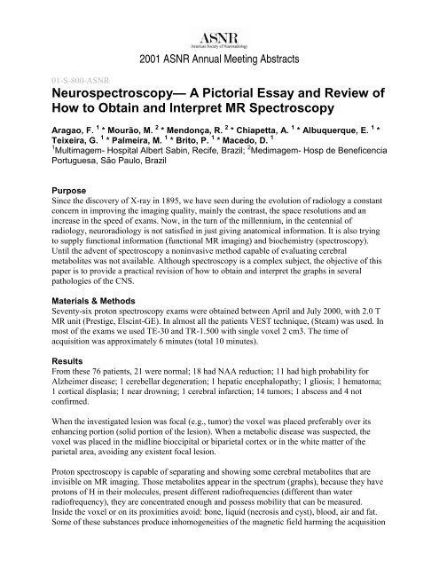
2001 ASNR Annual Meeting Abstracts01-S-800-ASNR<strong>Neurospectroscopy—</strong> A <strong>Pictorial</strong> <strong>Essay</strong> <strong>and</strong> <strong>Review</strong> ofHow to Obtain <strong>and</strong> Interpret MR SpectroscopyAragao, F. 1 * Mourão, M. 2 * Mendonça, R. 2 * Chiapetta, A. 1 * Albuquerque, E. 1 *Teixeira, G. 1 * Palmeira, M. 1 * Brito, P. 1 * Macedo, D. 11 Multimagem- Hospital Albert Sabin, Recife, Brazil; 2 Medimagem- Hosp de BeneficenciaPortuguesa, São Paulo, BrazilPurposeSince the discovery of X-ray in 1895, we have seen during the evolution of radiology a constantconcern in improving the imaging quality, mainly the contrast, the space resolutions <strong>and</strong> anincrease in the speed of exams. Now, in the turn of the millennium, in the centennial ofradiology, neuroradiology is not satisfied in just giving anatomical information. It is also tryingto supply functional information (functional MR imaging) <strong>and</strong> biochemistry (spectroscopy).Until the advent of spectroscopy a noninvasive method capable of evaluating cerebralmetabolites was not available. Although spectroscopy is a complex subject, the objective of thispaper is to provide a practical revision of how to obtain <strong>and</strong> interpret the graphs in severalpathologies of the CNS.Materials & MethodsSeventy-six proton spectroscopy exams were obtained between April <strong>and</strong> July 2000, with 2.0 TMR unit (Prestige, Elscint-GE). In almost all the patients VEST technique, (Steam) was used. Inmost of the exams we used TE-30 <strong>and</strong> TR-1.500 with single voxel 2 cm3. The time ofacquisition was approximately 6 minutes (total 10 minutes).ResultsFrom these 76 patients, 21 were normal; 18 had NAA reduction; 11 had high probability forAlzheimer disease; 1 cerebellar degeneration; 1 hepatic encephalopathy; 1 gliosis; 1 hematoma;1 cortical displasia; 1 near drowning; 1 cerebral infarction; 14 tumors; 1 abscess <strong>and</strong> 4 notconfirmed.When the investigated lesion was focal (e.g., tumor) the voxel was placed preferably over itsenhancing portion (solid portion of the lesion). When a metabolic disease was suspected, thevoxel was placed in the midline bioccipital or biparietal cortex or in the white matter of theparietal area, avoiding any existent focal lesion.Proton spectroscopy is capable of separating <strong>and</strong> showing some cerebral metabolites that areinvisible on MR imaging. Those metabolites appear in the spectrum (graphs), because they haveprotons of H in their molecules, present different radiofrequencies (different than waterradiofrequency), they are concentrated enough <strong>and</strong> possess mobility that can be measured.Inside the voxel or on its proximities avoid: bone, liquid (necrosis <strong>and</strong> cyst), blood, air <strong>and</strong> fat.Some of these substances produce inhomogeneities of the magnetic field harming the acquisition
of the spectroscopy.The secret of spectroscopy is to always use the same technique (Steam or Press) <strong>and</strong> parameters,because any change in them, will correspond to alterations in the relationship between the peaks,being more difficult to know what is normal or pathologic. Another important thing is to knowthe normal pattern of the cerebral place examined for the technique used <strong>and</strong> for the patient’sage. In a patient more than 2 years old we consider the adult normal pattern in the gray matter aswell as in the white matter.In the analysis of the spectroscopy we consider that NAA would be a neuronal marker. Cholineis used as a marker for cell wall synthesis <strong>and</strong> cell viability. Creatine is a marker of energymetabolism. Myoinositol is an astrocitic marker.ConclusionIn several pathologies of CNS, spectroscopy can offer valuable additional information, wheninterpreted together with MR imaging, CT <strong>and</strong> clinical data. It has been used mainly in theevaluation of CNS neoplasias <strong>and</strong> its differentiation with radionecrosis <strong>and</strong> other types of focallesions. It also has been used quite frequently in the investigation of the dementias due to its highsensibility, specificity <strong>and</strong> predictive value in differentiating Alzheimer disease from otherdementias <strong>and</strong> from the normal.References1. Brian Ross. Magnetic resonance spectroscopy diagnosis of neurological diseases. MarcelDekker, Inc. 19992. Castillo M, Kwock L, Scatliff J, Mukherji SK. Proton MR spectroscopy in neoplastic <strong>and</strong>nonneoplastic brain. MR Imag Clin N Am. Saunders, Feb/19983. Hirsch JA, Lenkinsk RE, Grossman RI. MR spectroscopy in the evaluation of enhancinglesion in the brain in mmultiple sclerosis. AJNR Am J Neuroradiol Nov 19964. Castillo M, Kwock L, Kwock L. Proton MR spectroscopy of common brain tumors.Neuroimag Clin N Am. Saunders Nov 19985. Kwock L. Localized MR spectroscopy: Basic principles. Neuroim Clin N Am. Saunders Nov1998


