Wound Healing & Limb Salvage - New Jersey Center for Biomaterials
Wound Healing & Limb Salvage - New Jersey Center for Biomaterials
Wound Healing & Limb Salvage - New Jersey Center for Biomaterials

Create successful ePaper yourself
Turn your PDF publications into a flip-book with our unique Google optimized e-Paper software.
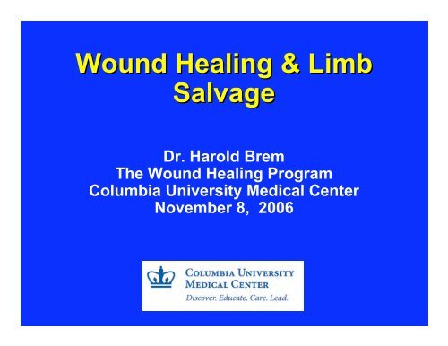
<strong>Wound</strong> <strong>Healing</strong> & <strong>Limb</strong><strong>Salvage</strong>Dr. Harold BremThe <strong>Wound</strong> <strong>Healing</strong> ProgramColumbia University Medical <strong>Center</strong>November 8, 2006
February 12, 2006The <strong>Wound</strong>ed; <strong>Healing</strong>, With <strong>New</strong> <strong>Limb</strong>s and Fragile DreamsBy JULIET MACUR; Correction AppendedIt was a victory <strong>for</strong> Lance Cpl. Matthew Schilling to walk into the upper gallery of the House of Representatives onJan. 31 <strong>for</strong> the State of the Union address. He wore his dress blues and a prosthetic leg. Five months earlier, hehad been carried on a stretcher, wounded and bleeding, into a hospital in Iraq after a roadside bomb exploded 10feet from him…. The blast tore through his right foot and calf……Corporal Schilling's Marine Corps unit and a victimof the same blast, Lance Cpl. Mark Beyers, wheeled up to him at the Walter Reed physical therapy clinic. CorporalBeyers's right arm and leg were amputated in Iraq . Since Aug. 26, when they were wounded, the two marines eachhave endured some 20 operations in three countries. Charting their care over the ensuing months, beginning justhours after the blast, has revealed a journey of medical setbacks and emotional turmoil. Among the more than ….About an hour later, Corporals Schilling and Beyers were in surgery at the nearby Al Asad Military Base. It was thefirst of 13 operations they would endure in eight days, during stays at five hospitals in three countries. A doctoramputated Corporal Schilling's right leg below the knee. Corporal Beyers, with severe lung injuries, was in worseshape. Doctors amputated his right lower leg and his entire right arm, including the shoulder; shrapnel haddestroyed his shoulder joint and just missed slicing his carotid artery, doctors said. For the next week he would bein an induced coma …After 19 operations and 63 days in hospitals, Corporal Beyers went home in October <strong>for</strong> avisit..
…16,653 Americans wounded in Iraq are 387 amputees,including 62 who, like Corporal Beyers, have lost more than onelimb, said Lt. Col. Paul Pasquina, chief of physical medicine andrehabilitation at Walter Reed. The amputations, traumaticthough they are, are often accompanied by painfulcomplications. "It's not as easy as putting on even the mosthigh-tech prosthetic and just walking off," Colonel Pasquinasaid….
What is the amputation rate in battleinjuries?WarRawpercentage# amputees% multilimbamputationCivil War12~50,000UnknownWW I1.7261027WW II1.214,9127KoreanWar1.414778Vietnam3.4528320Iraq2.338116Potter BK, Scoville CR. Amputation Is Not Isolated: An Overview of the US Army Amputee Patient Care Program and Associated Amputee Injuries.J Am Acad Orthop Surg. 2006 Oct;14(10):S188-90
441 limbs lost (337 lower extremity) in waron terrorism alone!WHAT CAN BE DONE?Potter BK, Scoville CR. Amputation Is Not Isolated: An Overview of the US Army Amputee Patient Care Program and Associated Amputee Injuries.J Am Acad Orthop Surg. 2006 Oct;14(10):S188-90
<strong>Wound</strong> <strong>Healing</strong> Programat Columbia UniversityDedicated Inpatient Unit: <strong>New</strong> YorkPresbyterian HospitalOutpatient <strong>Wound</strong> <strong>Center</strong>: Case ManagementClinical Research, Mailman School of PublicHealth: OutcomesResearch Laboratory: College of Physicians& Surgeons
<strong>Healing</strong> traumatic wounds : Could thishave been avoided?
Can a leg ulcer heal with humancell therapy ?Brem H, Gill K, Tarnovskaya A, Ehrlich HP, Carasa M, Weinberger S, Baskin-BeyE, Tomic-Canic M, Vladeck B.. Surgery Technology International. 2003; XI: 161-167
YES!Ulcer healed at 7 weeks.Brem H, Gill K, Tarnovskaya A, Ehrlich HP, Carasa M, Weinberger S, Baskin-Bey E, Tomic-Canic M, Vladeck B. <strong>Healing</strong> ofElderly Patients with Diabetic Foot Ulcers, Venous Stasis Ulcers, and Pressure Ulcers. Surgery TechnologyInternational. 2003; XI: 161-167
Leg Ulcer ProtocolVenous ulcers are commonly on the ankle, but include any area below the knee that has + reflux (e.g. the foot)PrimaryCareMDCommunication Medical RecordInitial Visit<strong>Wound</strong>CareProviderLaboratory TestsCBC with Manual DiffPlateletsHemoglobin A1CESRPT/PTTLFT’s12.0Albumin10.0Prealbumin8.0Na + Cl -6.0BUNK + CO 2CreatenineGlucose 4.02.00.0Weekly DigitalPhotography,Objective Measurement*Drainage is often a sign4/24/84/144/204/265/25/8PathologyEvaluate <strong>for</strong>:(1) Tumor(2) Infection(3) Necrosis(4) Scarof infection, in which casecompression is not advisable.Topical TherapyBrem H, Kirsner RS, Falanga V. Am J Surg 2004; 188:1-8FungalNails(1) Evaluate(2) TreatDebridementPhysical ExamCellulitusOrDrainage*Or>2 months durationDeep CultureAntibioticsNo cellulitusMinimal/decreased drainage*4-Layer Compression
What is Cell Therapy?cell therapy inPetri DishSalineIrrigationLiftedwithForcepsFibroblastKeratinocyteLayer
What Quality Control is There toAssure the Cells Are Living?Note thekeratinocytesNote thefibroblastsHematoxylin and eosin (H&E) stained is done as part ofquality assurance be<strong>for</strong>e shipping <strong>for</strong> use in a patient.Brem H, Young J, Tomic-Canic M, Isaacs C, Ehrlich, H.P. Clinical Efficacy and Mechanism of Bilayered Living Human SkinEquivalent in the Treatment of Diabetic Foot Ulcers. Surgery Technology International. 2003; XI: 23-31.
Use of Tissue Engineered Skin Already ShowsClinical Success in Promoting <strong>Wound</strong> <strong>Healing</strong>Brem H, Balledux J, Sukkarieh T, Falanga V. <strong>Healing</strong> of Venous Ulcers of long Duration with a BilayeredLiving Skin Substitute: Results from a General Surgery and Dermatology Department. Dermatologic Surgery. 2001 Nov;27(11):915-9.
What is Clinical Presentation of the non-healing<strong>Wound</strong>?Initial presentation ofdiabetic foot ulcerAppears healthy,significant underminingCallous is indication <strong>for</strong>debridementRapid healing with earlytreatment after CelltherapyBrem H, Sheehan P, Boulton AJM: Protocol <strong>for</strong> the treatment of diabetic foot ulcers. American Journal of Surgery.2004;187:1-10.
Figure 1ACan toe ulcers be a source ofamputation?Non-neuropathic“DFU”Clinical Efficacy and Mechanism of Human Skin Equivalent in theTreatment of Diabetic Foot UlcersHarold Brem , MD; Jan Young, PhD ; Cary Isaacs , MS; Marjana Tomic -Canic , PhD ; and H. Paul Ehrlich , PhDImmediate treatmentwith cell therapyEarly treatmentprevents amputationBrem H, Young J, Tomic-Canic M, Isaacs C, Ehrlich, H.P. Clinical Efficacy and Mechanism of Bilayered Living Human SkinEquivalent in the Treatment of Diabetic Foot Ulcers. Surgery Technology International. 2003; XI: 23-31.
What is the mechanism of celltherapy? Clue comes from woundswith large deficitscelltherapy5 – 0 AbsorbableSutureFour Days After celltherapyHealed at 49 DaysBrem H, Balledux J, Bloom T, Kerstein M, Hollier L: <strong>Healing</strong> of diabetic ulcers and pressure ulcers w+ith human skinequivalent: a new paradigm in wound healing. Archives of Surgery 2000;135:627-634.
Mechanism of Cell Therapy:Keratinocytes and Fibroblasts release atleast 17 growth factorsBrem H, Young J, Tomic-Canic M, Isaacs C, Ehrlich, H.P. Clinical Efficacy and Mechanism of Bilayered Living Human SkinEquivalent in the Treatment of Diabetic Foot Ulcers. Surgery Technology International. 2003; XI: 23-31.
Importance of protocol <strong>for</strong> FootUlcer x 17 years?: Looks “good”butPhysiologicallyimpairedSewing celltherapy to <strong>New</strong><strong>Wound</strong> EdgeSutured 1-mm inner<strong>Wound</strong> Edge soKeratinocytes arestimulatedApplication ofRedundant celltherapy<strong>New</strong> EpithelialEdgeHealed at 20Weeks
Is Amputation Necessary?TendonTendonBrem H, Tomic-Canic M, Tarnovskaya A, Gill K, Ehrlich HP, Carasa M, Weinberger S, Baskin-Bey E,Entero. <strong>Healing</strong> of Elderly Patients with Diabetic Foot Ulcers, Venous Stasis Ulcers, and PressureUlcers. Surg Technol Int 2003;XI:161-167.
<strong>Healing</strong> rate of cell therapy incomplicated leg ulcers?Age (years) <strong>Center</strong> A (n=32) <strong>Center</strong> B (n=18)<strong>Wound</strong> Size (cm 2 )<strong>Wound</strong> Duration (years)Median (years)Number of ApplicatoinsTime to <strong>Healing</strong> (days)Mean 39 ± 68Median 12Mean 8.2 ± 9.4Median 52 ± 1Median 2Median 61 ± 12Median 5565 ± 2197.6 ± 7.14.21.7 ± 0.5255 ± 1431H. Brem, MD, Balledux J, Falanga V, et alDermatologic Surgery 2001, p. 915-921
Can Telemedicine Accelerate <strong>Wound</strong>Closure: WEMR
What is the importance of the <strong>Wound</strong>Electronic Medical Record (WEMR) <strong>for</strong>accurate data?
How do you interpret data? Percentage of <strong>Healing</strong>PatientsProtocol Only. Not Compliance Driven.703 <strong>Wound</strong>s, 431 Patient VisitsQ/A Non <strong>Healing</strong> <strong>Wound</strong>sVisit DateNumberof<strong>Wound</strong>sPUtotalPU IPU IIPU IIIPU IVDFVSUArtSurgOtherNon-<strong>Healing</strong><strong>Wound</strong>Percentage ofHEALING07_01_051553111071321742522696.10%07_08_0513924131197831834894.20%07_15_0513423141171269163111092.50%07_22_0512812012958020471191.40%07_29_05147280432112762146894.60%JULYTOTALS70311832214795738210016304393.90%
What are the clinical signs of awound infection?8/6/06 8/9/06
How much does a non healing woundcost? The James Peters VA experience250,000200,000PublishedAverage CostsCost ($)150,000100,00050,0000Hospital AcquiredPressure UlcersCommunity AcquiredPressure UlcersActual chargesdirectly relatedto HospitalStage IVpressure ulcersBrem H, Vladeck B, Nierman D, et al. <strong>Wound</strong> Repair and Regeneration 2001
Can large ulcers heal?1098765432101 0 / 1 2 / 01 0 / 2 8 / 0 1 1 / 1 3 / 01 1 / 2 9 / 01 2 / 1 5 / 0 1LengthWidthDepthArea1. Zimny S, Schatz H, Pfohl M. Exp Clin Endocrinol Diabetes 2004; 112:191-42. Kantor J, Margolis DJ. Arch Dermatol 1998; 134:1571-4
Demographics:145 Consecutive Hospitalized Patients Institutional Review Boardconsent to measure rate ofhealing and take photographs Age: 62.52 year Albumin 2.46 ± 0.58 g/dl Prealbumin 14.56 ± 6.92 mg/dlWallenstein S, Brem H. Am J Surg 2004; 188:73-8
<strong>Healing</strong> of Pressure UlcersOpen <strong>Wound</strong> Area100%90%80%70%60%50%40%30%20%10%0%ALL PATIENTS All Patients (n=145)= 130)0 1 2 3 4 5 6 7 8Time in Weeks145 consecutive pressure ulcers treatedWallenstein S, Brem H. Am J Surg 2004; 188:73-8
What are theexpected healing rates? Diabetic foot ulcers - 100%100% of diabetic footulcers will heal in absence of ischemia andosteomyelitis Venous ulcers - 100%100% healing of venous ulcersless than a year duration; 78% healing of venousulcers greater than a year duration Pressure Ulcer - Preventstages 2 and 3Prevent stage IV; 100% healing
Future: Can we make blood vesselsgrow in wounds? VEGFFigure 2
Adenovirus Delivery System
Mechanical properties of healing wounds aremeasured by tensile strength
Human cell bankThrough partnership with Coriell Cell Bank. All cellcultures are grown and frozen in antibiotic-free mediato aid in the detection and prevention of contamination.Cultures are tested and found free of mycoplasma,bacteria, and fungi during expansion, at the time offrozen storage, and after recovery of stock <strong>for</strong>distribution from liquid nitrogen.
How can we grow skin ?1. Molecular tools allow <strong>for</strong> purification and isolation ofEpidermal Stem Cells from which one can recreate fullydifferentiated epidermisIRSBulgeORSDermalpapillaStem Cell+ Niche TransitAmplifyingCellsDifferentiatingCellsEpidermisMorasso MI and Tomic-Canic, M. Biol Cell. 200597(3):173-83.
Opportunities3. Epidermal stem cells (ESC) can be geneticallyengineered to sustain persistent expression of a transgenein tissue engineered skinCultured ESC transduced with b-galgene sustain its expression inxenograft <strong>for</strong> more than a yearGhazizadeh S and Taichman LB : EMBO J (2001) 20, 1215–1222;1222;J Invest Dermatol. (2005) 124(2):367-72
Cell RepositoryProtocol
Human Skin Equivalent (HSE) Reconstructed invitro from Normal Human Primary Keratinocytesand Fibroblasts Heals in a Similar Manner toHuman Skin24hrsDay248hrsDay472hrsDay6HSENormal HumanSkin
What is the Molecular Basis of <strong>Healing</strong>?
c-myc IS INDUCED IN NON HEALING WOUNDSUntreated<strong>Wound</strong> <strong>Healing</strong>Inhibitor GCCHRONIC ULCERStojadinovic O, Brem H, Vouthounis C, Lee B, Fallon J, Stallcup M, Merchant A, Galiano RD, Tomic-Canic M. Molecularpathogenesis of chronic wounds: the role of beta-catenin and c-myc in the inhibition of epithelialization and woundhealing.Am J Pathol. 2005 Jul;167(1):59-69.
Identification of the Chronic <strong>Wound</strong> TranscriptomeLocationPatient1Patient2Patient3Gene ExpressionProfiles201645_at 219454_at 1.62 7.37 2.01 7.23 1.59 1.81 2.29 2.15 2.25 1.09 1.39 6.79 1.72 6.67 1.36 1.66 1.95 1.98 1.93 1.00 AdhesionCa binding201561_s_at 61734_at -2.34 2.45 -2.26 1.92 -2.06 1.12 -1.61 3.32 -2.12 1.19 -1.72 2.38 -1.66 1.87 -1.51 1.09 -1.18 3.22 -1.56 1.15 AdhesionCa binding201681_s_at 200755_s_at -1.90 3.04 -1.53 1.43 -1.36 1.34 -1.39 3.20 -1.54 1.93 -1.96 2.65 -1.59 1.25 -1.41 1.17 -1.44 2.79 -1.60 1.68 AdhesionCa binding219197_s_at -4.75 -4.34 -8.64 -2.90 -9.01 -3.74 -3.42 -6.81 -2.29 -7.10 Ca binding213369_at -9.14 -2.99 -4.15 -4.67 -5.35 -5.59 -1.83 -2.54 -2.85 -3.27 Adhesion, cadherin202870_s_at 4.25 5.28 8.28 4.01 7.78 3.80 4.72 7.41 3.58 6.96 Cell cycle208153_s_at 204170_s_at -2.46 2.40 -2.11 1.86 -2.50 2.69 -2.00 1.94 -2.51 2.57 -2.00 3.24 -1.72 2.51 -2.03 3.62 -1.62 2.62 -2.04 3.47 Adhesion, Cell cyclecadherin208407_s_at 204026_s_at -1.27 2.43 -1.81 2.11 -1.72 3.28 -1.55 2.00 -1.38 2.94 -1.27 2.39 -1.82 2.07 -1.73 3.23 -1.55 1.96 -1.38 2.90 Adhesion, Cell cyclecadherin204029_at 202388_at -2.18 1.76 -1.95 5.18 -3.09 2.07 -1.40 1.49 -1.59 3.22 -1.80 1.43 -1.61 4.19 -2.55 1.68 -1.16 1.21 -1.32 2.61 Adhesion, Cell cyclecadherin204750_s_at 201853_s_at 12.77 1.14 3.99 1.60 8.96 1.49 5.41 1.82 13.93 1.67 5.38 1.27 1.68 1.78 3.78 1.66 2.28 2.03 5.87 1.87 Adhesion, Cell cycledesmosomal207324_s_at 201371_s_at -61.89 -1.95 -2.80 -2.04 -6.70 -2.20 -1.33 -2.06 -3.31 -2.27 -52.50 -1.77 -2.37 -1.85 -5.69 -2.00 -1.13 -1.87 -2.81 -2.07 Adhesion, Cell cycledesmosomal211382_s_at -4.35 -2.17 -3.35 -2.08 -2.63 -3.31 -1.65 -2.55 -1.58 -2.00 Cell cycle211075_s_at 1.73 1.21 1.96 1.58 2.09 1.57 1.10 1.77 1.44 1.90 Adhesion, integrin201482_at 1.37 1.77 1.75 1.45 2.34 1.33 1.72 1.71 1.41 2.28 Cell cycle inhibitor204455_at -1.78 -1.87 -2.29 -1.37 -1.14 -2.13 -2.22 -2.73 -1.63 -1.36 Adhesion, integrin218346_s_at -3.58 -2.16 -4.65 -2.36 -3.88 -2.59 -1.57 -3.37 -1.71 -2.81 Cell cycle inhibitor210869_s_at 200920_s_at -2.13 3.45 -1.51 1.84 -1.79 2.22 -1.09 2.89 -1.49 2.41 -2.18 2.87 -1.55 1.53 -1.84 1.84 2.40 -1.12 2.00 -1.53 Adhesion, Cell cycle junctional inhibitor203757_s_at 201236_s_at 46.88 -1.77 10.21 -1.13 16.03 -2.54 -1.59 1.38 21.42 -1.79 41.65 -1.57 9.08 1.00 14.24 -2.25 1.23 -1.41 19.04 -1.58 Adhesion, Cell cycle junctional inhibitor201615_x_at 202192_s_at -1.89 2.84 -1.30 1.31 -2.30 1.99 -1.52 2.42 -2.17 1.67 -1.89 2.67 -1.29 1.23 -2.30 1.86 2.27 -1.51 1.57 -2.17 Adhesion, Cell cycle junctional inhibitor205490_x_at 201289_at -2.03 6.77 -2.09 3.52 -1.83 4.17 10.89 -2.30 -1.46 5.94 -1.79 3.71 -1.85 1.93 -1.61 2.28 -2.03 5.97 -1.28 3.26 Adhesion, Cell growth, junctionalproliferation201540_at -2.43 -1.90 -2.81 -1.60 -3.99 -1.55 -1.22 -1.80 -1.02 -2.56 Cell growth, proliferation201470_at 1.91 2.24 3.12 1.55 2.06 1.67 1.96 2.73 1.36 1.80 Antioxidant217733_s_at 2.48 1.95 2.58 1.75 2.30 2.24 1.76 2.33 1.58 2.07 Cytoskeletal202967_at -3.13 -1.93 -2.48 -1.92 -2.27 -3.21 -1.98 -2.55 -1.97 -2.33 Antioxidant205547_s_at 4.79 2.24 1.16 7.06 3.27 5.07 2.37 1.23 7.48 3.46 Cytoskeletal201427_s_at -1.66 -1.61 -3.85 -1.62 -3.20 -1.55 -1.50 -3.59 -1.51 -2.98 Antioxidant202565_s_at -1.22 -1.92 -1.84 -1.71 -1.84 -1.26 -1.98 -1.90 -1.76 -1.90 Cytoskeletal204168_at 204083_s_at -2.57 5.03 -1.94 3.68 -2.14 2.79 -1.81 5.78 -2.22 2.70 -1.85 5.16 -1.39 3.77 -1.53 2.86 -1.30 5.93 -1.59 2.77 AntioxidantCytoskeletal, actin209276_s_at 201954_at -2.60 2.19 -1.53 2.78 -3.26 3.10 -1.09 1.84 -3.43 2.38 -4.62 1.60 -2.72 2.03 -5.80 2.26 -1.94 1.34 -6.10 1.74 AntioxidantCytoskeletal, actin201432_at 211160_x_at -2.41 2.27 -1.99 -2.83 1.98 -1.32 1.74 -1.71 2.89 -2.05 1.86 -1.69 1.64 -2.41 1.63 -1.12 1.43 -1.45 2.38 AntioxidantCytoskeletal, actin202831_at 60528_at -3.46 1.21 -1.25 -1.46 -2.31 -1.80 -2.31 -1.55 -2.94 -1.75 -1.11 -3.66 -1.69 -1.54 -3.11 -1.90 -3.11 -1.64 -3.97 -1.85 AntioxidantCytoskeletal, actin209046_s_at -1.47 -1.57 -1.81 -1.22 -1.51 -1.65 -1.76 -2.02 -1.37 -1.70 Cytoskeletal, actin206662_at -2.32 -1.11 -2.24 -1.34 -2.85 -3.08 -1.48 -2.98 -1.78 -3.79 Antioxidant38340_at -2.34 -1.26 -2.10 -1.80 -1.73 -2.00 -1.08 -1.80 -1.54 -1.48 Cytoskeletal, actin211922_s_at -1.93 -2.25 -3.20 1.06 -1.54 -2.54 -2.95 -4.20 -1.24 -2.02 Antioxidant211776_s_at 5.89 2.13 4.06 1.74 3.04 4.11 1.49 2.84 1.21 2.12 Cytoskeletal, actin, membrane218856_at 200974_at 2.94 3.12 2.23 1.59 2.30 1.30 1.96 2.86 2.23 1.93 3.14 3.49 2.38 1.78 2.45 1.45 2.09 3.19 2.38 2.15 ApoptosisCytoskeletal, actin, motility209230_s_at 200801_x_at -4.09 1.87 -1.94 1.52 -2.97 1.47 -1.52 1.39 -2.56 1.77 -3.19 1.85 -1.51 1.50 -2.32 1.46 -1.19 1.37 -1.99 1.75 ApoptosisCytoskeletal, actin, motility
Location ABench to Bedside: From Microarray Analyses toPatient BiopsiesLocation BControlLocation ALocation BLocation BLocation AGene Expression Profiles
0hrs.Cells from Location A do not Respond to WH Stimuliwith VEGFControlLocation ALocation BLocation BLocation A4hrs.8hrs.24hrs.% <strong>Wound</strong> (scratch) area120100806040200VEGFControl Location A Location B0hrs 4hrs 8hrs 24hrs
Amphibian <strong>Limb</strong>Regeneration<strong>Wound</strong> <strong>Healing</strong> (Days 1-2)De-differentiation (Days 3-12)Blastema <strong>for</strong>mation (Days 13-21)Re-differentiation& PatternFormation (Days22-40)
<strong>Limb</strong> RegenerationAmphibiansHumans?
Human <strong>Limb</strong>Regeneration1. <strong>Healing</strong> Veterans’ wounds with current therapy2. Education3. Telemedicine4. <strong>New</strong> Delivery systems :cells, antibiotics5. Focus on developing tissue engineeredcomponents comparable to human tissue: Skin,blood vessels, neurons, cartilage, bone,muscles.