capsule experience - MG Lorenzatto
capsule experience - MG Lorenzatto
capsule experience - MG Lorenzatto

You also want an ePaper? Increase the reach of your titles
YUMPU automatically turns print PDFs into web optimized ePapers that Google loves.
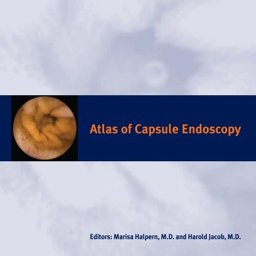
Atlas of Capsule Endoscopy<br />
Editors: Marisa Halpern, M.D. and Harold Jacob, M.D.
Atlas of Capsule Endoscopy<br />
Wireless <strong>capsule</strong> endoscopy allows painless endoscopic<br />
imaging of the entire small bowel. In addition the<br />
examination of the intestine takes place in the physiological<br />
state. Artifact induction does not occur as in push<br />
enteroscopy as there is no need to push the device. These<br />
differences between wireless endoscopy and push<br />
endoscopy are the major factors governing the physiological<br />
and bio-optical principles of image acquisition, resulting<br />
in clear and detailed visualization of intestinal structures.
Atlas of Capsule Endoscopy<br />
Marisa Halpern, M.D.<br />
First Edition<br />
Senior Pathologist<br />
Director of Gastrointestinal Pathology Unit<br />
Rabin Medical Center, Golda Campus<br />
Petach Tiqva, Israel<br />
Harold Jacob, M.D. Director of Medical Affairs<br />
Given Imaging Ltd.<br />
Yoqneam, Israel
Preface<br />
We are proud to present this first Atlas of Capsule Endoscopy. With the rapid adoption<br />
of Capsule Endoscopy by practicing gastroenterologists, a new spectrum of<br />
pathological images are being generated. New dimensions of the common diseases<br />
we treat are coming to light. In response to the intense interest and growth in the<br />
area of Capsule Endoscopy, we have produced the first Atlas of Capsule Endoscopy.<br />
By placing these images in your hand, we know that it will enhance<br />
your ability to diagnose and treat patients with gastrointestinal disorders.<br />
In this book we present the spectrum of disease that has been seen to<br />
date by Capsule Endoscopy. Where possible, we have correlated the<br />
M2A ® Capsule images with other diagnostic modalities including endoscopy, radiology<br />
and histopathology findings. This first edition of the Atlas of Capsule<br />
Endoscopy also contains information on normal <strong>capsule</strong> endoscopic anatomy,<br />
how to perform Capsule Endoscopy and principles of physiological image<br />
acquisition. Some consideration is also given to Capsule Endoscopy findings in other<br />
parts of the GI tract as well.<br />
The companion CD to the Atlas will enable you to load the M2A ® Capsule images<br />
onto your computer for easy reference in your practice.<br />
With the increased use and expanding indications of Capsule Endoscopy,<br />
we know that the Atlas of Capsule Endoscopy will continue to develop and grow.<br />
We hope this Atlas will be a useful tool for physicians who care for patients<br />
with gastrointestinal disease.<br />
Harold Jacob, M.D.<br />
Marisa Halpern, M.D.
Acknowledgments<br />
We would like to thank all the worldwide contributors to the "Atlas of<br />
Capsule Endoscopy" for their devoted efforts (see Contributors list).<br />
We would like to thank Sharon Besser for her immense devotion to this<br />
publication. Without her dedication, this project would not be completed.<br />
Dr Halpern would like to acknowledge her colleagues at the Department of<br />
Pathology and especially Professor Rivka Gal who understood the meaning<br />
of this project.<br />
Finally, we would like to thank our families for their understanding,<br />
moral support and patience during this period of long hours and hard work.
Given Imaging Inc.<br />
Oakbrook Technology Center<br />
5555 Oakbrook Parkway #355<br />
Norcross, GA, 30093, USA<br />
Managing Editor and Production...............Sharon S. Besser<br />
This first edition of the Atlas of Capsule Endoscopy does not integrate Capsule Endoscopy Standard Terminology.<br />
For comments or suggestions, please contact terminology@givenimaging.com<br />
Copyright © 2002 Given Imaging, Ltd. All rights reserved.<br />
Given,M2A, RAPID and/or other products and/or services referenced herein are either registered trademarks,<br />
trademarks or service marks of Given Imaging, Ltd. All other names are or may be registered trademarks or<br />
trademarks of their respective owners.<br />
This publication and its content are for your personal and non-commercial use. You may not modify, copy,<br />
distribute, transmit, display, perform, reproduce, publish, license, create derivative works from, transfer or sell<br />
any part of this publication and/or its content, without prior written permission from Given Imaging, Ltd.<br />
Given Imaging Ltd. Is not and will not be responsible or liable for any damage or loss caused or alleged to be<br />
caused due to inaccuracies or typographical errors in this publication, or for any action taken in reliance thereon.<br />
Contents are subject to change without notification.<br />
Please send all inquiries to sbesser@givenimaging.com<br />
Graphic Design......... Studio Rosinger Ltd., Haifa, Israel<br />
Printing ................... Rahash Offset Printing Ltd., Haifa, Israel<br />
Expanding the scope of GI
Table of contents<br />
Development of the Swallowable Video Capsule (G. Meron, Ph.D.) . . . . . . . . . . . . . . . . . . . . . . . . . . . . . . . . . . . . 3<br />
Notes from the Inventor (G. J. Iddan, D.Sc.) . . . . . . . . . . . . . . . . . . . . . . . . . . . . . . . . . . . . . . . . . . . . . . . . . . . . . . 7<br />
Chapter 1 - Physiological Endoscopy (A. Glukhovsky, D.Sc., H. Jacob, M.D., P. Halpern, B.Sc.) . . . . . . . . . . . . . . . . . . . . . . . . 9<br />
Chapter 2 - Performance of the Capsule Endoscopy (B. Lewis, M.D., C. J. Gostout, M.D.) . . . . . . . . . . . . . . . . . . . . . . . . . 15<br />
Chapter 3 - Normal M2A ® Anatomy (P. Swain, M.D., M. Appleyard, M.D.) . . . . . . . . . . . . . . . . . . . . . . . . . . . . . . . . . . . . . . . . . . 23<br />
Chapter 4 - Inflammatory Diseases of the Small Intestine (A. L. Buchman, M.D.) . . . . . . . . . . . . . . . . . . . . . . . . . . . . . . . . . . . . 33<br />
Chapter 5 - Neoplastic Diseases (F.P. Rossini, M.D., M. Pennazio, M.D.) . . . . . . . . . . . . . . . . . . . . . . . . . . . . . . . . . . . . . . . . . . 47<br />
Chapter 6 - Iatrogenic Diseases (D. Cave, M.D.) . . . . . . . . . . . . . . . . . . . . . . . . . . . . . . . . . . . . . . . . . . . . . . . . . . . . . . . . . . . . . . . . 63<br />
Chapter 7 - Vascular Abnormalities (M. Hahne, M.D., J. F. Riemann, M.D.) . . . . . . . . . . . . . . . . . . . . . . . . . . . . . . . . . . . . . . . . 73<br />
Chapter 8 - Malabsorption (G. Gay, M.D., I. Fassler, M.D., Ch. Florent, M.D., M. Delvaux, M.D.) . . . . . . . . . . . . . . . . . . . . . . . 83<br />
Chapter 9 - Pediatrics (E. Seidman, M.D., G. L. de Angelis, M.D., Ana Maria Sant Anna, M.D.) . . . . . . . . . . . . . . . . . . . . . . . . . 103<br />
Chapter 10 - Transplantation (R. de Franchis, M.D., E. Rondonotti, M.D., C. Abbiati, M.D., G. Beccari, M.D., E. Villa, M.D., A. Merighi, M.D., A. Pinna, M.D.) . . 111<br />
Chapter 11 - Non Small Bowel Pathology (S. Adler, M.D., S. Kadish, M.D.) . . . . . . . . . . . . . . . . . . . . . . . . . . . . . . . . . . . . . . . 119<br />
Contributors List . . . . . . . . . . . . . . . . . . . . . . . . . . . . . . . . . . . . . . . . . . . . . . . . . . . . . . . . . . . . . . . . . . . . . . . . . . . . . . . . . . . . . . . . . 125
Development of the Swallowable Video Capsule<br />
Gavriel Meron, Ph.D.<br />
It was in 1981 while on a sabbatical leave from his regular<br />
position as Senior Engineer at the Electro-Optical Design Section<br />
of Rafael, the R&D group of the Israeli Ministry of Defense, that<br />
Dr. Gavriel Iddan, a talented mechanical engineer, started his<br />
long term interest in medical imaging. The idea started as a<br />
seed of an invention in the midst of defense projects, particularly<br />
the development of electro-optical imaging devices for missiles.<br />
During the sabbatical, Dr. Iddan relocated to Boston to perform<br />
research for another Israeli company, Elscint Inc., doing x-ray<br />
and ultrasound medical imaging R&D. In Boston, Dr. Gavriel<br />
Iddan befriended Professor Eitan Scapa, an Israeli<br />
gastroenterologist, who was, at that time, at the local medical<br />
center. They had many mutual interests and discussions, which<br />
made Dr. Iddan wonder how he could "help" gastroenterologists.<br />
It was then that he dreamed up the idea of a miniature "missile"<br />
that could be easily swallowed and passed through the GI tract,<br />
transmitting images along the way. Dr. Iddan was specifically<br />
interested in "opening up a new frontier" by enabling direct<br />
imaging of the small intestine, which was until then, terra<br />
incognito, due to the inability of the other imaging methods to<br />
reach the small intestine.<br />
Dr. Iddan and a team of engineers and technicians in Rafael<br />
worked on the development of an initial prototype in the<br />
laboratories, and performed some feasibility trials of imaging<br />
and transmission through animal tissue developing some of the<br />
basic technologies. Based on the initial research performed in<br />
the Rafael laboratories, the first of a number of patents were<br />
written up and submitted by Rafael in January 1994.<br />
At the same time and totally unbeknownst to Dr. Iddan, in the<br />
United States, Dr. Paul Swain presented the possibility of wireless<br />
endoscopy during the Los Angeles World Congress of<br />
Gastroenterology, in an invited talk entitled Microwaves in<br />
Gastroenterology in September 1994.<br />
In London, Dr. Swain had made some progress in making several<br />
(rather large) prototype wireless endoscopy devices from<br />
commercially available components in 1995 and 1996. It was<br />
in 1996 that his team achieved the first live transmissions from<br />
the stomach of a pig.<br />
The first two abstracts published by Swain's team on this topic<br />
were:<br />
Wireless transmission of a color television moving image from<br />
the stomach using a miniature CCD camera, light source and<br />
microwave transmitter. Swain CP, Gong F, Mills TN. Gut<br />
1996;39:A26<br />
3
4<br />
Development of the Swallowable Video Capsule<br />
The Evolution of the Capsule. (from top to bottom). Components of the prototype <strong>capsule</strong> in a container. In the<br />
center, the prototype <strong>capsule</strong> is shown. Finally, the existing M2A Capsule.
Wireless transmission of a color television moving image from<br />
the stomach using a miniature CCD camera, light source<br />
and microwave transmitter. Swain CP, Gong F, Mills TN.<br />
Gastrointest Endosc 1997;45:AB40.<br />
Meanwhile, back in Israel, Dr. Iddan knew that if there was<br />
to be a future for the <strong>capsule</strong> for small intestine imaging, it<br />
would have to be championed by a commercial organization.<br />
He began to arrange meetings with different organizations<br />
in the hope that they would take the challenge and invest<br />
in the business.<br />
It was one of these meetings that brought Dr. Iddan to me.<br />
We had first met in 1995 when I was CEO of Applitec Ltd., an<br />
Israeli company that had developed and was selling video<br />
cameras for endoscopy.<br />
In 1997, the patent in the US was approved, and the available<br />
technologies needed for the <strong>capsule</strong>'s development had<br />
moved in the right direction. It was at this time that I<br />
approached the Rafael Development Corporation (RDC), who<br />
has the right of first refusal to commercialize technologies<br />
coming out of Rafael, in order to found together a start-up<br />
that would develop the <strong>capsule</strong> and bring it to market.<br />
I left my position at Applitec and set out to raise funds and<br />
develop a business model and strategy for the new company,<br />
which was named Given (GastroIntestinal Video Endoscopy)<br />
Imaging Ltd., and established in January 1998.<br />
At that time, I defined the fledgling company's mission as "to<br />
develop, produce, and achieve worldwide leadership in the<br />
marketing and sales of swallowable disposable electronic<br />
<strong>capsule</strong>s, for diagnostics and therapy of the gastrointestinal<br />
(GI) tract". This was clearly a much wider mandate than the<br />
Development of the Swallowable Video Capsule<br />
initial small intestine <strong>capsule</strong>, and was based on the<br />
development of a technological platform that would then be<br />
further developed by listening to gastroenterologists,<br />
understanding the barriers in small intestine imaging, and<br />
implementing solutions to overcome them.<br />
By the end of 1998, the initial team, that included Dr. Gavriel<br />
Iddan, Dr. Paul Swain, and Dr. Arkady Glukhovsky, was in<br />
place and serious research & development went underway<br />
to transform the idea into reality. Successfully overcoming<br />
the enormous obstacles of size, transmission strength, battery<br />
power and image resolution, among many others, working<br />
prototypes were produced in January 1999. In May 2000, at<br />
the DDW 2000 meeting, Dr. Swain, together with Given<br />
Imaging, presented the results of the animal trials performed<br />
with the prototype system that was developed. [Wireless<br />
Capsule Endoscopy, Nature, Vol. 405, 25 May 2000].<br />
During 2001, Given achieved major milestones with the<br />
completion of successful clinical trials, receipt of FDA<br />
clearance, CE Mark certification, and launch of the Given ®<br />
Diagnostic Imaging System worldwide. The initial clinical<br />
results have been excellent, and the feedback from patients<br />
and physicians has been remarkable.<br />
The idea of publishing this Atlas of Capsule Endoscopy came<br />
in recognition of the need that was expressed by many<br />
physicians to see with their own eyes specific pathologies<br />
and findings, and compare the images between the different<br />
available modalities. We hope the content of this Atlas assists<br />
in educating gastroenterologists and furthers the<br />
understanding and acceptance of the M2A ® as a standard<br />
tool of GI diagnostics in the clinical path.<br />
5
Notes From the Inventor<br />
Gavriel J. Iddan, D.Sc.<br />
Approximately 20 years ago, I took a sabbatical from my<br />
position as an electro-optical systems engineer at Rafael, the<br />
Armaments Development division of the Israeli Ministry<br />
of Defense, and went to work for a medical instrument<br />
manufacturer in Boston, Mass. Coincidentally, I discovered<br />
that my next-door neighbor was a gastroenterologist, Prof.<br />
Eitan Scapa, who was also on sabbatical and working at a<br />
local hospital. After getting to know each other, we would<br />
spend time discussing our respective professions. Among<br />
other things, I learned about the field of endoscopy and was<br />
exposed to some of its challenges.<br />
After returning to my work at Rafael, I became deeply<br />
interested in medical devices and did some design work in<br />
this area. During my next sabbatical, and after further<br />
discussions with Professor Scapa, I decided to focus on the<br />
specific problems of imaging of the small intestine. After<br />
consulting with a number of gastroenterologists, I finally<br />
decided to attempt the development of a wireless camera.<br />
The major obstacles that I encountered and needed to<br />
overcome included: attaining an adequate field of view,<br />
achieving power requirements for the CCD, and the challenge<br />
of performing a diagnostic study that required close contact<br />
with the patient for many hours. A major breakthrough came<br />
in 1993 when I realized that the system could be separated<br />
into 3 major units: <strong>capsule</strong>/transmitter, receiver/recorder<br />
and a workstation. The separation of these components would<br />
allow the patient to be ambulatory without the need to be<br />
connected to a monitor.<br />
During this period, the scientific literature started to report<br />
a breakthrough in video imaging. This was the introduction<br />
of the CMOS imager which consumed only a fraction of the<br />
energy required by a CCD, had all the required circuits on the<br />
same chip, and was not expensive to manufacture. To complete<br />
the idea, a special self-cleaning optical configuration was<br />
added and detailed design and experiments commenced.<br />
(One of the first experiments was done on a frozen chicken<br />
purchased in the supermarket.)<br />
By the second half of 1993, I began writing a patent<br />
application, which was submitted in January 1994. While<br />
searching urgently for financing to create this device, I<br />
approached Dr Gavriel Meron, who at that time was the<br />
manager of a company specializing in small endoscopic<br />
cameras. Taking my invention and developing a viable<br />
business plan, Dr. Gavriel Meron established Given Imaging<br />
Ltd in 1998, thus launching the new field of <strong>capsule</strong> endoscopy<br />
and making the dream a reality.<br />
7
Chapter 1<br />
Physiological Endoscopy<br />
Arkady Glukhovsky, D.Sc.<br />
Harold Jacob, M.D.<br />
Pablo Halpern, B.Sc.<br />
INTRODUCTION<br />
Wireless <strong>capsule</strong> endoscopy allows painless endoscopic<br />
imaging of the entire small bowel, but more significantly, the<br />
examination of the intestine takes place in the physiological<br />
state. Since there is no need to push the device, artifact<br />
induction does not occur as in push enteroscopy. In addition,<br />
<strong>capsule</strong> endoscopy is wireless, obviating the need for air<br />
insufflation and the rate of the propulsion is determined by<br />
peristalsis. These differences between wireless endoscopy<br />
and push endoscopy are the major factors governing the<br />
physiological and bio-optical principles of image acquisition,<br />
resulting in clear and detailed visualization of intestinal<br />
structures.<br />
CAPSULE EXPERIENCE<br />
PHYSIOLOGICAL ENDOSCOPY<br />
The M2A ® wireless <strong>capsule</strong> endoscope is shown in Figure 1A.<br />
A schematic cross-section appears in Figure 1B. The wireless<br />
<strong>capsule</strong> endoscope has a cylindrical shape, with a diameter<br />
of 11mm, and a length of 26mm. It has two convex domes,<br />
one of them being the optical dome (1). The intestine is<br />
illuminated through the optical dome by white Light Emitting<br />
Diodes (LEDs) (3).<br />
The acquired image is focused by a short focal aspherical lens<br />
(2), on the Complementary Metal-Oxide Silicone (CMOS)<br />
imager (4). The optical dome has a shape that prevents light<br />
reflected by the dome to reach the imager, thus enhancing<br />
the image quality. The <strong>capsule</strong> is powered by two silver-oxide<br />
batteries (5). An Application Specific Integrated Circuit (ASIC)<br />
transmitter (6) is located in the rear dome. The Radio Frequency<br />
(RF) signal is transmitted by the antenna (7).<br />
Figure 1. Wireless Capsule Endoscope<br />
A - External view B - Schematic cross-section<br />
11mm<br />
1 2 4<br />
Legend:<br />
1 - Optical dome<br />
2 - Short focal aspheric lens<br />
3 - White LEDs<br />
4 - CMOS imager<br />
5 - Watch batteries<br />
6 - ASIC transmitter<br />
7 - Antenna<br />
There are two major factors that are thought to govern image<br />
acquisition by of the M2A ® <strong>capsule</strong>. These factors are: the<br />
proprietary optical dome and the fact that image acquisition<br />
occurs in the physiological state.<br />
3<br />
3<br />
5 5 6 7<br />
26mm<br />
9
10<br />
Chapter 1<br />
THE OPTICAL DOME<br />
The optical dome and its relationship to the other optical<br />
components create two important advantages in <strong>capsule</strong><br />
endoscopy:<br />
a. Improved illumination efficiency.<br />
b. Favorable imaging geometry within the field of view.<br />
Another minor factor which may be contributing to image<br />
quality is the presence of a fluid interface between the optical<br />
dome and tissue at the time of examination improving the<br />
resolution of different anatomical structures such as villi.<br />
Finally, another advantage of the optical dome is that it lends<br />
itself to cleaning by the GI tract mucosa.<br />
Geometric and optical differences between image acquisition<br />
in wired and wireless endoscopy are compared in Figure 2A<br />
and Figure 2B. Figure 2A depicts the geometrical relationship<br />
for a push enteroscope inserted into the intestine. Geometrical<br />
relationship for the M2A ® <strong>capsule</strong> endoscope is depicted in<br />
Figure 2B. The standard endoscope (Figure 2A) includes an<br />
illumination source (2), and a lens (3), producing the field of<br />
illumination (4), and the field of view (5) shown in Figure 2A.<br />
Collapse of the intestinal wall (6) may obscure either the field<br />
of view or field of illumination. This technical problem is<br />
resolved in standard endoscopy by insufflating the intestine<br />
with air (7), resulting in the distancing of the intestinal muco<br />
from the tip of the endoscope, clearing both the field of view<br />
and the field of illumination. Air insufflation also enables the<br />
operator to orient the tip of the endoscope in the proper<br />
luminal direction, and to advance the endoscope tip accordingly.<br />
Figure 2B shows the wireless <strong>capsule</strong> endoscope (1) in the<br />
intestine. The <strong>capsule</strong> includes its illumination sources (2),<br />
and the lens (3), comprising a field of illumination (4), and a<br />
field of view (5) respectively. In order to prevent collapse of<br />
Physiological Endoscopy<br />
the intestinal wall (6), and resultant obscuring of either the<br />
field of illumination or the field of view, a specially designed<br />
optical dome (8) covers both the source of illumination and<br />
the lens. The space remaining between the dome and the<br />
intestinal wall may at times be occupied by fluid.<br />
Figure 2. Geometrical and optical interrelationships for<br />
the enteroscope and <strong>capsule</strong> endoscope in the<br />
intestine.<br />
A - Push enteroscope<br />
2 - Illumination source<br />
1 - Endoscope inside<br />
intestine<br />
6 - Intestinal wall<br />
B - Capsule endoscope<br />
6 - Intestinal wall<br />
3 - Lens<br />
4 - Field of illumination 5 - Field of view<br />
3 - Lens<br />
1 - Wireless <strong>capsule</strong><br />
endoscope inside<br />
intestine<br />
7 - Air filling, due to insufflation<br />
8 - Optical dome<br />
The field of view of most commercially available endoscopes<br />
is within the range of 120˚-140˚, similar to the M2A ® <strong>capsule</strong><br />
endoscope. Depth of field of the standard endoscope starts<br />
from 3-5mm from the endoscope tip and extends to a distance<br />
of 100 mm. Unlike in the push endoscope, depth of view of<br />
the <strong>capsule</strong> endoscope starts on the optical dome itself.<br />
b<br />
2 - Illumination source<br />
4 - Field of illumination<br />
5 - Field of view<br />
a<br />
7 - Liquid filling,<br />
intestinal<br />
liquids
Chapter 1<br />
The illumination efficiency in <strong>capsule</strong> endoscopy is higher<br />
due to the fact that the illumination angles are not sharp,<br />
thereby allowing the illumination to be returned back to the<br />
imager. Fig 3a shows that some of the illumination is not<br />
being returned to the imager in standard endoscopy as a<br />
result of air insufflation. Fig 3b demonstrates the effectivenes<br />
of the illumination provided by airless endoscopy.<br />
Figure 3. Efficiency of illumination in air insufflating<br />
and airless endoscopy.<br />
A - In air insufflating case the illumination is less efficient<br />
due to the fact that some of the rays are reaching the<br />
intestine at flat edges, and are not returned back to the<br />
lens and to the camera.<br />
B - In airless endoscopy, most of the illumination is<br />
returned back to the lens due to sharp edges of<br />
illumination, close to perpendicular.<br />
2<br />
PHYSIOLOGICAL ENDOSCOPY<br />
Performing endoscopy in the physiological state is a paradigm<br />
shift in the approach to endoscopic diagnosis. The elements<br />
that are present in physiological <strong>capsule</strong> endoscopy include<br />
the use of peristalsis to propel, orient and steer the M2A ®<br />
<strong>capsule</strong> in the intestine.<br />
1<br />
Physiological Endoscopy<br />
The elements that are absent in physiological <strong>capsule</strong><br />
endoscopy, thereby creating an advantageous environment<br />
include:<br />
a. Sedation and its resultant change in the<br />
physiological state.<br />
b. Air insufflation resulting in increased pressure on<br />
the intestinal wall.<br />
An additional factor that is integral to push endoscopy and<br />
does not play a role in <strong>capsule</strong> endoscopy is forceful insertion<br />
of the endoscope which impacts on the intestinal wall.<br />
Physiological changes may develop due to insufflation and<br />
increasing air pressure in the intestine:<br />
a. Under normal physiological conditions blood pressure<br />
in the blood vessels of the GI tract may be within the<br />
following range: arterioles 40-80 mmHg, capillaries<br />
20-40 mmHg, and venules 15-30 mmHg. During push<br />
enteroscopy, the intraluminal pressure within the<br />
intestine may reach values above 300 mmHg 2 ,<br />
significantly higher than the blood pressure. We may<br />
speculate that this increase in pressure may decrease<br />
the blood flow to small vessels, and in some cases<br />
even temporarily arrest the flow. Although we have<br />
not found reference of the tamponade effect in<br />
endoscopy, this phenomena in well-known in<br />
laparoscopy for more than a century. In rare cases,<br />
air insufflation may even cause a fatal air embolism<br />
during gastrointestinal endoscopy.<br />
b. Insufflation of the small intestine, and insertion<br />
of a long flexible tube (the endoscope), affects the<br />
pressure receptors embedded in the small intestinal<br />
wall. In the case of wireless endoscopy, the intestine's<br />
physiological or ‘natural’ conditions remain undisturbed.<br />
11
12<br />
Chapter 1<br />
c. Conscious sedation is usually administered during<br />
push enteroscopy. The adminisration of sedation to<br />
patients alters systemic physiology. This may have<br />
an effect on the ability of the endoscope to detect<br />
vascular or inflammatory lesions.<br />
d. Examination of the intestine under physiological<br />
conditions may enable measurement of additional<br />
physiological parameters, some of them unrelated<br />
to the image, e.g. gastric emptying, small and large<br />
bowel passage times, and peristaltic contraction<br />
cycles (rhythms).<br />
CONCLUSION<br />
Wireless <strong>capsule</strong> endoscopy acquires detailed images of the<br />
GI tract, which allows identification of the spectrum of<br />
pathologies present within the small bowel. Specific design<br />
elements of the <strong>capsule</strong>, coupled with the fact that the M2A ®<br />
device acquires images in the physiological state are the<br />
major contributing factors to this breakthrough in GI<br />
endoscopy.<br />
References and Suggested Readings<br />
1 Litynski GS. The history of laparoscopy. Frankfurt/M: Bernert Ve<br />
2 Katzgraber F, Glenewinkel F, Fischler S. Mechanism of fatal air<br />
after gastrointestinal endoscopy. Int J Legal Med 1998; 111(3):<br />
Physiological Endoscopy
Chapter 2<br />
The Performance of Capsule Endoscopy<br />
Blair S. Lewis, M.D.<br />
Christopher J. Gostout, M.D.<br />
Since the inception of gastrointestinal endoscopy, physicians<br />
have wanted to obtain direct visualization of the entire GI<br />
tract. Standard endoscopic and colonoscopic exams view<br />
only small amounts of the proximal and distal ends of the<br />
small bowel. Endoscopic examination of the entire small<br />
bowel has remained elusive. Push enteroscopy was the first<br />
step in the endoscopic evaluation of the intestine. Initially,<br />
colonoscopes, both adult and pediatric, were used to evaluate<br />
the entire duodenum and proximal jejunum. On average, a<br />
160 cm long instrument can be advanced 40 cm beyond the<br />
ligament of Treitz. Present 2.1-2.5 meter long push<br />
enteroscopes have greatly improved the depth of insertion<br />
and visualization of the small bowel and it is now possible to<br />
inspect the jejunum in its entirety. Examination of the distal<br />
small bowel has previously been achieved using Sonde and<br />
Rope-way techniques. Both exams are lengthy and quite<br />
uncomfortable, even painful. The medical community has<br />
largely abandoned these exams.<br />
An endoscopic <strong>capsule</strong> (Given<br />
Imaging Limited, Yoqneam, Israel)<br />
has been developed to obtain<br />
images from the entire small<br />
bowel. Developed by Dr. Gavriel<br />
Iddan in 1981, the <strong>capsule</strong>, which<br />
measures 11 x 26 mm, contains 4<br />
LEDs (light emitting diodes), a lens, a color camera chip, two<br />
batteries, a radio frequency transmitter and an antenna. The<br />
camera is a CMOS (complementary metal oxide sensor) chip.<br />
This chip requires less power than present CCD (charged<br />
coupled device) chips found on video endoscopes and digital<br />
cameras, and it can operate at very low levels of illumination.<br />
The <strong>capsule</strong> obtains two images per second and transmits<br />
the data via radio frequency to a recording device worn about<br />
a patient's waist. Once the acquisition time is reached, the<br />
data from the recording device is downloaded to a computer<br />
workstation whose software processes the images to be<br />
viewed on the computer screen.<br />
The <strong>capsule</strong> is disposable and does not need to be retrieved<br />
by the patient. It is passed naturally. An average of 50,000<br />
images are obtained during an eight-hour exam. Thus <strong>capsule</strong><br />
endoscopy appears to be the answer to the long-standing<br />
desire for the complete endoscopic examination of the entire<br />
small bowel and it accomplishes this goal in a non-invasive way.<br />
The <strong>capsule</strong> is indicated as an adjunctive tool for evaluation<br />
of suspected diseases of the small intestine. It is<br />
contraindicated in patients with known or suspected small<br />
bowel obstruction since the <strong>capsule</strong> may become lodged<br />
within the intestinal tract. The <strong>capsule</strong> has not been approved<br />
for use in patients with pacemakers or implanted defibrillators.<br />
15
16<br />
Chapter 2<br />
Typical timing of a <strong>capsule</strong> exam is to have a patient swallow<br />
the <strong>capsule</strong> at 8 am and disconnect them from the recorder<br />
at 4 pm. This allows 8 hours of acquisition of images during<br />
the day. Capsule endoscopy is performed after the patient<br />
follows a 12 hour fast. Patients are told to have nothing to<br />
eat or drink after dinner on the evening before the examination.<br />
Patients should not smoke cigarettes, since this may cause<br />
a change in the color of the stomach lining. They are also<br />
told not to take medications or antacids. Medications such<br />
as iron and sucralfate can coat the intestinal lining limiting<br />
visualization. Narcotics and antispasmodics can delay both<br />
gastric and intestinal emptying making it difficult to visualize<br />
the entire small bowel during the 8-hour acquisition time.<br />
Patients are told to bring their medications with them to take<br />
accordingly during the day if necessary. If a patient is diabetic,<br />
insulin doses need to be adjusted. Patients are also told to<br />
wear loose clothing on the day of the exam. Dresses should<br />
be avoided. A buttoned shirt and loose fitting pants work<br />
best. During the evening prior to the exam, the recorder's<br />
battery pack is trickle charged through a standard outlet.<br />
Initially on the day of the exam, the patient's personal data<br />
is entered into the computer workstation (Figure 1). The<br />
recording device is then initialized to the patient. This ensures<br />
that once completed the recording device and the data<br />
contained within cannot be confused with any other patient.<br />
At this point, patients may be asked to drink a small glass of<br />
water containing simethicone. This surfactant eliminates any<br />
bubbles inside the<br />
stomach.<br />
The patient's abdomen is<br />
marked with a surgical<br />
marker using a template<br />
for accurate placement<br />
of sensors.<br />
The Performance of Capsule Endoscopy<br />
The markings are best removed at the end of the exam using<br />
rubbing alcohol. The sensor array leads are attached by<br />
adhesive to the patient's abdomen. Some patients may need<br />
to shave their abdomen prior to sensor attachment.<br />
The empty belt is placed around the patient's waist.<br />
The recording device and battery pack are then placed into<br />
the belt pouches. The sensor leads are attached to the<br />
recording device which is then attached to the battery pack.<br />
The powered recorder will illuminate its light for a short period<br />
of time. This light will go out once the hard drive has<br />
successfully booted. The <strong>capsule</strong> is then removed from its<br />
blister pack. Removing the <strong>capsule</strong> from the magnet in the<br />
pack turns the <strong>capsule</strong> on and it begins to flash twice per<br />
second and transmit images. It is important to look at the<br />
recorder and ascertain that its light flashes in synchrony with<br />
the <strong>capsule</strong> verifying successful transmission.<br />
The patient then<br />
swallows the <strong>capsule</strong><br />
followed by a full glass of<br />
water. We ask patients<br />
to drink two additional<br />
glasses of water to assure<br />
that the <strong>capsule</strong><br />
impasses through the esophagus into the stomach. The<br />
patient is then told that she/he can leave the facility and carry<br />
about a normal day. Patients are told to refrain from exercising<br />
and heavy lifting during the exam. They should avoid large<br />
transmitters and MRI machines. They may walk, sit and lay<br />
down. They can drive a car. They can return to work. They<br />
may use a computer, radio, stereo or cell phone. They should<br />
not stand directly next to another patient undergoing <strong>capsule</strong><br />
endoscopy. They should not touch the recorder or the antenna<br />
array leads, nor should they remove the leads.
Chapter 2<br />
Patients may loosen the Velcro on the belt to allow them to<br />
go to the bathroom. They are told not to take the belt off and<br />
that the shoulder straps should never come off. Patients are<br />
also told to be very careful when bringing up underwear over<br />
the sensors to avoid disconnection. Patients can eat beginning<br />
4 hours after swallowing the <strong>capsule</strong>. They can take their<br />
medications at this time as well.<br />
Capsule transit times have been reported in several studies.<br />
The average gastric time is approximately 60 minutes, the<br />
average time in the small bowel is 240 minutes, and the<br />
average passage time to the colon is 300 minutes. An 8-hour<br />
acquisition time assures that most <strong>capsule</strong>s will reach the<br />
colon allowing for complete inspection of the small bowel.<br />
Patients return to the facility after this amount of time to have<br />
the recorder, belt and sensor array removed. They are<br />
instructed to avoid MRI machines for at least 3 days or until<br />
the <strong>capsule</strong> is seen to pass. An x-ray can be obtained should<br />
there be a question if the <strong>capsule</strong> has remained within the<br />
patient and not excreted.<br />
Downloading begins with clearing the download memory<br />
in the workstation. Once accomplished, the recorder is<br />
attached to the workstation. Generally, download of a<br />
complete patient study lasts 2 and one-half hours. Once<br />
downloaded, the recorder can be disconnected from the<br />
workstation or it can be initialized for a new patient.<br />
The images of the <strong>capsule</strong> exam are then reviewed on the<br />
workstation. A sample working endoscopy report form can<br />
be seen in Figure 5.<br />
Review of the images should be<br />
performed by individuals who are<br />
<strong>experience</strong>d in viewing and<br />
interpreting endoscopic images.<br />
References and Suggested Readings<br />
The Performance of Capsule Endoscopy<br />
There is a brief learning curve to achieve competency in<br />
reviewing, as the images are somewhat different than<br />
conventional endoscopic images. The ideal environment for<br />
review is a darkened quiet room. The computer controls are<br />
similar to using a videotape machine and images may be<br />
viewed singly or as a video stream. In the following chapters<br />
the images and diagnoses that can be made using <strong>capsule</strong><br />
endoscopy will be described.<br />
1 Lewis B. Enteroscopy. Gastrointest Endosc Clin N Am; 2000;1:101-16.<br />
2 ® Meron G. The development of the swallowable video <strong>capsule</strong> (M2A ).<br />
Gastrointest Endosc 2000;6:817-9.<br />
3 Appleyard Glukhovsky A, Jacob H, et al. Transit times for the <strong>capsule</strong><br />
endoscope. Gastrointest Endosc 2001;53:AB122<br />
17
18<br />
Chapter 2<br />
Figure 1.<br />
Intializing Information Form<br />
Prior to swallowing a <strong>capsule</strong> endoscope, we need certain<br />
information to initialize the computer for your study.<br />
Please complete the following:<br />
All information is confidential.<br />
Please print.<br />
1. First name:__________________<br />
2. Middle name:________________<br />
3. Last name:__________________<br />
4. Social security number: _______-_______-________<br />
5. Gender: Male Female<br />
6. Birthdate:______/________/_______<br />
7. Weight (lbs):_________________<br />
8. Height (inches):______________<br />
9. Waist (inches):_______________<br />
For Office Use<br />
P (Time pill swallowed) -<br />
L (Time of first eating) -<br />
D (Time of disconnection) -<br />
Recorder # -<br />
Pt. # _<br />
Thank You<br />
Figure 2.<br />
The Performance of Capsule Endoscopy<br />
During the Examination Instructions Form<br />
You have just swallowed a <strong>capsule</strong> endoscope. This sheet<br />
contains information about what to expect over the next 8<br />
hours.<br />
Please call our office if you have severe or persistent<br />
abdominal or chest pain, fever, difficulty swallowing, or if<br />
you just have a question.<br />
Our phone number is __________.<br />
Ask to speak with ____________ .<br />
1. Do not eat for 4 hours after swallowing the <strong>capsule</strong>.<br />
After _____ PM, you may eat or drink normally. You may<br />
take your medications at this time as well.<br />
2. Do not exercise. Avoid heavy lifting. You may walk, sit<br />
and lay down. You can drive a car. You can return to<br />
work.<br />
3. Avoid going near MRI machines and radio transmitters.<br />
You may use a computer, radio, stereo, or cell phone.<br />
4. Do not stand directly next to another patient undergoing<br />
<strong>capsule</strong> endoscopy.<br />
5. Do not touch the recorder or the antenna array leads.<br />
Do not remove the leads.<br />
6. You may loosen the belt to allow yourself to go to the<br />
bathroom. Do not take the belt off.<br />
7. Return to the office at _____ PM for disconnection and<br />
removal of the equipment.
Chapter 2<br />
Figure 3.<br />
Post Examination Instructions Form<br />
You have just had a <strong>capsule</strong> endoscopy. This sheet contains<br />
information about what to expect over the next two days.<br />
Please call our office if you have severe or persistent<br />
abdominal or chest pain, fever, difficulty swallowing,<br />
or if you just have a question.<br />
1. Pain: Pain is uncommon following <strong>capsule</strong> endoscopy.<br />
Should you feel sharp or persistent pain, please call<br />
our office.<br />
2. Nausea: This is also very uncommon and should it occur,<br />
please notify the office.<br />
3. Diet: You may eat. There are no dietary restrictions.<br />
4. Activities: You may resume normal activities including<br />
exercise tomorrow.<br />
5. Medications: You may resume all medications<br />
immediately. Do not make up for doses you have<br />
missed, but rather just begin your normal dosage.<br />
6. Further Testing: Until the <strong>capsule</strong> passes, further testing<br />
that includes any type of MRI should be avoided. If you<br />
have a MRI scheduled for the next 3 days, this should<br />
be postponed.<br />
7. The Capsule: The <strong>capsule</strong> passes naturally in a bowel<br />
movement, typically in 24 hours. Most likely you will<br />
be unaware of its passage. It does not need to be<br />
retrieved and can safely be flushed down the toilet.<br />
Occasionally, the <strong>capsule</strong> lights will still be flashing<br />
when it passes.<br />
This is of no importance. Should you be concerned that<br />
the <strong>capsule</strong> did not pass, in the absence of symptoms,<br />
an abdominal x-ray can be obtained after 3 days to<br />
confirm its passage.<br />
Figure 4.<br />
Capsule Endoscopy Report Form<br />
The Performance of Capsule Endoscopy<br />
Patient Name:_______________________________<br />
Recorder ID#:________________<br />
Date:_______________________<br />
Capsule ID#_________________<br />
Pre-Exam Checklist:<br />
1. Overnight fast confirmed<br />
2. Consent obtained<br />
3. Battery pack fully charged<br />
4. Recorder powered and connected to workstation<br />
5. Recorder initialized<br />
6. Simethicone administered (4 drops in cup of water)<br />
7. Skin marked with stencil<br />
8. Sensor array applied to skin<br />
9. Belt applied<br />
10. Recorder and battery pack installed in belt pack<br />
11. Sensor array attached to Recorder<br />
12. Recorder connected to battery pack<br />
13. Continuous light appears and then stops<br />
14. Capsule blinking on removal from holder<br />
15. Recorder flashes<br />
16. Patient told no drinking for 4 hrs, no eating for 5 hrs<br />
Post-Exam Checklist:<br />
8. Recorder disconnected from battery<br />
9. Recorder disconnected from sensor array<br />
10. Belt removed<br />
11. Sensor array removed<br />
12. Recorder powered and connected to workstation<br />
13. Download begun<br />
14. Battery pack powered for charging<br />
19
20<br />
Chapter 2<br />
Figure 5.<br />
Time Capsule Swallowed:____________<br />
Recorder Disconnect Time:___________<br />
Review<br />
Length of Review<br />
Start: Start: Start: Start: Start:<br />
Stop: Stop: Stop: Stop: Stop:<br />
Time: Time: Time: Time: Time: Total Time:<br />
Colon Reached: Yes No<br />
Time in Stomach:_____ Time to Colon:_____ Time in SB:_____<br />
Findings: (Give time codes for all findings/Use additional pages if necessary)<br />
Signature:______________________<br />
The Performance of Capsule Endoscopy
Chapter 3<br />
Normal M2A ® Anatomy<br />
Paul Swain, M.D.<br />
Mark Appleyard, M.D.<br />
INTRODUCTION<br />
It is important to be familiar with normal M2A ® anatomy. This<br />
knowledge will serve as a basis while evaluating images with<br />
potential abnormalities. After placing the M2A ® <strong>capsule</strong> in<br />
the oral cavity there may be some transient condensation<br />
that rapidly clears as the <strong>capsule</strong> reaches body temperature.<br />
As the <strong>capsule</strong> is manipulated with the tongue, excellent<br />
views of the tongue and oral anatomy are obtained.<br />
CAPSULE EXPERIENCE<br />
ESOPHAGUS<br />
The M2A ® esophageal transit time is usually rapid, thereby<br />
limiting the number of images transmitted from the esophagus.<br />
Typically one or two frames of the esophagus are acquired.<br />
There is a tendency for slight hold up at the lower esophageal<br />
sphincter so good images of the Z line at the gastroesophageal<br />
junction can be acquired. Subjects are more likely to swallow<br />
the <strong>capsule</strong> with the optical dome pointing down, which may<br />
help with acquisition of images of the lower esophageal sphincter.<br />
STOMACH<br />
Average M2A ® <strong>capsule</strong> gastric emptying time is approximately<br />
one hour, the range being very wide. The closed pylorus has<br />
a more pleated clover leaf like appearance than at conventional<br />
endoscopy because the antrum is not distended with air. The<br />
most common characteristic appearance in the stomach is of<br />
the large folds and intermittent movement often with repetitive<br />
viewing of various areas of the stomach. Detailed physiological<br />
images of the gastric mucosa can be seen.<br />
Gastric movements can be divided into propulsive and nonpropulsive.<br />
Propulsive movements force the <strong>capsule</strong> into the<br />
antrum and the pylorus may be seen if the optical dome is<br />
pointing in that direction. Non-propulsive contractions are<br />
much commoner and may be due to the contraction of the<br />
abdominal wall. The RAPID ® (the software used to review the<br />
M2A ® images) viewer should keep in mind that the stomach<br />
as well as the other images displayed are being viewed at<br />
a much faster speed than in real time.<br />
DUODENUM<br />
As the <strong>capsule</strong> is propelled forward into the duodenal bulb,<br />
the pylorus may be well seen. There is usually a color change<br />
since the 11 mm <strong>capsule</strong> fits the small intestine more snugly<br />
so the images are brighter. Bile can at times be seen streaming<br />
proximally from the third portion of the duodenum. In the<br />
bulb a nodular appearance due to the presence of Brunner's<br />
glands may be seen. The <strong>capsule</strong> usually passes quickly into<br />
the second part of the duodenum where the villous pattern<br />
23
24<br />
Chapter 3<br />
becomes very obvious. Because the wireless images are<br />
acquired without distension with air, and because there is<br />
usually some liquid present, the villi often appear to be sticking<br />
up and are more easily seen than at conventional endoscopy.<br />
The ampulla is rarely seen because it is concealed by folds<br />
and lies below a linear fold. The transit in the second part of<br />
the duodenum is usually rapid. Sometimes serpiginous linear<br />
white lines can be seen which are an artifact caused by the<br />
<strong>capsule</strong> pressing on and parting the villi. Another unusual<br />
artifact, which can be seen in the normal small intestine, is<br />
an appearance reminiscent of the convolutions of the brain.<br />
This is probably due to folds of small intestine being flattened<br />
by pressure, either due to gravity with the optical dome<br />
pressing downwards, or due to a small intestinal wave of<br />
contraction pressing the <strong>capsule</strong> against the folds.<br />
The vascular pattern of the small bowel becomes easier to<br />
identify once the <strong>capsule</strong> has entered the distal jejunum.<br />
Sometimes quite large veins with their accompanying arteries<br />
can be seen in normal subjects. White spots are sometimes<br />
apparent especially in the proximal jejunum, which are<br />
probably dilated lymphatic vessels. Lymphangiectatic cysts<br />
are very commonly seen in normal subjects. Bile becomes<br />
concentrated, darkening the images further down the small<br />
intestine. Villi may become less obvious as the <strong>capsule</strong><br />
progresses into the ileum. Backwards and forwards movement<br />
is not uncommon.<br />
TERMINAL ILEUM<br />
The transition from the terminal ileum to cecum is usually<br />
apparent. The ileum usually includes lymphoid follicles that<br />
appear as small white nodules in its villous pattern. There<br />
may be a delay if the valve fails to relax and retains the <strong>capsule</strong><br />
for a while. The <strong>capsule</strong> then drops into a large lumen as it<br />
enters the cecum. The pattern of movement changes,<br />
becoming much slower. The different and more marked<br />
vascular pattern of the colonic mucosa will become apparent.<br />
COLON<br />
Even without preparation, some views of colonic mucosa and<br />
its vascular pattern are seen. Usually the <strong>capsule</strong> remains in<br />
the cecal pole for an extended period of time without moving.<br />
Because the lumen of the colon is larger than that of the small<br />
bowel, the views may be slightly darker, but if the colon is<br />
clean, usually good views are obtained. The appendiceal<br />
orifice can sometimes be seen. The classic triangulated<br />
appearance of the transverse colon may be seen. It is often<br />
possible to see a bluish color, which may feature a meniscuslike<br />
edge through the wall of the colon. This may be due to<br />
transillumination of the liver or spleen. At times blood can be<br />
seen pulsing through the colonic arteries.<br />
The more vascular appearance of rectal mucosa can be<br />
distinguished from that of the more proximal colonic mucosa.<br />
The normal hemorrhoidal vasculature is sometimes seen<br />
clearly if the <strong>capsule</strong> is still transmitting images. When the<br />
<strong>capsule</strong> passes through the anus, images change and turn<br />
whiter and brighter.<br />
CONCLUSION<br />
Normal M2A ß Anatomy<br />
M2A ® <strong>capsule</strong> endoscopy gives detailed physiological images<br />
of the normal GI tract. To achieve competence in interpreting<br />
M2A ® <strong>capsule</strong> abnormalities, it is important to become first<br />
familiar with normal M2A ® anatomy.
Chapter 3<br />
M2A ® M2A ®<br />
Figure 3.1 As M2A ® <strong>capsule</strong> is being ingested, a well<br />
papillated normal appearing tongue is seen.<br />
M2A ® M2A ®<br />
Figure 3.2b Optical dome of <strong>capsule</strong> exerting slight<br />
pressure on esophageal tissue as it is passing through.<br />
Figure 3.2a Pharynx.<br />
Figure 3.2c EG junction.<br />
Normal M2A ß Anatomy<br />
25
26<br />
Chapter 3<br />
M2A ® M2A ®<br />
Figure 3.2d Detailed view of the Z line.<br />
M2A ® M2A ®<br />
Figure 3.3a Details of gastric folds seen by physiological<br />
<strong>capsule</strong> endoscopy in the mid-gastric region.<br />
Figure 3.2e View of normal proximal gastric folds.<br />
Figure 3.3b View of gastric antrum.<br />
Normal M2A ß Anatomy
Chapter 3<br />
M2A ®<br />
Figure 3.4a Typical stellate-like appearance of normal<br />
pyloric opening as seen by <strong>capsule</strong>.<br />
M2A ®<br />
Figure 3.4c Normal proximal small bowel.<br />
M2A ®<br />
M2A ®<br />
Figure 3.5a Normal jejunum.<br />
Normal M2A ß Anatomy<br />
Figure 3.4b Brunner’s gland hyperplasia with suspected<br />
ectopic gastric mucosa within duodenal bulb.<br />
27
28<br />
Chapter 3<br />
M2A ®<br />
Figure 3.5b Normal jejunum with normal villi as seen<br />
in former figure.<br />
M2A ® M2A ®<br />
Figure 3.6a The Ampulla of Vater. Not commonly<br />
visualized by the M2A ® <strong>capsule</strong>.<br />
Normal M2A ß Anatomy<br />
Figure 3.5c Histological appearance of normal villi.<br />
Figure 3.6b Detailed view of the normal vasculature<br />
of the small bowel.
Chapter 3<br />
Figure 3.7a Small bowel follow through showing<br />
evidence of nodular lymphoid hyperplasia in the<br />
terminal Ileum.<br />
M2A ®<br />
Figure 3.7c Capsule view of the same area showing<br />
lymphoid hyperplasia.<br />
Normal M2A ß Anatomy<br />
Figure 3.7b Ileoscopy done on same patient revealing<br />
nodular lymphoid hyperplasia.<br />
M2A ®<br />
Figure 3.8 Normal Ampulla of Vater.<br />
29
30<br />
Chapter 3<br />
M2A ® M2A ®<br />
Figure 3.9a Lymphangiectasia of the small bowel.<br />
These are frequently seen in normal patients.<br />
M2A ®<br />
Figure 3.10 M2A ® <strong>capsule</strong> approaching the ileocecal<br />
valve.<br />
M2A ®<br />
Normal M2A ß Anatomy<br />
Figure 3.9b Lymphangiectasia of the small bowel.<br />
Sometimes referred to as Xanthomas of the small<br />
bowel and are rarely associated with GI bleeding.<br />
M2A ®<br />
Figure 3.11a Normal vascular pattern of the cecal wall.
Chapter 3<br />
M2A ®<br />
Figure 3.11b M2A ® <strong>capsule</strong> imaging the right colon.<br />
M2A ®<br />
Normal M2A ß Anatomy<br />
Figure 3.11c View of the anus by the M2A ® <strong>capsule</strong>.<br />
31
Chapter 4<br />
Inflammatory Diseases of the Small Intestine<br />
Alan L. Buchman, M.D.<br />
INTRODUCTION<br />
Development of the flexible enteroscope in the 1960's allowed<br />
the practitioner direct visualization of the intestinal tract for<br />
the first time. Insertion of these forward-viewing fiber-optic<br />
endoscopes (now supplanted by video endoscopes) permitted<br />
visualization of the complete duodenum. Subsequently,<br />
longer endoscopes were used and continue to be used today<br />
in order to visualize distally to the proximal or mid-jejunum.<br />
Enteroscopes have been developed over the last 10-15 years<br />
that permit visualization of the proximal and mid-jejunum.<br />
Such endoscopes utilize an overtube through which the<br />
endoscope is inserted through the stomach. This limits gastric<br />
looping of the endoscope, permitting more distal intubation.<br />
However, even the push enteroscope does not allow<br />
visualization of the majority of small intestine, although it<br />
does permit steering, re-visualization of lesions, and targeted<br />
biopsy and therapeutic maneuvers. In addition, conventional<br />
enteroscopes have a field of vision that approaches 110-120<br />
degrees, versus approximately 140 degrees with the M2A ®<br />
Capsule Endoscope.<br />
The Sonde enteroscope is a very thin endoscope that is passed<br />
transnasally into the gastrointestinal tract. This endoscope<br />
has a balloon at its tip which is propelled via peristalsis, to<br />
the distal extent of the small intestine. The endoscope<br />
position is monitored using fluoroscopy, and may often require<br />
4-6 hours until the terminal ileum is reached. The clinician<br />
then observes the intestinal mucosa as the endoscope is<br />
slowly withdrawn. Unfortunately, this instrument is expensive,<br />
cumbersome, and usually more than half of the intestine<br />
cannot be viewed as the tip of the instrument often becomes<br />
lodged in the intestinal folds and the image is obscured.<br />
The primary use for the enteroscope in inflammatory bowel<br />
disease is to diagnose celiac sprue, Crohn's disease as well<br />
as other, more uncommon forms of inflammatory bowel<br />
disease, including systemic lupus erythematosus (SLE),<br />
radiation enteritis, ischemic enteritis, eosinophilic<br />
gastroenteritis as well as infectious enteritis, including<br />
giardiasis, Whipple's disease, mycobacteria, and tropical<br />
sprue. Celiac sprue, giardiasis, and Whipple's disease typically<br />
involve the proximal intestine. Crohn's disease typically<br />
involves the terminal ileum and is rarely isolated to the<br />
duodenum or jejunum in the absence of involvement of other<br />
areas of the gastrointestinal tract. Rare cases of chronic, nongranulomatous<br />
jejunoileitis have been described. Tropical<br />
sprue typically involves both the proximal jejunum as well as<br />
the ileum. Mycobacterium Avium may involve any portion of<br />
the small intestine, although M. tuberculosis, histoplasmosis,<br />
Yersinia enterocolitis, and Behcet 's syndrome usually involve<br />
the ileocecal region.<br />
33
34<br />
Chapter 4<br />
CAPSULE EXPERIENCE<br />
Endoscopic appearance of celiac sprue, eosinophilic<br />
gastroenteritis, and infectious enteritis may be normal,<br />
although non-specific findings may be evident. These include<br />
mucosal thickening, erythema, nodularity, or even ulceration.<br />
The endoscopic appearance of celiac or topical sprue may<br />
include scalloping of the valvulae conniventes as well as a<br />
mosaic pattern of the mucosa.<br />
The endoscopic appearance of Crohn's disease may include<br />
erythema, apthoid and linear ulceration, thickening of mucosal<br />
folds, nodules, stenosis, and even fistula formation. The latter<br />
may be exceedingly difficult to visualize endoscopically.<br />
Ulcers may be linear, longitudinal or transverse, and may<br />
coalesce, forming a grid over non-ulcerated mucosa.<br />
Characteristically, there are areas of normal intervening<br />
mucosa in between areas of mucosal involved with Crohn's<br />
disease ("skip lesions"). Capsule Endoscopy has revealed an<br />
entirely new spectrum of inflammatory lesions allowing<br />
endoscopic diagnosis of small bowel inflammation before it<br />
is apparent by other diagnostic modalities.<br />
Barium contrast radiographic studies, complemented by<br />
computerized tomography (CT) have been the primary tool<br />
for the diagnosis of inflammatory lesions of the small intestine.<br />
The findings from these studies are often non-specific, and<br />
include dilated intestinal loops, separation of the intestinal<br />
loops, or mucosal spiculation which suggests the outline of<br />
mucosal ulceration. Although CT does not detect mucosal<br />
inflammation, marked transmural thickening and signs of<br />
extraintestinal inflammation such as peri-intestinal fat<br />
stranding and mesenteric lymphadenopathy may be evident;<br />
fistula formation may also be identified. Since barium fills<br />
the lumen, strictures are often readily identified, although<br />
may not always be evident. Whether the stricture is<br />
inflammatory, fibrotic, or carcinogenic, cannot be<br />
differentiated.<br />
In this chapter, a spectrum of new images reflecting<br />
inflammatory pathology is described. As more <strong>experience</strong><br />
is gained with <strong>capsule</strong> endoscopy, these abnormalities<br />
will redefine our approach to suspected inflammatory<br />
bowel disease.<br />
CONCLUSION<br />
Inflammatory Diseases of the Small Intestine<br />
Video <strong>capsule</strong> endoscopy permits the direct viewing of mucosa<br />
throughout the entire small intestine. Strictures and other<br />
mucosal abnormalities not evident on radiographic studies<br />
or beyond the reach of convention endoscopy can be<br />
visualized. This noninvasive technique avoids many of the<br />
pitfalls inherent in the use of standard push or Sonde<br />
enteroscopy, although mucosal biopsy sampling is not<br />
possible at the present time.<br />
References and Suggested Readings<br />
1 Sasamura H, Nakamoto H, Ryuzaki M, et al. Repeated intestinal ulcerations<br />
in a patient with systemic lupus erythematosus and high serum<br />
antiphospholipid antibody levels. South Med J 84:515- 517, 1991<br />
2 Mashako MN, Cezard JP, Navarro J, et al. Crohn's disease lesions in the<br />
upper gastrointestinal tract: Correlation between clinical, radiological,<br />
endoscopic, and histologic features in adolescents and children.<br />
J Pediatr Gastroenterol Nutr 8:442-446 1989<br />
3 Cameron D. Upper and lower gastrointestinal endoscopy in children and<br />
adolescents with Crohn's disease. J Gastroenterol Hepatol 6:355-358, 1991<br />
4 Jeffires GH, Steinberg H, Sleisenger MH. Chronic ulcerative<br />
(nongranulomatous) jejunitis. Am J Med 44:47-59, 1968<br />
5 Baer AN, Bayless TM, Yardley JH. Intestinal ulceration and malabsorption<br />
syndromes. Gastroenterology 79:754-765, 1980
Chapter 4<br />
6 Sayek I, Aran O, Uzunaliamoglu B, et al. Intestinal Behcet's disease: surgical<br />
<strong>experience</strong> in seven cases. Hepatogastroenterology 38:81-83, 1991<br />
7 Tawil S, Brandt LJ, Bernstein LH. Scalloping of the valvulae conniventes and<br />
mosaic mucosa in tropical sprue. Gastrointestinal Endosc 37:365-366, 1991<br />
8 Alcantara M, Rodriguez R, Potenciano JL, et al. Endoscopic and bioptic<br />
findings in the upper gastrointestinal tract in patients with Crohn's disease.<br />
Endoscopy 25:282-286, 1993<br />
9 Lescut D, Vanco D, Bonniere P, et al. Perioperative endoscopy of the whole<br />
small bowel in Crohn's disease. Gut 34:647-649, 1993<br />
Inflammatory Diseases of the Small Intestine<br />
35
36<br />
Chapter 4<br />
M2A ® M2A ®<br />
Figure 4.1a Villous erosion with fibrosis in the jejunal<br />
area. Presence of prominent whitish villi suggests<br />
submucosal fibrosis.<br />
M2A ®<br />
M2A ® M2A ®<br />
Figure 4.2 Ulceration in the terminal ileum in a patient<br />
with IBD.<br />
Inflammatory Diseases of the Small Intestine<br />
Figure 4.1b Area of edema, erythema and villous<br />
erosion in a patient with small bowel inflammatory<br />
disease.<br />
Figure 4.3 Superficial jejunal ulcer in a patient with<br />
patchy areas of moderate to severe enteritis.
Chapter 4<br />
M2A ® M2A ®<br />
Figure 4.4a Focal area of inflammation characterized<br />
by erythema, edema, dilated lymphatics and mucosal<br />
breakdown.<br />
Figure 4.4c The biopsy shows mild to moderate<br />
inflammation with partial villous atrophy and blunting.<br />
(H&E X 20).<br />
Inflammatory Diseases of the Small Intestine<br />
Figure 4.4b Jejunum with nodular area of inflammation<br />
and superficial ulceration.<br />
M2A ®<br />
Figure 4.5a Jejunum of patient with Crohn’s disease<br />
showing thickened infiltrated folds.<br />
37
38<br />
Chapter 4<br />
M2A ®<br />
Figure 4.5b Ongoing infiltration, ulceration and<br />
nodularity in Crohn’s disease.<br />
M2A ®<br />
Figure 4.5d Small bowel stricture in this patient with<br />
Crohn’s disease. Note the slit-like opening of the<br />
stricture. Capsule passed easily.<br />
M2A ®<br />
Inflammatory Diseases of the Small Intestine<br />
Figure 4.5c Inflammatory process infiltrating and<br />
thickening this small bowel fold.<br />
M2A ®<br />
Figure 4.5e Small bowel inflammation with edema,<br />
erythema and prominent folds.
Chapter 4<br />
M2A ®<br />
Figure 4.5f Pseudopolyp with surrounding<br />
cobblestoning in Crohn’s disease.<br />
M2A ®<br />
Figure 4.5h Irregular ulcer in IBD.<br />
M2A ®<br />
Inflammatory Diseases of the Small Intestine<br />
Figure 4.5g Near total obliteration of lumen secondary<br />
to inflammatory process.<br />
M2A ®<br />
Figure 4.6a M2A ® Capsule entering a narrowed area<br />
with surrounding geographic ulceration.<br />
39
40<br />
Chapter 4<br />
M2A ®<br />
Figure 4.6b M2A ® Capsule passing through the<br />
narrowed area as depicted in 4.6a.<br />
M2A ®<br />
Figure 4.6d Isolated ulcer in a normal surrounding<br />
area in a patient with known IBD.<br />
M2A ®<br />
Inflammatory Diseases of the Small Intestine<br />
Figure 4.6c Narrowed lumen with surrounding<br />
ulceration in Crohn’s disease.<br />
M2A ®<br />
Figure 4.6e Inflammation, ulceration and narrowing<br />
in a patient with IBD.
Chapter 4<br />
M2A ®<br />
Figure 4.6f Extensive linear ulceration in IBD.<br />
M2A ®<br />
Figure 4.8a Early lesion of inflammatory bowel disease<br />
revealing submucosal edema and ulceration.<br />
M2A ®<br />
Inflammatory Diseases of the Small Intestine<br />
Figure 4.7 Apthous ulcer of distal ileum.<br />
M2A ®<br />
Figure 4.8b Early inflammatory lesions with spectrum<br />
of abnormalities showing edema, villous erosion and<br />
ulceration.<br />
41
42<br />
Chapter 4<br />
M2A ®<br />
Figure 4.9a 18 year old male. CE revealed ulcer in the<br />
distal part of the small bowel.<br />
Figure 4.9c At surgery Crohn’s disease was diagnosed.<br />
A deep fissure can be seen in the histological<br />
examination. (H&E).<br />
M2A ®<br />
Inflammatory Diseases of the Small Intestine<br />
Figure 4.9b Same patient. View of additional ulcer<br />
with narrowing of lumen.<br />
Figure 4.9d Typical granulotoma can be seen in the<br />
wall of the small intestine. (H&E).
Chapter 4<br />
M2A ® M2A ®<br />
Figure 4.10a Lymphoid hyperplasia. This is a normal<br />
variant and should not be interpreted as pathological<br />
nodularity.<br />
M2A ®<br />
Figure 4.11 Early duodenal fissuring in a patient with<br />
early Crohn’s lesion.<br />
M2A ®<br />
Inflammatory Diseases of the Small Intestine<br />
Figure 4.10b Lymphoid hyperplasia.<br />
Figure 4.12 Ileal linear ulceration.<br />
43
44<br />
Chapter 4<br />
M2A ®<br />
Figure 4.13a Segment of proximal small bowel with<br />
edema and erythema.<br />
M2A ®<br />
Figure 4.13c Area of proximal jejunum with erythema,<br />
edema. Note the ulcer between 6 and 7 o’clock.<br />
M2A ®<br />
M2A ®<br />
Inflammatory Diseases of the Small Intestine<br />
Figure 4.13b Early proximal small bowel inflammation.<br />
Figure 4.14 Ileal lesion in Crohn’s disease.
Chapter 4<br />
M2A ®<br />
Figure 4.15a Jejunal linear ulcerations.<br />
M2A ®<br />
Figure 4.15c Linear ulceration in Crohn’s disease.<br />
M2A ®<br />
M2A ®<br />
Inflammatory Diseases of the Small Intestine<br />
Figure 4.15b Jejunal linear ulcerations.<br />
Figure 4.16 Crohn’s disease with mucosal fissure in<br />
proximal jejunum.<br />
45
Chapter 5<br />
Neoplastic Diseases<br />
Francesco P. Rossini, M.D.<br />
Marco Pennazio, M.D.<br />
INTRODUCTION<br />
Tumors of the small bowel comprise 5% to 7% of all<br />
gastrointestinal tumors. With the use of more accurate<br />
diagnostic methods, diagnosis of small bowel tumors has<br />
become more frequent and it is probable that the actual<br />
incidence is underestimated. The most important symptom<br />
in cases of small bowel neoplasia is undoubtedly obscure<br />
bleeding with secondary iron deficiency anemia. Indeed, small<br />
bowel tumors are the second most common cause of obscure<br />
gastrointestinal bleeding, accounting for 5% to 10% of all<br />
cases of chronic blood loss. Among patients with obscure<br />
gastrointestinal bleeding, small bowel tumors are the single<br />
most common lesion in patients below 50 years of age.<br />
Having excluded the upper and lower portions of the<br />
gastrointestinal tract, attention should be concentrated on<br />
the small bowel as being responsible for bleeding. This<br />
strategy probably affords the rapid identification of a tumor<br />
as a cause of the bleeding. The most frequent location both<br />
for epithelial tumors and for non-epithelial small bowel tumors<br />
is the jejunum rather than the ileum. Adenomas,<br />
adenocarcinomas, and gastrointestinal stromal tumors (GISTs)<br />
are much more frequent in the duodenum and jejunum.<br />
Metastatic tumors may occur in different parts of the small<br />
bowel and carcinoids are more common in the ileum.<br />
Adenomas are the most common benign small bowel tumors<br />
with malignant potential and adenocarcinoma is the most<br />
common malignant small bowel tumor. Carcinoid tumors are<br />
the second most frequent neoplasm encountered in the small<br />
bowel. Primary intestinal lymphoma accounts for about 20<br />
to 30% of malignant neoplasms of the small bowel and is the<br />
third most common small bowel neoplasm. Among vascular<br />
tumors, hemangiomas and lymphangiomas account for 3%<br />
to 8% of all benign small bowel neoplasms; Kaposi's sarcoma<br />
is the most frequent neoplasm in AIDS patients. GISTs are<br />
non-epithelial neoplasms that originate from cells located in<br />
the wall of the stomach and small bowel and are characterized<br />
by extreme variability of differentiaton potential. GISTs with<br />
smooth muscle differentiation (leiomyomas) are the secondcommonest<br />
benign tumors of the small bowel. GISTs with<br />
neural differentiation (schwannomas, gastrointestinal<br />
autonomic nerve tumors) are rare neoplasms that may be the<br />
cause of obscure gastrointestinal bleeding.<br />
CAPSULE EXPERIENCE<br />
Diagnostic methods for small bowel tumors include<br />
enteroclysis, CT, MR imaging, arteriography, enteroscopy and<br />
<strong>capsule</strong> endoscopy. Barium studies of the small bowel have<br />
low diagnostic yield. In our personal <strong>experience</strong> of 24 patients<br />
47
48<br />
Chapter 5<br />
with small bowel tumors identified enteroscopically, only 25%<br />
had enteroclysis compatible with the presence of a small<br />
bowel tumor. Although tumors may escape diagnosis even<br />
with enteroscopy, in any case it appears to be superior to<br />
barium studies of the small bowel in patients with obscure<br />
bleeding in whom tumor is suspected. The two methods are<br />
usually considered to be complementary, but it is hoped that<br />
recently introduced diagnostic methods such as helical CT<br />
enteroclysis, MR enteroclysis and, above all, <strong>capsule</strong><br />
endoscopy may modify the non invasive diagnostic approach<br />
to this important pathology.<br />
In the two clinical trials performed in the United States and<br />
in Italy to evaluate the use of <strong>capsule</strong> endoscopy in patients<br />
with obscure bleeding reported so far, 2 out of 36 patients<br />
(5%) were ultimately diagnosed to have a small bowel tumor<br />
and had curative surgery. This further stresses that <strong>capsule</strong><br />
endoscopy is an extremely promising tool for the diagnosis<br />
of small bowel tumors.<br />
Capsule endoscopy and push enteroscopy are also extremely<br />
important in the surveillance of groups of patients with<br />
increased risk of small bowel tumors, such as the following<br />
precancerous conditions: celiac disease, ulcerative jejunoileitis,<br />
familial adenomatous polyposis (FAP), Peutz-Jeghers<br />
syndrome (PJS), juvenile polyposis, immunodeficiency<br />
syndromes, alpha-chain disease, small bowel adenomas,<br />
hereditary non-polyposis colorectal cancer syndrome (HNPCC).<br />
In particular, in patients with FAP or PJS, whereas surveillance<br />
of the upper and lower gastrointestinal tract is easily achieved<br />
through esophagogastroduodenoscopy and total colonoscopy<br />
with terminal ileoscopy, the small bowel is still an important<br />
and challenging problem. Capsule endoscopy offers the great<br />
opportunity to identify polyps and map their distribution.<br />
It is to be hoped that this new technique will be able to replace<br />
Neoplastic Diseases<br />
the more invasive enteroclysis in the surveillance strategy for<br />
the small bowel. A follow-up program might be based on<br />
periodic <strong>capsule</strong> endoscopies, and the use of other techniques<br />
such as push enteroscopy and/or intraoperative enteroscopy<br />
could be targeted on the basis of the data acquired by<br />
the <strong>capsule</strong>.<br />
CONCLUSION<br />
Capsule endoscopy is highly innovative both from the<br />
technological and the clinical standpoint, since it provides<br />
non invasive visualization of areas of the small bowel that<br />
are not easily accessible using wired endoscopy. There is<br />
also the undoubted advantage of a simple, complication-free<br />
procedure that does not require hospitalization. Capsule<br />
endoscopy opens up new horizons for the diagnosis of small<br />
bowel tumors. It will very probably bring about the progressive<br />
abandonment of some currently used invasive and costly<br />
diagnostic methodologies (which also have a low diagnostic<br />
yield), which greatly increase the cost of managing<br />
gastroenterological patients.<br />
References and Suggested Readings<br />
1 Rossini FP, Risio M, Pennazio M. Small bowel tumors and polyposis<br />
syndromes. Gastrointest Endosc Clin N Am 1999; 9: 93-114<br />
2 Lewis BS, Swain P. Capsule endoscopy in the evaluation of patients with<br />
suspected small intestinal bleeding: the results of the first clinical trial.<br />
Gastrointest Endosc 2001; 53: 70<br />
3 Pennazio M, Santucci R, Rondonotti E, et al. Wireless <strong>capsule</strong> endoscopy<br />
in patients with obscure gastrointestinal bleeding: preliminary results of<br />
the Italian multicentre <strong>experience</strong>. Digest Liver Dis 2001; 33: 2<br />
4 Pennazio M, Rossini FP. Small bowel polyps in Peutz-Jeghers syndrome:<br />
management by combined push enteroscopy and intraoperative enteroscopy.<br />
Gastrointest Endosc 2000; 51: 304-8<br />
5 Rossini FP, Pennazio M. Small bowel endoscopy. Endoscopy 2002; 34:13-20
Chapter 5<br />
M2A ®<br />
Figure 5.1 Benign appearing polyp of small bowel.<br />
M2A ®<br />
Figure 5.3a Duodenal polyp in patient with familial<br />
polyposis.<br />
M2A ®<br />
Neoplastic Diseases<br />
Figure 5.2 80 year old patient with chronic GI blood<br />
loss requiring transfusion. CE revealed a large polypoid<br />
mass of small intestine<br />
Figure 5.3b Histology of polyp in figure 5.3a reveals<br />
a villous adenoma with high grade dysplasia. (H & E).<br />
49
50<br />
Chapter 5<br />
M2A ®<br />
Figure 5.4a Large hamartomatous polyp in Peutz-<br />
Jegher’s Syndrome.<br />
M2A ®<br />
Figure 5.5a Large jejunal polyp causing recurrent<br />
bleeding in a 70 year old patient.<br />
Neoplastic Diseases<br />
Figure 5.4b Small bowel series of same patient<br />
revealing the polyp.<br />
Figure 5.5b Surgical specimen of case 5.5a. Notice<br />
polyp at 6 o’clock.
Chapter 5<br />
M2A ®<br />
Figure 5.6a Diffuse lymphoid hyperplasia present<br />
throughout the entire GI tract in a patient with Common<br />
Variable Immunodeficiency.<br />
Figure 5.6c Small intestine. Lymphoid hyperplasia.<br />
There are hyperplastic B-cell follicles as well as a<br />
prominent interfollicular infiltrate.<br />
M2A ®<br />
M2A ®<br />
Neoplastic Diseases<br />
Figure 5.6b Detailed view of the lymphoid nodular<br />
hyperplasia in same case.<br />
Figure 5.7a Patient with PJS. CE revealed hamartomatous<br />
polyp in duodenum.<br />
51
52<br />
Chapter 5<br />
M2A ®<br />
Figure 5.7b Same patient with polyps in distal jejunum.<br />
M2A ®<br />
Figure 5.8a Carcinoid tumor of small bowel.<br />
M2A ®<br />
Neoplastic Diseases<br />
Figure 5.7c Same patient with hamartomatous polyp.<br />
Figure 5.8b Histology of carcinoid tumor of small bowel<br />
shows solid nests of tumor cells in the submucosa<br />
which were positive for neuroendocrine markers. (H&E).
Chapter 5<br />
M2A ®<br />
Figure 5.9 Ileal Carcinoid. A 45 year old patient with<br />
three episodes of GI hemmorrhage. Multiple evaluations<br />
did not reveal a bleeding source. M2A ® Capsule<br />
endoscopy revealed an ileal submucosal mass.<br />
M2A ®<br />
Figure 5.10b Same GIST as in former figure.<br />
M2A ®<br />
Neoplastic Diseases<br />
Figure 5.10a Gastrointestinal Stromal Tumor. The<br />
white mass at 5 o’clock is the submucosal portion of<br />
a 3 cm exophytic malignant GIST causing repeated GI<br />
bleeding. The vessel coursing at its apex is being<br />
eroded by the tumor.<br />
Figure 5.10c High magnification of GIST shows a dense<br />
cellular area with pleomorphic spindle-shaped cells. (H&E).<br />
53
54<br />
Chapter 5<br />
M2A ®<br />
Figure 5.11a A 49 year old male, suffering from weight<br />
loss, recurrent abdominal pain and diarrhea. Polypoid<br />
lesions in duodenum, with thickened mucosa and linear<br />
erosions. This lesion was proven to be a lymphoma of<br />
the small bowel.<br />
Figure 5.11c Upper GI series showing an irregularity in<br />
the 3rd and 4th portion of the duodenum corresponding<br />
to an infiltration of the wall by the lymphoma.<br />
M2A ®<br />
Neoplastic Diseases<br />
Figure 5.11b Same patient with lymphomatous<br />
polypoid mass in jejunum.<br />
Figure 5.11d Close up of tumor in distal duodenum<br />
showing filling defect and wall infiltration.
Chapter 5<br />
Figure 5.11e Histology reveals Non-Hodgkin Follicular<br />
Lymphoma (grade 1). Immunohistochemical staining<br />
showed: CD20, bcl-2 and CD10: (+). CD3, CD5, Cyclin D: (-).<br />
M2A ®<br />
Figure 5.12a 52 year old male with advanced cirrhosis<br />
(HCV), portal hypertension and esophageal varices.<br />
Capsule endoscopy reveals duodenal lymphoma.<br />
M2A ®<br />
Neoplastic Diseases<br />
Figure 5.11f Same patient with a small polyp in ileum.<br />
M2A ®<br />
Figure 5.12b Duodenal lesion in the same patient.<br />
55
56<br />
Chapter 5<br />
Figure 5.12c Biopsy of the duodenal mucosa showed<br />
lymphomatous polyposis. The lymphocytes infiltrating<br />
the lamina propria were B, CD5 positive. (H&E).<br />
Figure 5.12e Endoscopy reveals lymphoma of the<br />
duodenum.<br />
Neoplastic Diseases<br />
Figure 5.12d Higher magnification of the monotonous<br />
infiltrate of B lymphocytes. (H&E).<br />
Figure 5.12f Endoscopy reveals the same area in the<br />
duodenum.
Chapter 5<br />
M2A ®<br />
Figure 5.13a Images of proximal small bowel showing<br />
thickened folds with onset of ulceration.<br />
M2A ®<br />
Figure 5.14a Infiltrating adenocarcinoma of the<br />
jejunum with active bleeding.<br />
M2A ®<br />
Neoplastic Diseases<br />
Figure 5.13b A narrowed lumen with a key hole<br />
configuration is seen in this infiltrating Non-Hodgkin’s<br />
T-Cell Lymphoma, same patient.<br />
M2A ®<br />
Figure 5.14b Infiltrating adenocarcinoma of the<br />
jejunum with ulceration in the center.<br />
57
58<br />
Chapter 5<br />
M2A ®<br />
Figure 5.15a Adenocarcinoma of small bowel in a 57<br />
year old male with a pancreatic mass. The pancreatic<br />
mass proved to be a metastatic lesion from this primary.<br />
M2A ®<br />
Figure 5.16a CE reveals small bowel polyps in a patient<br />
who underwent resection for a small bowel<br />
adenocarcinoma two years prior to CE.<br />
M2A ®<br />
Figure 5.15b Additional view of same case.<br />
Neoplastic Diseases<br />
Figure 5.16b Push enteroscopy of the same case.
Chapter 5<br />
M2A ®<br />
Figure 5.16c Same case revealing an elongated<br />
polypoid lesion.<br />
M2A ®<br />
Figure 5.17a Submucosal jejunal mass in a patient<br />
with GI bleeding.<br />
Neoplastic Diseases<br />
Figure 5.16d Histological examination of one of the<br />
polyps showing normal mucosa with no signs of<br />
malignancy. These polyps proved to be normal tissue<br />
in a polypoid configuration.<br />
M2A ®<br />
Figure 5.17b As <strong>capsule</strong> endocope passes the mass,<br />
an ulcer is seen in the center of it.<br />
59
60<br />
Chapter 5<br />
M2A ®<br />
Figure 5.18a Nodular lesion of small bowel in AIDS<br />
patient with biopsy proven Kaposi’s.<br />
M2A ®<br />
Figure 5.19a Nodular bluish lesion in a patient with<br />
suspected Kaposi’s sarcoma.<br />
M2A ®<br />
Neoplastic Diseases<br />
Figure 5.18b Submucosal nodular lesion of small<br />
bowel in Kaposi’s sarcoma.<br />
M2A ®<br />
Figure 5.19b Nodular bluish lesion in a patient with<br />
suspected Kaposi’s sarcoma, same case.
Chapter 5<br />
M2A ®<br />
Figure 5.19c Nodular bluish lesion in a patient with<br />
suspected Kaposi’s sarcoma, same case.<br />
M2A ®<br />
Neoplastic Diseases<br />
Figure 5.20 AIDS patient with recurrent unexplained<br />
GI bleeding. Note bluish nodular lesion suspected to<br />
be Kaposi’s sarcoma of the small bowel.<br />
61
Chapter 6<br />
Iatrogenic Diseases<br />
David R. Cave, M.D.<br />
INTRODUCTION<br />
Capsule endoscopy [CE] has provided a revolutionary and<br />
sensitive method for the visualization of the small intestinal<br />
mucosa at an 8:1 magnification with excellent resolution. This<br />
technology allows us for the first time to view non-invasively<br />
the majority of the intestinal mucosa. Any technology is<br />
subject to the Heisenberg uncertainty principle. In brief, this<br />
states that, in the process of making an observation, the<br />
measurement alters the behavior of the object of study. In<br />
the case of CE, the <strong>capsule</strong> behaves as a large particle of food.<br />
Because of this, the mucosal folds are in their near natural<br />
state unlike during conventional push enteroscopy where the<br />
insufflation of air opens the lumen and flattens the mucosa.<br />
Furthermore, the process of mucosal flattening may obscure<br />
subtle villous changes. Similarly diverticula are generally not<br />
seen with CE, because the lumen is collapsed and are therefore<br />
unlikely to trap a <strong>capsule</strong>.<br />
It is important to understand that succus entericus and partially<br />
digested food are liquid and rarely contain large food particles.<br />
Therefore quite tight strictures may only be revealed by<br />
passage or retention of the video <strong>capsule</strong> that has a diameter<br />
of 11mm. The implications of this are, that the pros and cons<br />
of the procedure should be carefully considered, by both<br />
patient and physician, prior to <strong>capsule</strong> ingestion. This is<br />
particularly true in patients who use NSAIDs and who have<br />
had radiation, because these strictures may not be<br />
demonstratable by conventional technology including<br />
enteroclysis. Therefore, CE should not be performed on<br />
patients who are an unacceptable operative risk, since<br />
retention of a <strong>capsule</strong> in such a patient could put all concerned<br />
in a serious predicament. Retained <strong>capsule</strong>s have remained<br />
in patients for up to 14 weeks without causing symptoms,<br />
but more long-term data regarding retained <strong>capsule</strong>s is<br />
needed. The small bowel may be injured by a variety of<br />
medications, ionizing radiation and surgery with short and/or<br />
long-term consequences. This chapter will review these<br />
conditions and provide images that demonstrate each of<br />
these situations.<br />
CAPSULE EXPERIENCE<br />
NSAIDS AND OTHER MEDICATIONS<br />
NSAIDs are well known to cause gastro-duodenal injury. Less<br />
commonly, injury to the more distal small bowel and colon<br />
has been reported. Well described, but rare, are NSAID<br />
associated webs or strictures. How common these latter<br />
lesions are is unknown, as they will only draw attention to<br />
themselves if they cause bleeding or iron deficiency anemia.<br />
Generally small bowel series or enteroclysis will not show<br />
them, as they are often only more rigid versions of the normal<br />
63
64<br />
Chapter 6 Iatrogenic Diseases<br />
plicae circulares. A history of NSAIDs or aspirin use should<br />
be a caveat to users of CE that a normal small bowel series<br />
or enteroclysis does not preclude the presence of a stricture<br />
tight enough to cause retention of the <strong>capsule</strong>. Usually the<br />
<strong>capsule</strong> will tumble around proximal to the stricture,<br />
asymptomatically, and eventually pass spontaneously.<br />
However it may produce pain and transient obstruction and<br />
even require surgical retrieval. The stricture may or may not<br />
be ulcerated. Ulcers may occur without strictures. It is<br />
not clear as to whether the process of stricturing and<br />
ulceration continues after the cessation of NSAID use. Other<br />
medications have been implicated in small intestinal<br />
strictures such as slow release potassium tablets. This<br />
is very rare. Chemotherapy induced mucositis is quite<br />
common, but usually easily detected with an endoscope<br />
in the duodenum.<br />
RADIATION INJURY<br />
Radiation therapy for a variety of neoplastic conditions,<br />
especially cervical and endometrial lesions, may unavoidably<br />
radiate the small intestine, despite the radio therapist having<br />
taken measures to avoid this problem. The small intestine<br />
is moderately resistant to long-term injury but may<br />
nevertheless develop chronic radiation injury including<br />
mucosal changes, strictures and ulcerations leading to<br />
obscure gastrointestinal bleeding and bacterial overgrowth.<br />
Careful choice of patient for CE is mandatory in patients who<br />
have received radiation. Only those who are operative<br />
candidates should be considered for study, since <strong>capsule</strong><br />
retention and subsequent retrieval may entail a difficult<br />
dissection of matted loops of small bowel and a resection<br />
of a long length of irradiated bowel. Anastomoses may heal<br />
poorly in this setting. Small bowel series may reveal strictures<br />
but may miss them. Limited <strong>experience</strong> using intra-operative<br />
enteroscopy has demonstrated tight strictures not seen<br />
on small bowel series.<br />
SURGICAL INTERVENTION<br />
Small bowel resective surgery, unless for Crohn's disease, is<br />
uncommon. The blood supply to the small bowel is usually<br />
excellent and hence small intestinal anastomoses rarely heal<br />
with stricturing. Ileo-colonic anastomoses also usually heal<br />
well. Anastomoses as well as staples and sutures can be<br />
visualized by CE. Development of small bowel adhesions is<br />
quite common after any abdominal surgery. The role of CE in<br />
this setting remains to be determined. In any patient who<br />
presents with presumed adhesions and intermittent or partial<br />
small bowel obstruction, a small bowel series should be a<br />
necessary prelude to CE. The patient and physician should<br />
clearly understand the potential for retention of the <strong>capsule</strong><br />
and possible need for surgical retrieval of the <strong>capsule</strong>, prior<br />
to embarking on the study.<br />
CONCLUSION<br />
Iatrogenic small intestinal mucosal abnormalities, ulcers and<br />
strictures may be found with much higher frequency than was<br />
possible with conventional endoscopy and radiology. These<br />
often-unanticipated small intestinal strictures may lead to<br />
<strong>capsule</strong> retention. The <strong>capsule</strong> passes the majority of<br />
strictures uneventfully within a few hours or days. A small<br />
proportion of these patients may need surgical removal of<br />
the <strong>capsule</strong> and the related lesions.
Chapter 6 Iatrogenic Diseases<br />
M2A ®<br />
Figure 6.1a Ulcer secondary to NSAID use. Patient<br />
was on COX-1 inhibitors.<br />
M2A ®<br />
Figure 6.1c NSAID induced membranous stricture,<br />
same case.<br />
M2A ®<br />
Figure 6.1b Membranous stricture caused by NSAID<br />
use, same case.<br />
M2A ®<br />
Figure 6.1d NSAID induced small intestinal ulcer, same<br />
case.<br />
65
66<br />
Chapter 6 Iatrogenic Diseases<br />
M2A ®<br />
Figure 6.1e Membranous stricture with ulceration<br />
secondary to NSAID use.<br />
M2A ®<br />
Figure 6.3 Surgical staples at an anastomosis.<br />
M2A ®<br />
Figure 6.2 NSAID induced small bowel stricture.<br />
M2A ®<br />
Figure 6.4 NSAID induced small bowel stricture.
Chapter 6 Iatrogenic Diseases<br />
M2A ®<br />
Figure 6.5 NSAID associated ileal ulcer.<br />
M2A ®<br />
Figure 6.7 Small erosion in a patient on COX-2 inhibitors.<br />
Patient was not symptomatic from this lesion.<br />
M2A ®<br />
Figure 6.6 Ulcerated stricture secondary to NSAIDs.<br />
M2A ®<br />
Figure 6.8 Small sub-clinical erosions and mild<br />
erythema were found in a patient on COX-2 inhibitors.<br />
67
68<br />
Chapter 6 Iatrogenic Diseases<br />
M2A ®<br />
Figure 6.9a NSAID stricture with active bleeding.<br />
Figure 6.9c Microscopic examination showing<br />
ulceration of stricture depicted in Fig. 6.9a (H&E).<br />
Figure 6.9b Histology showing fibromuscular<br />
hyperplasia of stricture depicted in Fig. 6.9a (H&E).<br />
M2A ®<br />
Figure 6.10a Minor erosion secondary to NSAID use.
Chapter 6 Iatrogenic Diseases<br />
M2A ®<br />
Figure 6.10b NSAID induced erosions. Patient was on<br />
COX-2 inhibitors.<br />
M2A ®<br />
Figure 6.11a Radiotherapy induced stricture. Note<br />
coffee grounds.<br />
M2A ®<br />
Figure 6.10c Mucosal erythema and edema secondary<br />
to NSAIDs.<br />
M2A ®<br />
Figure 6.11b Radiotherapy induced stricture. Note<br />
abnormal villous pattern.<br />
69
70<br />
Chapter 6 Iatrogenic Diseases<br />
M2A ®<br />
Figure 6.12a CE reveals edematous erythematous<br />
mucosa with early neo-vascularization. This patient<br />
underwent abdominal radiotherapy within the last 12<br />
months.<br />
M2A ®<br />
Figure 6.12c Above patient showing active bleeding.<br />
M2A ®<br />
Figure 6.12b Same case as previous image.
Chapter 7<br />
Vascular Abnormalities<br />
Margit Hahne, M.D.<br />
Jürgen F. Riemann, M.D.<br />
INTRODUCTION<br />
Vascular abnormalities in the small bowel have been<br />
increasingly recognized as important causes of bleeding.<br />
They may affect any section of the small bowel and in some<br />
patients with hereditary conditions are associated with<br />
vascular anomalies elsewhere, particularly in the skin. Terms<br />
used to describe these lesions include telangiectasias,<br />
phlebectasias, angioectasias and angiodysplasias or<br />
arteriovenous malformations (AVMs).<br />
Obscure bleeding typically refers to recurrent or persistent<br />
iron deficiency anemia, positive FOBT, or visible bleeding<br />
with no bleeding source found at original endoscopy (EGD<br />
and colonoscopy). These cases pose difficult diagnostic<br />
and management problems. They require numerous<br />
transfusions, repeated hospital admissions and multiple<br />
endoscopic procedures. Up to 25% of lower intestinal<br />
bleeding causes remain undiagnosed after initial and<br />
sometimes exhaustive investigation. The main problem is,<br />
that not all parts of the small bowel can be reached by<br />
conventional endoscopy and flat lesions like AVMs<br />
angioectasia cannot be detected by radiological<br />
examinations. Invasive intraoperative enteroscopy with its<br />
risk for complications was the only possibility for total<br />
visualization of the small bowel. Today, the M2A ® <strong>capsule</strong><br />
allows diagnostic exploration of the whole small intestine<br />
with very low risk and high comfort for the patient.<br />
CAPSULE EXPERIENCE<br />
AVMs of the small bowel are lesions that occur with increasing<br />
frequency secondary to aging. These lesions are presumed<br />
to be degenerative in nature, secondary to either intermittent<br />
obstruction of the submucosal veins or hypoxemia of the<br />
microcirculation resulting from cardiac or pulmonary disease.<br />
They can appear as small red spots, sometimes slightly<br />
elevated. They may be large and flat or even spider like. In<br />
contrast to congenital or neoplastic vascular lesions such as<br />
hemangioma and arteriovenous malformations they are not<br />
associated with dermal angiomas. The differential diagnoses<br />
of AVMs of the GI tract should include post-radiation<br />
telangiectasia and lesions of Hereditary Hemorrhagic<br />
Telangiectasia and Osler-Weber-Rendu.<br />
Small bowel AVMs are a source of significant morbidity from<br />
bleeding and are the most common cause of obscure GI<br />
bleeding, regardless of presentation (obscure-occult or<br />
obscure-overt) or mode of investigation. They were identified<br />
as the source of bleeding by push-enteroscopy in 8%-45%<br />
by sonde enteroscopy in 7%-27% and by combination of both<br />
73
74<br />
Chapter 7<br />
diagnostic tools in 31% of obscure bleeding cases. In large<br />
studies involving intraoperative enteroscopy angiodysplasias<br />
were identified in 34%-40% of patients.<br />
We performed <strong>capsule</strong> endoscopy in 28 patients with obscureoccult<br />
or obscure-overt bleeding and we found bleeding<br />
sources in 72% of these patients. In 11 cases the <strong>capsule</strong><br />
found AVMs: one or multiple flat telangiectasias in 10 patients<br />
and jejunal varices in one case. 7 patients showed sites of a<br />
bleeding source, in these cases AVMs are also a probable<br />
origin of bleeding. Many authors have reported on the<br />
increased incidence of AVMs in patients with aortic stenosis<br />
(Heyde-syndrome), renal failure, von Willebrand’s disease,<br />
cirrhosis and pulmonary disease. Association with aortic<br />
stenosis and decrease of bleeding after aortic valve<br />
replacement is widely described, but because of<br />
methodological flaws of these mostly retrospective studies<br />
a clear relationship has not yet be confirmed. Three of our 10<br />
patients with small bowel telangiectasias suffered from<br />
additional aortic stenosis. Not all of the other associations<br />
have been subjected to critical analysis, but available evidence<br />
does not support a strong relationship in most instances.<br />
Therapeutic options for vascular abnormalities of the GI tract<br />
include interventional-endoscopic procedures like electro- or<br />
lasercoagulation, argon plasma coagulation, sclerotherapy<br />
and ligation. According to a recent multicenter, randomized<br />
clinical trial hormonal replacement therapy does not seem to<br />
be useful in the prevention of rebleeding from gastrointestinal<br />
angiodysplasia.<br />
Vascular abnormalities of the small bowel other than acquired<br />
AVMs are rare. Some case reports describe phlebectasia or<br />
varices of the small bowel, solitary jejunal or ileal vascular<br />
abnormalities or jejunal Dieulafoy’s lesions as gastrointestinal<br />
bleeding sources. In one of our younger patients (36 years<br />
old) with recurrent obscure-overt bleeding <strong>capsule</strong><br />
examination could reveal a short part of the jejunum with<br />
extensive varices. After successful surgery the patient did not<br />
bleed any more.<br />
Syndromes like Hereditary Hemorrhagic Telangiectasia (HHT),<br />
von Willebrand’s disease, the Blue Rubber Bleb Nevus<br />
Syndrome or Klippel-Trenaunay syndrome can be associated<br />
with gastrointestinal vascular malformations. Hereditary<br />
hemorrhagic telangiectasia is transmitted in an autosomal<br />
dominant way and is characterized by multiple telangiectasias<br />
of the skin, mucous membranes, extremities, lung and brain.<br />
The prevalence of typical telangiectases throughout the<br />
gastrointestinal tract is estimated to be about 15 to 44% of<br />
persons affected with HHT. Intestinal bleeding occurs in 10<br />
- 40% of these patients, mostly in the older patients with this<br />
disorder. Epistaxis is found more frequently in the young.<br />
CONCLUSION<br />
Vascular Abnormalities<br />
Vascular abnormalities are important causes of gastrointestinal<br />
bleeding, especially obscure bleeding. The lesions can occur<br />
throughout the whole intestine and can often not be reached<br />
by conventional endoscopic examinations. In these cases<br />
visualization of the total intestine as provided by the M2A ®<br />
Capsule is a crucial addition to our diagnostic approach to<br />
these cases.
Chapter 7<br />
References and Suggested Readings<br />
1 Zuckerman GR, Prakash C, Askin MP, Lewis BS. AGA technical review on the<br />
evaluation and management of occult and obscure gastrointestinal bleeding.<br />
Gastroenterology 2000;118:201-221.<br />
2 Zuckerman GR, Prakash C. Acute lower intestinal bleeding. Part II; etiology,<br />
therapy and outcomes. Gastrointest Endosc 1999;49:228-238<br />
3 Boley SJ, Sprayregen S, Sammartano RJ, Adams A, Kleinhaus S.<br />
The pathophysiologic basis for the angiographic signs of vascular ectasis<br />
of the colon. Radiology 1977;125:615-621<br />
4 Rogers BHG. Endoscopic diagnosis and therapy of mucosal vascular<br />
abnormalities of the gastrointestinal tract occuring in elderly patients<br />
and associated with cardiac, vascular and pulmonary disease.<br />
Gastrointest Endosc 1980;26:134-138<br />
5 Foutch PG, Sawyer R, Sanowski RA. Push-enteroscopy for diagnosis<br />
of patients with gastrointestinal bleeding of obscure origin.<br />
Gastrointest Endosc 1990;36:337-341<br />
6 Landi B, Tkoub M, Gaudric M, Guimbaud R, Cervoni JP, Chaussade S, Couturier<br />
D, Barbier JP, Cellier C. Diagnostic yield of push-type enteroscopy in relation<br />
to indication. Gut 1998;42:421-425<br />
7 Schmit A, Gay F, Adler M, Cremer M, van Gossum A. Diagnostic efficacy of<br />
push-enteroscopy and long-term follow-up of patients with small bowel<br />
angiodysplasias. Dig Dis Sci 1996;41:2348-2352<br />
8 Lewis BS, Waye JD. Chronic gastrointestinal bleeding of obscure origin:<br />
role of small bowel enteroscopy. Gastroenterology 1998;94:1117-1120<br />
9 Berner JS, Mauer K, Lewis BS. Push and Sonde enteroscopy for the diagnosis<br />
of obscure gastrointestinal bleeding. Am J Gastroenterol 1994;89:2139-<br />
2142<br />
10 Szold A, Katz LB, Lewis BS. Surgical approach to occult gastrointestinal<br />
bleeding. Am J Surg 1992;163:90-92<br />
11 Ress AM, Benacci JC, Sarr <strong>MG</strong>. Efficacy of intraoperative enteroscopy in<br />
diagnosis and prevention of recurrent, occult gastrointestinal bleeding.<br />
Am J Surg 1992;163:94-98<br />
12 Imperiale TF, Ransohoff DF. Aortic stenosis, idiopathic gastrointestinal<br />
bleeding and angiodysplasia: is there an association, Gastroenterology<br />
1988;95:1670-1676<br />
13 Sharma R, Gorbien MJ. Angiodysplasia and lower gastrointestinal tract<br />
bleeding in elderly patients. Arch Intern Med 1995;155:807-812<br />
14 Krevsky B. Detection and treatment of angiodysplasias. Gastrointest Endosc<br />
Clin N Am 1997;7:509-524<br />
Vascular Abnormalities<br />
15 Junquera F, Feu F, Papo M, Videla S, Armengol JR, Bordas JM, Saperas E,<br />
Piqué JM, Malagelada J-R. A multicenter, randomized, clinical trial of hormonal<br />
therapy in the prevention of rebleeding from gastrointestinal angiodysplasia.<br />
Gastroenterology 2001;121:1073-1079<br />
16 Kumar P, Salcedo J, al-Kawas FH. Enteroscopic diagnosis of bleeding jejunal<br />
phlebectasia: a case report and review of literature. Gastrointest Endosc<br />
1997;46:185-7<br />
17 Chen JJ, Changchien CS, Lin CC. Dieulafoy’s lesion of the jejunum.<br />
Hepatogastroenterology 1999;46:1699-701<br />
18 Saunders MP. A solitary jejunal vascular abnormality: a source of massive<br />
rectal bleeding. Postgrad Med J 1991;67:683-6<br />
19 Baba R, Hashimoto E, Yashiro K. Multiple abdominal telangiectases and<br />
lymphangiectases. A limited form of Osler-Weber-Rendu disease, J Clin<br />
Gastroenterol 1995;21:154-157<br />
75
76<br />
Chapter 7<br />
M2A ®<br />
Figure 7.1a Patient with phlebactasia. Work-up for<br />
varices, including angiogram was negative.<br />
M2A ®<br />
Figure 7.1c Same case as above.<br />
M2A ®<br />
Figure 7.1b Phlebactasia of small intestine.<br />
M2A ®<br />
Figure 7.1d Additional view of same case.<br />
Vascular Abnormalities
Chapter 7<br />
M2A ®<br />
Figure 7.2 74 year old female with severe<br />
gastrointestinal bleeding. A repeat deep ileocolonoscopy<br />
revealed the vascular malformation. It<br />
was treated by argon plasma coagulation. No bleeding<br />
episodes occurred in follow-up.<br />
M2A ®<br />
Figure 7.4a Arteriovenous malformation of jejunum<br />
as a source of recurrent bleeding.<br />
M2A ®<br />
Vascular Abnormalities<br />
Figure 7.3 36 year old male with lower gastrointestinal<br />
bleeding and asplenia. Capsule endoscopy revealed<br />
short jejunal segment with varices. Section was<br />
successfully resected.<br />
Figure 7.4b Histology of AVM (same case as 7.4a)<br />
showing mucosal and submucosal dilated vessels with<br />
fresh superficial bleeding (H&E).<br />
77
78<br />
Chapter 7<br />
M2A ®<br />
Figure 7.5a Duodenal angiodysplasia in 73 year old<br />
male with recurrent massive bleeding.<br />
M2A ®<br />
Figure 7.6 Angiodysplasia of small intestine.<br />
M2A ®<br />
Vascular Abnormalities<br />
Figure 7.5b Same case as Fig. 7.5a showing ileocecal<br />
angiodysplasia at 6 o’clock.<br />
M2A ®<br />
Figure 7.7 Angiodysplasia of small bowel.
Chapter 7<br />
M2A ®<br />
Figure 7.8a Small angiodysplasia of jejunum.<br />
M2A ®<br />
Figure 7.9 Venous malformation in a patient with Blue<br />
Rubber Bleb Nevus Syndrome.<br />
Vascular Abnormalities<br />
Figure 7.8b Histology of Fig. 7.8a revealing prominent<br />
and congested vessels (PAS stain, 50 X).<br />
M2A ®<br />
Figure 7.10 Giant ileal angiodysplasias.<br />
79
80<br />
Chapter 7<br />
M2A ®<br />
Figure 7.11a Venous malformation in Blue Rubber<br />
Bleb Nevus Syndrome.<br />
M2A ®<br />
Figure 7.12 Jejunal Dieulafoy’s lesion with active<br />
bleeding.<br />
M2A ®<br />
M2A ®<br />
Vascular Abnormalities<br />
Figure 7.11b Venous malformation in Blue Rubber<br />
Bleb Nevus Syndrome, same case.<br />
Figure 7.13 Dilated and tortouous vein in a patient<br />
with portal hypertension. This lesion may represent<br />
an early stage of small bowel varices.
Chapter 8<br />
Malabsorption<br />
Gerard Gay, M.D.<br />
Isaac Fassler, M.D.<br />
Christian Florent, M.D.<br />
Michel Delvaux, M.D.<br />
INTRODUCTION<br />
The clinical work-up of patients with intestinal malabsorption<br />
needs a precise medical history and detailed physical<br />
examination. In addition, laboratory tests for biochemistry,<br />
hematology and immunology will provide the physician<br />
with an initial diagnostic orientation. In most cases, this<br />
initial set of tests will allow the choice of morphological<br />
investigations that will firmly establish the diagnosis.<br />
Gastroduodenoscopy with duodenal biopsies and now,<br />
Capsule Endoscopy (CE) help in determining the cause of<br />
malabsorption. The wireless endoscopic video <strong>capsule</strong> (Given<br />
Imaging M2A ® , Yoqneam, Israel) allows a safe and complete<br />
examination of the small bowel, not requiring<br />
patient sedation.<br />
CAPSULE EXPERIENCE<br />
Available reports are mainly isolated clinical cases of patients<br />
with intestinal diseases that are responsible for marked<br />
malabsorption of nutrients.<br />
PARTIAL (HYPOBETALIPOPROTEINEMIA) OR COMPLETE<br />
(ABETALIPOPROTEINEMIA) DEFICIENCY IN ß-LIPOPROTEIN.<br />
Hypobetalipoproteinemia (HBL) and abetalipoproteinemia<br />
(ABL) are the consequence of various genetic disorders and<br />
are characterized by low, almost undetectable plasma level<br />
of Apo-B or Apo-B containing lipoproteins: chylomicrons, very<br />
low density lipoproteins (VLDL) and light density lipoproteins<br />
(LDL) and consequently, of triglycerides and cholesterol.<br />
In ABL, the genetic defect affects the synthesis and secretion<br />
of VLDL in the liver and of the chylomicrons in the intestine<br />
due to the absence of microsomal triglyceride transfer protein<br />
(MTP). In HBL, the disorder is caused by a mutation or deletion<br />
of the apo-B gene, which produces a truncated Apo-B protein.<br />
The biological and clinical pictures are similar to those in<br />
abetalipoproteinemia. The diagnosis can be evoked in patients<br />
with hypocholesterolemia. Endoscopic examination of the<br />
small intestine shows a whitish or yellow discoloration of the<br />
intestinal mucosa (Figures 8.1a, 8.1b, 8.1c). This aspect is<br />
similar when observed with the wireless endoscopic <strong>capsule</strong><br />
and is characterized by a marked mucosal swelling that<br />
involves the whole intestine (Figures 8.1d, 8.1e, 8.1f). The<br />
advantage of the wireless <strong>capsule</strong> is that it investigates<br />
83
84<br />
Chapter 8<br />
the ileum and does not need to be followed by retrograde<br />
ileoscopy (Figures 8.1f, 8.1g).<br />
CELIAC DISEASE<br />
The diagnosis of celiac disease is usually based upon the<br />
presence of villous atrophy on endoscopic duodenal biopsies.<br />
The endoscopic pattern is characterized by a decrease in<br />
height or a disappearance of Kerckring folds, the presence<br />
of swollen mucosal folds, a mosaic pattern and a marked<br />
vascular pattern (Figures 8.2a, 8.2b, 8.2c). Again, the wireless<br />
endoscopic <strong>capsule</strong> will observe the same aspect (Figure<br />
8.2d) and the precise extent of the lesions in the jejunum<br />
(Figure 8.2e).<br />
CHRONIC NON GRANULOMATOUS ULCERATIVE<br />
JEJUNO-ILEITIS<br />
Chronic non granulomatous ulcerative jejuno-ileitis was first<br />
described by Jeffries as an idiopathic ulcerative sprue of<br />
unknown origin. The condition might be a pre-lymphomatous<br />
state like refractory sprue. The clinical picture is one of a<br />
malabsorption syndrome, possibly associated with a proteinlosing<br />
enteropathy. Its evolution is characterized by a high<br />
mortality rate, more than 70 %, due to chronic bleeding,<br />
intestinal perforation or obstruction. Gluten-free diet,<br />
corticosteroids and other immunosuppressive medications<br />
are usually ineffective. VPE shows a swollen inflammatory<br />
pattern of the mucosa, ulcerations, and plaques of atrophic<br />
mucosa (Figures 8.3a, 8.3b). Pathological examination of<br />
intestinal biopsies is not specific and shows a thickened wall,<br />
inflammatory infiltrate and variable mucosal atrophy but no<br />
specific granuloma. The wireless endoscopic <strong>capsule</strong> shows<br />
patterns similar as those described during PE (Figures 8.3c,<br />
8.3d, 8.3e, 8.3f, 8.3g, 8.3h, 8.3i, 8.3j). As these conditions<br />
may become pre-lymphomatous, as shown by<br />
immunohistochemical and molecular biology studies,<br />
patients will require repeated examinations for surveillance.<br />
The <strong>capsule</strong> could make these repeated procedures less<br />
invasive and thus, more acceptable to the patient.<br />
Repeated investigations with the <strong>capsule</strong> could then allow<br />
monitoring of the response to treatment in patients on<br />
immunosuppressive therapy or specific diet, without repeating<br />
PE (Figures 8.3k, 8.3l, 8.3m, 8.3n, 8.3o, 8.3p).<br />
CONCLUSION<br />
Malabsorption<br />
Although it does not allow the collection of mucosal biopsies,<br />
the wireless endoscopic M2A ® Capsule will play an important<br />
role in the diagnostic strategy and follow-up of patients with<br />
intestinal malabsorption. Advantages that need to be<br />
evaluated in clinical trials include its ease of use, excellent<br />
tolerance, and the possibility of investigation of the whole<br />
small intestine. Presently one may outline two clinical<br />
situations where Capsule Endoscopy is indicated: 1) in<br />
malabsorptive disease patients with complex clinical<br />
conditions like gastrointestinal manifestations of systemic<br />
diseases involving the whole small bowel, 2) follow-up of<br />
patients with celiac disease to exclude ulcerative jejunitis<br />
and potential lymphoma. In any case, one should carefully<br />
exclude an intestinal obstruction that might impede<br />
progression of the <strong>capsule</strong> before it is used.
Chapter 8<br />
References and Suggested Readings<br />
1 RICEY SA, MARSH MN. Maldigestion and malabsorption in : Sleisenger MH,<br />
Fordtran JS Eds. Gastrointestinal disease. Pathophysiology, diagnosis,<br />
management, Philadelphia, WB Saunders 1998 : 1501-1522<br />
2 GAY G, PENNAZIO M, DELMOTTE JS, ROSSINI FP. Push enteroscopy in Atlas<br />
of enteroscopy, Eds FP Rossini, G Gay. Springer Verlag Milan 1998 : 51-55<br />
3 SCOAZEC JG, BOUMA ME, ROCHE JF, BLACHE D, VERTHIER N, FELDMANN G,<br />
GAY G. Liver fibrosis in a patient with familial homozygous<br />
hypobetalipoproteinemia : possible role of vitamin supplementation.<br />
Gut 1992 ; 32 : 414-417<br />
4 WETTEREAU JR, AGGERBECK LP, BOUMA ME, EISENBERG C, MUNCK A,<br />
HERMIER, SCHMITZ J, GAY G, RADER DJ, GREGG RE. Absence of microsomal<br />
triglyceride transfer proteins in individuals with abetalipoproteinemia.<br />
Science 1992 ; 258 : 99-101<br />
5 GAY G, DELMOTTE JS. Abeta and hypobetalipoproteinemias in Atlas of<br />
Enteroscopy. Eds FP ROSSINI, G. GAY. Springer Verlag Eds 1998 : 119-120<br />
6 CORAZZA GR, DI STEPHANO M, PISTOIA MA. Celiac disease in Atlas of<br />
enteroscopy, Eds FP Rossini, G Gay. Springer Verlag Milan 1998 : 93-96<br />
7 DAUMS S, WEISS D, HUMMEL M, ULRICH R, HEISE W, STEIN H, RIECKEN E<br />
O, FOSS HD and the intestinal lymphoma group. Frequency of clonal<br />
intraepithelial T lymphocyte proliferations in enteropathy-type intestinal T<br />
cell lymphoma, coeliac disease, and refractory sprue. Gut 2001 ; 49 : 804-812<br />
Malabsorption<br />
85
86<br />
Chapter 8<br />
Intestinal diseases and pathological conditions responsible for malabsorption<br />
Lactose and other disaccharide intolerance<br />
Specific intestinal infections<br />
Giardiasis<br />
HIV<br />
Other immuno-deficiency syndromes<br />
Lymphomas and IPSID<br />
Abeta- and hypobetalipoproteinemias<br />
Mastocytosis<br />
Whipple's disease<br />
Eosinophilic enteropathy<br />
Crohn's disease<br />
Waldenstrom's disease<br />
Amyloidosis<br />
Malabsorption
Chapter 8<br />
Endoscopic management of patients with intestinal malabsorption<br />
Abnormal<br />
Specific treatment<br />
Suspected intestinal disease<br />
e.g. celiac disease<br />
EGD + Duodenal Biopsies<br />
Abnormal<br />
Push video enteroscopy +<br />
duodenal and jejunal biopsies<br />
if jejunal lesions<br />
Inconclusive<br />
Malabsorption<br />
Inconclusive<br />
Repeat examinations ?<br />
87
88<br />
Chapter 8<br />
Figure 8.1a Yellow and snowy jejunal mucosa<br />
and blood acanthocytosis in a patient with<br />
Hypobetalipoproteinemia.<br />
Figure 8.1c Yellow and snowy ileal mucosa in same<br />
patient.<br />
Figure 8.1b Yellow and snowy jejunal mucosa without<br />
atrophic area in same patient.<br />
M2A ®<br />
Malabsorption<br />
Figure 8.1d CE shows yellow and snowy mucosa with<br />
lymphatic stasis without atrophic area of the duodenal<br />
and jejunal mucosa in this same patient.
Chapter 8<br />
M2A ® M2A ®<br />
Figure 8.1e CE reveals yellow and snowy mucosa with<br />
lymphatic stasis without atrophic area of the duodenal<br />
and jejunal mucosa in same patient.<br />
M2A ®<br />
Figure 8.1g Additional view of same case.<br />
Malabsorption<br />
Figure 8.1f CE reveals yellow and snowy mucosa with<br />
lymphatic stasis without atrophic area of the ileal<br />
mucosa in same patient.<br />
Figure 8.2a Enteroscopy reveals scalloped folds,<br />
atrophic area, mosaic pattern in the duodenum and<br />
jejunum in patient with celiac disease.<br />
89
90<br />
Chapter 8<br />
Figure 8.2b Enteroscopy reveals scalloped folds,<br />
atrophic area, mosaic pattern in the duodenum and<br />
jejunum in same patient.<br />
M2A ® M2A ®<br />
Figure 8.2d CE reveals scalloped folds, atrophic area,<br />
mosaic pattern in the duodenum in this patient.<br />
Figure 8.2c Close-up of same diagnosis as in Fig. 8.2b.<br />
Figure 8.2e Same diagnosis as Fig. 8.2d.<br />
Malabsorption
Chapter 8<br />
Figure 8.3a Enteroscopy performed reveals<br />
pathological aspect before treatment: ulcers, lymphatic<br />
stasis, and atrophic area in a patient with Chronic Non<br />
Granulomatous Ulcerative Jejuno-ileitis.<br />
M2A ® M2A ®<br />
Figure 8.3c CE reveals same aspect as PE before<br />
treatment along the jejunum but also along the length<br />
of the ileum.<br />
Figure 8.3b Enteroscopy in same patient as in previous<br />
figure.<br />
Figure 8.3d CE reveals same diagnosis as in previous<br />
image.<br />
Malabsorption<br />
91
92<br />
Chapter 8<br />
M2A ® M2A ®<br />
Figure 8.3e CE reveals same aspect as PE before<br />
treatment along the jejunum but also along the length<br />
of the ileum.<br />
M2A ® M2A ®<br />
Figure 8.3g Note presence of small bowel diverticula<br />
and telangiectasia, same case.<br />
Figure 8.3f Chronic Non Granulomatous Ulcerative<br />
Jejuno-ileitis, same case.<br />
Figure 8.3h Additional view of same case.<br />
Malabsorption
Chapter 8<br />
M2A ® M2A ®<br />
Figure 8.3i CE reveals same aspect as PE before<br />
treatment.<br />
Figure 8.3k Enteroscopy performed shows normal<br />
aspect of the jejunum after treatment.<br />
Figure 8.3j Different view of same case.<br />
Malabsorption<br />
Figure 8.3l Additional view of enteroscopy performed<br />
in same patient.<br />
93
94<br />
Chapter 8<br />
M2A ®<br />
Figure 8.3m CE post-therapy reveals persistance of<br />
lesions as ulcers, atrophic area with no bleeding.<br />
No lymphatic stasis along the jejunum and the ileum<br />
is seen.<br />
M2A ®<br />
Figure 8.3o Additional view of same case.<br />
M2A ®<br />
Figure 8.3n Additional view of same case.<br />
M2A ®<br />
Figure 8.3p Additional view of same case.<br />
Malabsorption
Chapter 8<br />
M2A ®<br />
Figure 8.4a Celiac sprue with active jejunal bleeding.<br />
Note AVM at 4 o’clock.<br />
M2A ®<br />
Figure 8.4c Villous atrophy, same case as Fig. 8.4a.<br />
Note AVM at 6 o’clock.<br />
M2A ®<br />
Figure 8.4b Note AVM at 5 o’clock, same case as<br />
previous Fig. 8.4a.<br />
M2A ®<br />
Figure 8.5 Primary lymphangiectasia of GI tract with<br />
poor villous formation and disarray.<br />
Malabsorption<br />
95
96<br />
Chapter 8<br />
M2A ®<br />
Figure 8.6a Eosinophilic enteritis in a 32 year old<br />
female with asthma, iron deficiency, anaemia and<br />
hypoproteinaemia. Patient has eosinophilia in peripheral<br />
blood and positive anti nuclear antibodies. No diarrhea.<br />
M2A ®<br />
Figure 8.6c Eosinophilic enteritis. Note thickened<br />
infiltrated folds, same case as in Fig. 8.6a.<br />
M2A ®<br />
Malabsorption<br />
Figure 8.6b Eosinophilic enteritis. Note thickened<br />
infiltrated folds, same case as in Fig. 8.6a.<br />
M2A ®<br />
Figure 8.6d Eosinophilic enteritis. Note thickened<br />
infiltrated folds, same case as in Fig. 8.6a.
Chapter 8<br />
Figure 8.6e Histological overview of jejunal mucosa<br />
with prominent lymphofollicular hyperplasia. (x10,<br />
H&E), same case.<br />
Figure 8.6g Tip of the villi of jejunum showing discrete<br />
eosinophilic infiltration in lamina propria consistent<br />
with eosinophilic enteritis (x500, H&E), same case.<br />
Malabsorption<br />
Figure 8.6f Higher magnification of the base of the<br />
crypts of the jejunum with Paneth cells, lymphoid cells<br />
and eosinophils (x500, H&E), same case.<br />
M2A ®<br />
Figure 8.7a 70 year old male, transfusion dependent GI bleeding.<br />
Was given oral steroids, bleeding ceased. Histology from biopsy<br />
was compatible with celiac disease. Villous atrophy of jejunum.<br />
97
98<br />
Chapter 8<br />
M2A ®<br />
Figure 8.7b Diffuse bleeding was observed during<br />
<strong>capsule</strong> endoscopy procedure, same case.<br />
M2A ®<br />
Figure 8.7d Note lymphangiectasia, same case.<br />
M2A ®<br />
Malabsorption<br />
Figure 8.7c Distally in ileum villi reappear slowly,<br />
same case.<br />
Figure 8.7e Histologic view of atrophic villi. Note the<br />
hypertrophic crypts, the “normal¢ basal membrane and<br />
the dense lymphoid cell infiltrate. (Reticulin stain, x50).
Chapter 8<br />
Figure 8.7f Higher magnification. Hyperplastic crypts<br />
and dense lymphoid infiltrate (Reticulin, x500),<br />
same case.<br />
Figure 8.7h Sprue-like picture with hyperplastic crypts,<br />
lymphoid cellular infiltrate (PAS stain, x500),<br />
same case.<br />
Malabsorption<br />
Figure 8.7g Microscopic overview showing villous<br />
atrophy, hypertrophic crypts and lymphoid infiltrate<br />
(PAS stain, x100), same case.<br />
Figure 8.7i Sprue-like picture with hyperplastic crypts,<br />
lymphoid cellular infiltrate in lamina propria (PAS stain,<br />
x500), same case.<br />
99
100<br />
Chapter 8<br />
M2A ®<br />
Figure 8.8a 42 year old male with primary amyloidosis<br />
(Light chain Type) of GI tract. Note thickened infiltrated<br />
folds with poor villous formation.<br />
M2A ®<br />
Figure 8.8c Thickened infiltrated folds with erythema<br />
and early mucosal breakdown, same case.<br />
M2A ®<br />
Malabsorption<br />
Figure 8.8b Infiltration of the GI tract with ulceration,<br />
same case.<br />
M2A ®<br />
Figure 8.8d Small erosion seen in same case.
Chapter 8<br />
M2A ®<br />
Figure 8.9a 38 year old female with AIDS and chronic<br />
diarrhea. M2A ® Capsule examination reveals significantly<br />
thickened infiltrated folds with villous blunting.<br />
Malabsorption<br />
Figure 8.9b UGI series of small bowel follow through<br />
of same case showing thickened small bowel folds and<br />
erosions.<br />
101
Chapter 9<br />
Pediatrics<br />
Ernest Seidman, M.D.<br />
Gian L. de’ Angelis, M.D.<br />
Ana Maria Sant Anna, M.D.<br />
INTRODUCTION<br />
Since its introduction over four decades ago, flexible fiberoptic<br />
endoscopy has become an indispensable tool for the diagnosis<br />
of a wide variety of gastrointestinal disorders in children, as<br />
in adults. In addition to enabling direct visualization of much<br />
of the gastrointestinal tract, endoscopic procedures afford<br />
biopsy sampling of lesions. As well, endoscopy can be used<br />
therapeutically to localize and treat bleeding, dilate strictures,<br />
and to remove polyps. Despite advances in other imaging<br />
techniques, endoscopy remains the most cost-effective<br />
strategy to determine the type, extent and severity of<br />
inflammatory bowel disease, particularly for colitis. A major<br />
limitation of current endoscopic procedures is the inability to<br />
evaluate small bowel disorders beyond the range of currently<br />
available endoscopes. Experience with enteroscopy or small<br />
bowel endoscopy has been limited in children. This technique<br />
requires a large overtube (15mm), limiting its applicability in<br />
the pediatric age group. Intraoperative enteroscopy is an<br />
alternative technique, albeit much more invasive. Although<br />
the entire small bowel can be visualized, this approach<br />
necessitates abdominal laparotomy or laparoscopy.<br />
The recent development of wireless video endoscopy using<br />
a <strong>capsule</strong> provides the opportunity to evaluate the entire<br />
small bowel in a completely non-invasive manner.<br />
CAPSULE EXPERIENCE<br />
The first study to examine the diagnostic value of the video<br />
<strong>capsule</strong> in comparison with other imaging techniques in<br />
children with obscure small bowel disease is currently<br />
underway in our Montreal center.<br />
Pediatric patients (more than 10 years of age) suspected of<br />
having small bowel disorders were included in the study<br />
in one of the following study groups: a) occult GI<br />
bleeding/vascular malformations, b) polyposis, c) obscure<br />
Crohn's disease. The first group was evaluated using<br />
abdominal arteriography as the "gold standard" examination.<br />
The second group was evaluated either with a barium<br />
small bowel follow through and colonoscopy/gastroscopy.<br />
The third group consisted of patients clinically suspected,<br />
but not confirmed to have Crohn's disease. They had all been<br />
investigated with barium small bowel follow through<br />
and colonoscopy/gastroscopy before the <strong>capsule</strong> procedure,<br />
without a confirmed diagnosis. Strictures of the bowel<br />
were first excluded in view of their potential to preclude<br />
passage of the <strong>capsule</strong>. The investigators reviewing the video<br />
<strong>capsule</strong> results were blinded to the patients' case records or<br />
imaging results.<br />
103
104<br />
Chapter 9<br />
Our preliminary data includes evaluations on 14 patients<br />
between 10 and 16 years. Among the group with occult<br />
bleeding, the <strong>capsule</strong> exam confirmed a diagnosis of<br />
arteriovenous malformations in two cases. The one remaining<br />
case stopped bleeding spontaneously, and no diagnosis was<br />
made with any modality of investigation. Among the three<br />
with polyposis disorders, the video <strong>capsule</strong> confirmed the<br />
presence of small bowel polyps in all cases. Among the eight<br />
patients suspected to have Crohn's disease, the diagnosis<br />
was confirmed with the video <strong>capsule</strong> in five. The exam was<br />
negative in one other case with a functional bowel disorder.<br />
Among the two others, one was ultimately diagnosed with an<br />
eosinophilic gastroenteropathy. The <strong>capsule</strong> revealed edema<br />
and erythema of the small bowel mucosa, with pit-like lesions.<br />
In the one remaining case with a history of perianal abscess<br />
two years ago, no evidence of small bowel Crohn's disease<br />
was found.<br />
All the video <strong>capsule</strong> studies (M2A ® ) were well tolerated,<br />
without any adverse events. All patients were able to resume<br />
their usual activities during the examination.<br />
The results of this preliminary study confirm the high diagnostic<br />
accuracy of this extraordinary, novel diagnostic technique.<br />
Moreover this non-invasive exam was well tolerated in all<br />
cases. The children were able to return to school during the<br />
exam. This imaging procedure is likely to become one of the<br />
initial diagnostic procedures to be carried out where small<br />
bowel pathologies are suspected, but not otherwise<br />
documented.<br />
CONCLUSION<br />
References and Suggested Readings<br />
Pediatrics<br />
Wireless <strong>capsule</strong> endoscopy appears to permit a more accurate<br />
and non-invasive approach for diagnosing occult lesions in<br />
the small bowel distal to duodenum in children.<br />
1 Seidman EG. Role of endoscopy in inflammatory bowel disease.<br />
Gastrointest Endosc N Am 2001; 11: 641-57.<br />
2 Deutsch DE, Olson AD. Colonoscopy or sigmoidoscopy as the initial evaluation<br />
of pediatric patients with colitis: a survey of physician behavior and a cost<br />
analysis. J Pediatr Gastroenterol Nutr 1997; 25: 26-31.<br />
3 Lewis BS. Enteroscopy. Gastrointest Endosc Clin N Am 2000; 10: 101-16.<br />
4 Mackenzie JF. Push enteroscopy. Gastrointest Endosc Clin N Am 1999; 9:<br />
29-36.<br />
5 Seidman E. Wireless Capsule Video-endoscopy: An Odyssey Beyond the<br />
End of The Scope. J Pediatr Gastroenterol Nutr 2002; 34: 333-4.
Chapter 9<br />
M2A ® M2A ®<br />
Figure 9.1a Celiac disease in the descending duodenum<br />
of a 10 year old girl.<br />
M2A ® M2A ®<br />
Figure 9.1c Ileum in this 10 year old girl. The villi are<br />
present but blunted, same case.<br />
Figure 9.1b Jejunum in this patient begins to show<br />
recovery of villi.<br />
Figure 9.1d Suspected intestinal duplication in the<br />
jejunum of a 10 year old girl, same case.<br />
Pediatrics<br />
105
106<br />
Chapter 9<br />
M2A ® M2A ®<br />
Figure 9.2a Capsule endoscopy reveals gastric fundic<br />
polyps in a 9 year old child with Peutz-Jegher’s.<br />
M2A ® M2A ®<br />
Figure 9.2c Small hamartomatous polyp in ileum in<br />
patient with Peutz-Jegher’s.<br />
Figure 9.2b Same patient with PJ. Note gastric fundic<br />
polyps.<br />
Figure 9.2d Multiple small hamartomatous polyps in<br />
proximal ileum in same patient with Peutz-Jegher’s.<br />
Pediatrics
Chapter 9<br />
M2A ® M2A ®<br />
Figure 9.2e Hamartomatous polyps in Peutz-Jegher’s. Figure 9.2f Large hamartomatous polyp in mid-ileum<br />
in patient with Peutz-Jegher’s.<br />
M2A ® M2A ®<br />
Figure 9.3a 15 year old male with FAP post colectomy.<br />
Underwent CE for surveillance. Images show nodularity<br />
of bulbar mucosa.<br />
Figure 9.3b Same case as 9.3a. Adenomatous polyps<br />
are seen in the mid ileum.<br />
Pediatrics<br />
107
108<br />
Chapter 9<br />
M2A ®<br />
Figure 9.4a 11 year old girl, frequent episodes of melena.<br />
Required repeated transfusions and previous surgery. Capsule<br />
endoscopy diagnosed vascular malformation observed in<br />
the terminal ileum. This is a normal area in this patient.<br />
M2A ®<br />
Figure 9.5a 14 year old male with 8 months of chronic abdominal<br />
pain and negative evaluation. Capsule endoscopy reveals triangular<br />
ulcer in mid jejunum consistent with Crohn’s disease.<br />
M2A ®<br />
Figure 9.4b Note abnormal venous pattern in the<br />
terminal ileum of this same patient.<br />
M2A ®<br />
Figure 9.5b Same patient as in Figure 9.5a. A collar<br />
button ulcer in mid small bowel is present at 4 o’clock.<br />
Pediatrics
Chapter 10<br />
Transplantation<br />
Roberto de Franchis, M.D.<br />
Emanuele Rondonotti, M.D.<br />
Carla Abbiati, M.D.<br />
Gizela Beccari, M.D.<br />
Erica Villa, M.D.<br />
Alberto Merighi, M.D.<br />
Antonio Pinna, M.D.<br />
INTRODUCTION<br />
Small bowel transplantation (SBTx) has recently become a<br />
clinical reality, owing to major progress in harvesting and<br />
preservation procedures, surgical techniques and<br />
immunosuppression. However, major immunological or<br />
infectious complications still continue to pose threatening<br />
problems in SBTx recipients. As a consequence, post-operative<br />
monitoring and management of these patients require a very<br />
aggressive and multidisciplinary approach. In particular, postoperative<br />
monitoring is crucial for the early detection of posttransplant<br />
complications and for the assessment of the graft's<br />
anatomical and functional integrity. In this setting, intestinal<br />
graft enteroscopy plays a key role. The indications for<br />
enteroscopy in SBTx recipients are routine surveillance, and<br />
the onset of clinical symptoms or physical signs suggestive<br />
of the occurrence of major complications. Routine surveillance<br />
enteroscopies are done twice a week for the first month after<br />
SBTx, once a week for the next 2 months, monthly for the<br />
next 3 months and every 3-6 months thereafter.<br />
Standard trans-stomal terminal ileoscopy or jejunoscopy<br />
performed in SBTx recipients are invasive, and may be unsafe<br />
in frail patients. In addition, they allow only incomplete<br />
exploration of the transplanted graft, which may be<br />
unsatisfactory, since the distribution of the immunological or<br />
infectious lesions is often patchy or segmental. The<br />
swallowable M2A ® endoscopic <strong>capsule</strong> developed by Given ® ,<br />
allows noninvasive examination of the entire small bowel.<br />
The technique has been proven to be safe and extremely well<br />
tolerated by the patients. In this chapter, we present the first<br />
images obtained by preliminary <strong>experience</strong> with the use of<br />
the Given ® <strong>capsule</strong> endoscope in SBTx recipients.<br />
CAPSULE EXPERIENCE<br />
Five patients who underwent isolated small bowel transplant<br />
or multivisceral transplant between December, 2000 and<br />
September, 2001 were studied at various intervals post<br />
111
112<br />
Chapter 10<br />
transplant. Indications for SB transplantation were: intestinal<br />
pseudo-obstruction, post-surgical short bowel syndrome and<br />
radiation enteritis. All patients had both ileostomy and natural<br />
canalization and were on immunosuppressive, antibiotic and<br />
antimycotic drugs.<br />
Ileoscopy is poorly tolerated in these patients, as they<br />
complain of bloating and discomfort. The <strong>capsule</strong> is swallowed<br />
easily and passed naturally in all patients without adverse<br />
events. Standard ileoscopy revealed that the ileal mucosa<br />
was normal in all patients. Ileal histology showed mild<br />
inflammatory infiltrate and edema in the lamina propria in all<br />
cases. Using <strong>capsule</strong> endoscopy, it was demonstrated that<br />
the mucosa in the ileal segments reached by ileoscopy was<br />
normal. However, mucosal changes were observed in more<br />
proximal segments in 3 of the 4 patients in whom <strong>capsule</strong><br />
enteroscopy yielded small bowel images. These changes<br />
were: diffuse blunted or ridge-shaped villi and isolated<br />
hyperemic spots in a patient examined 20 days after<br />
transplantation (Figure 10.1a); small areas with blunted and<br />
ridge-shaped villi and isolated petechiae (Figure 10.2a, 10.2b)<br />
in a patient examined at 6 weeks; diffuse blunted edematous<br />
villi and isolated small erosions in a patient examined at 2<br />
months. In the fourth patient, studied 6 months after small<br />
bowel transplantation, normal well-shaped long villi were<br />
observed (Figure 10.5a).<br />
CONCLUSION<br />
Capsule enteroscopy is better tolerated than retrograde<br />
ileoscopy, and allows a complete examination of the<br />
transplanted small bowel. Proximal mucosal changes missed<br />
by retrograde ileoscopy were identified in 3/4 patients.<br />
Whether these findings seen at different time intervals after<br />
Transplantation<br />
transplantation represent the normal evolution of the graft<br />
over time, or were early signs of immunological or infectious<br />
complications, or represent the normal evolution of the graft<br />
is unknown. Owing to its excellent tolerability, <strong>capsule</strong><br />
enteroscopy could become the first step in the endoscopic<br />
monitoring of patients after SBTx. Standard ileoscopy<br />
with biopsy could be reserved for further assessment<br />
in the patients in whom mucosal lesions are identified by<br />
<strong>capsule</strong> enteroscopy.<br />
References and Suggested Readings<br />
1 Hassainen T, Schade RR, Soldevilla-Pico C, Tabasco-Minguillan J, Abu-Elmagd<br />
K, Furukawa K, Kadry Z, Demetris A, Tzakis A, Todo S. Endoscopy Is Essential<br />
for Early Detection of Rejection in Small Bowel Transplant Recipients.<br />
Transplantation Proceedings 1994;26:1414-15<br />
2 Scotti-Foglieni T, Tinozzi SD, Abu-Elmagd K, Starzl TE. Enteroscopy of the<br />
transplanted small bowel. In Rossini FP, Gay G (editors) Atlas of Enteroscopy.<br />
Springer, Milan, 1998:151-169<br />
3 Meron G The development of the swallowable video-<strong>capsule</strong> (M2A ® ).<br />
Gastrointestinal Endoscopy 2000;52:817-819<br />
4 Lewis BS, Swain P Capsule endoscopy in the evaluation of patients with<br />
suspected small intestinal bleeding: the results of the first clinical trial.<br />
Gastrointestinal Endoscopy 2001;53:AB70<br />
5 Pennazio M, Santucci R, Rondonotti E, Abbiati C, Beccari G, Luchetti R, Dezi<br />
A, Capurso L, de Franchis R, Rossini FP. Wireless <strong>capsule</strong> endoscopy in<br />
patients with obscure gastrointestinal bleeding: preliminary results of the<br />
Italian multicentre <strong>experience</strong>. Digest Liver Dis. 2001;33 (Suppl. 1): A2
Chapter 10<br />
M2A ®<br />
Figure 10.1a 20 days post-transplant.<br />
Blunted and ridge-shaped whitish villi and hyperemic<br />
spot in a patient examined 20 days after transplantation.<br />
Figure 10.1c Histology 20 days post-transplant.<br />
Normal ileal mucosa (H&E).<br />
M2A ®<br />
Figure 10.1b 20 days post-transplant.<br />
Same patient as Figure 10.1a.<br />
M2A ®<br />
Transplantation<br />
Figure 10.2a 46 days post-transplant.<br />
Blunted villi in a patient examined at 6 weeks after<br />
transplantation.<br />
113
114<br />
Chapter 10<br />
M2A ®<br />
Figure 10.2b 46 days post-transplant.<br />
Same patient as figure 10.2a.<br />
M2A ®<br />
Figure 10.2d 46 days post-transplant.<br />
Villous architecture more preserved here, same patient<br />
as before.<br />
M2A ®<br />
Figure 10.2c 46 days post-transplant.<br />
Same patient as previous figure.<br />
M2A ®<br />
Figure 10.2e 2 months post-transplant.<br />
Normal distal ileum.<br />
Transplantation
Chapter 10<br />
M2A ®<br />
Figure 10.3a 64 days post-transplant.<br />
Slight villous blunting with ulcer at 8 o’clock.<br />
M2A ®<br />
Figure 10.4a 183 days post-transplant.<br />
Normal villi in a patient examined 6 months after small<br />
bowel transplantation.<br />
M2A ®<br />
M2A ®<br />
Figure 10.4b 183 days post-transplant.<br />
Normal small bowel with two small areas of<br />
lymphangiectasia.<br />
Transplantation<br />
Figure 10.3b 64 days post-transplant.<br />
This segment shows erythema and edematous club<br />
shaped blunted villi. The patient was asymptomatic.<br />
115
116<br />
Chapter 10<br />
M2A ®<br />
Figure 10.4c 183 days post-transplant.<br />
Normal appearing small bowel.<br />
M2A ®<br />
Figure 10.5a 192 days post-transplant.<br />
Normal appearing small bowel.<br />
M2A ®<br />
Figure 10.4d 183 days post-transplant.<br />
Normal appearing small bowel.<br />
M2A ®<br />
Figure 10.5b 192 days post-transplant.<br />
Normal appearing small bowel.<br />
Transplantation
Chapter 11<br />
Non Small Bowel Pathology<br />
Samuel Adler, M.D.<br />
Steven Kadish, M.D.<br />
INTRODUCTION<br />
Capsule Endoscopy has revolutionized the gastroenterologist's<br />
ability to diagnose and treat small bowel disease. However,<br />
it has become apparent that in the process of performing<br />
small bowel <strong>capsule</strong> endoscopy, high quality, clinically<br />
meaningful images can be recorded from the esophagus,<br />
stomach and colon as well. It is important for<br />
gastroenterologists to recognize these abnormalities as they<br />
may have implications in the treatment of their patients. In<br />
this chapter, we highlight some of the findings outside the<br />
limits of the small bowel that may be encountered in the<br />
course of Capsule Endoscopy.<br />
CAPSULE EXPERIENCE<br />
As the <strong>capsule</strong> is swallowed, it rapidly enters the esophagus.<br />
The striated muscle of the proximal esophagus contracts and<br />
rapidly propels the <strong>capsule</strong> to the mid and distal part of the<br />
esophagus. For this reason with the present design of the<br />
video <strong>capsule</strong>, one can expect to see just a few images of the<br />
distal esophagus. However the <strong>capsule</strong> may at times be<br />
significantly delayed in the distal esophagus allowing a number<br />
of clear images of the distal esophagus and of the z-line. In<br />
these cases, findings of esophagitis are readily identified.<br />
Just prior to swallowing the <strong>capsule</strong>, the patient should be<br />
asked to drink a glass of water slowly with repeated swallows<br />
to clear the distal esophagus from accumulated saliva.<br />
The <strong>capsule</strong> transmits images from the stomach before it<br />
traverses the pylorus into the small bowel. Presently it is<br />
difficult to visualize the fundus with adequate reliability.<br />
Nevertheless, the gastroenterologist should remain alert<br />
to the possibility of pathology in the fundus and body of<br />
the stomach as that area of the RAPID video is scanned.<br />
The <strong>capsule</strong> is far more trustworthy in reference to pathology<br />
in the distal body, antrum and pylorus. It is noteworthy that<br />
the acquisition of images with the stomach in its normal<br />
physiologic state (i.e., not altered by the insufflation of air<br />
that occurs during endoscopy) may allow for resolution<br />
and reproduction of mucosal detail that is superior to the<br />
images obtained by standard commercial endoscopes.<br />
It is therefore not surprising that cases of occult GI bleeding<br />
were identified as actively bleeding lesions on <strong>capsule</strong><br />
endoscopy whereas they were missed on conventional<br />
endoscopy. One frequently observes gastric mucosal detail<br />
such as the areae gastricae. The ability to observe the area<br />
gastricae likely corresponds to a six to eight fold magnification.<br />
Minute focal erythema, mini erosions and even early atrophic<br />
gastritis have been identified on <strong>capsule</strong> endoscopy and<br />
missed on video endoscopy .<br />
119
120<br />
Chapter 11<br />
CONCLUSION<br />
During the course of small bowel <strong>capsule</strong> endoscopy,<br />
gastroenterologists may be provided with important diagnostic<br />
information in areas outside the small bowel. The RAPID<br />
viewer should scan the area of the GI tract outside the small<br />
bowel and report any abnormal findings.<br />
Non Small Bowel Pathology
Chapter 11<br />
M2A ®<br />
Figure 11.1 Distal esophagitis seen as <strong>capsule</strong> passes<br />
through.<br />
M2A ®<br />
Figure 11.3a Low-grade-malt lymphoma, confined to<br />
stomach.<br />
M2A ®<br />
Non Small Bowel Pathology<br />
Figure 11.2 CE revealed Watermelon stomach in a<br />
patient with chronic bleeding.<br />
Figure 11.3b Gastroscopy in a patient with low-grademalt<br />
lymphoma, confined to stomach, same case.<br />
121
122<br />
Chapter 11<br />
M2A ®<br />
Figure 11.4a Kaposi’s sarcoma of the stomach in an<br />
HIV positive patient.<br />
M2A ®<br />
Figure 11.5 Capsule endoscopy in a patient with GI<br />
bleeding of unknown etiology, Cameron’s lesion seen<br />
in a hiatal hernia.<br />
M2A ®<br />
Figure 11.4b Same case as before.<br />
M2A ®<br />
Non Small Bowel Pathology<br />
Figure 11.6 Mild atrophic changes of gastric folds.
Chapter 11<br />
M2A ®<br />
Figure 11.7a Colon polyp seen as <strong>capsule</strong> passes<br />
through cecum.<br />
M2A ®<br />
Figure 11.8 Appearance of stomach mucosa after<br />
biopsy was taken.<br />
Non Small Bowel Pathology<br />
Figure 11.7b Colon polyp. After the patient underwent<br />
prep and colonoscopy, the polyp was confirmed.<br />
123
Contributors<br />
Carla Abbiati, M.D.<br />
University of Milan and IRCCS<br />
Ospedale Maggiore Policlinico<br />
Milan, Italy<br />
Samuel Adler, M.D.<br />
Bikur Cholim Hospital<br />
Jerusalem, Israel<br />
Mark Appleyard, M.D.<br />
Royal Brisbane Hopital<br />
Herston, Queensland, Australia<br />
Jeffrey P. Baker, M.D.<br />
St. Michael's Hospital<br />
Toronto, ON, Canada<br />
Jamie Barkin, M.D.<br />
University of Miami<br />
Mount Sinai Medical Center<br />
Miami, Florida, USA<br />
Gizela Beccari, M.D.<br />
University of Milan and IRCCS<br />
Ospedale Maggiore Policlinico<br />
Milan, Italy<br />
Brett Bernstein, M.D.<br />
Beth Israel Medical Center<br />
New York, NY USA<br />
Luigi Berreta, M.D.<br />
Ospedaliera S.Gerardo<br />
Monza , Italy<br />
Faisal Bhinder, M.D.<br />
St Elizabeth s Medical Center<br />
Boston, MA , USA<br />
Edmund Bini, M.D.<br />
NYU Medical Center<br />
New York, NY, USA<br />
Alain Bitoun, M.D.<br />
CHU Paris<br />
Paris, France<br />
Peter D. Bloom, M.D.<br />
University School of Medicine &<br />
Atlanta Veterans Admin. Med. Ctr.<br />
Atlanta, GA, USA<br />
Alan L. Buchman, M.D.<br />
Northwestern University<br />
Chicago, IL, USA<br />
Carol A. Burke, M.D.<br />
The Cleveland Clinic Foundation<br />
Cleveland, OH, USA<br />
Marcus J. Burnstein, M.D.<br />
St. Michael's Hospital<br />
Toronto, ON, Canada<br />
Lucio Capurso, M.D.<br />
S. Filippo Neri Hospital<br />
Rome, Italy<br />
Angel Caunedo, M.D.<br />
Virgen Macarena University Hospital<br />
Seville, Spain<br />
David R. Cave, M.D.<br />
St. Elizabeth’s Medical Center<br />
Boston, MA , USA<br />
Rhonda Cole, M.D.<br />
Veterans Affairs Medical Center<br />
Houston, Texas, USA<br />
Felice Consentino, M.D.<br />
S. Paolo Hospital<br />
Milan, Italy<br />
Guido Costamagna, M.D.<br />
Catholic University of Rome<br />
Rome, Italy<br />
Ingrid Davies, R.N.<br />
South Shore Gastroenterology PC &<br />
South Nassau Communities Hospital<br />
Oceanside, NY , USA<br />
Gian L. de’Angelis, M.D.<br />
Instituto Di Clinica Pediatrica<br />
Parma, Italy<br />
Roberto De Franchis, M.D.<br />
University of Milan and IRCCS<br />
Ospedale Maggiore Policlinico<br />
Milan, Italy<br />
125
126<br />
Contributors (continued)<br />
Michel Delvaux, M.D.<br />
CHU Nancy<br />
Nancy, France<br />
Ingrid Demedts, M.D.<br />
University Hospital of Gasthuisberg<br />
Leuven, Belgium<br />
Jacques Deviere, M.D.<br />
Hopital Erasme<br />
Brussels, Belgium<br />
Angelo Dezi, M.D.<br />
S. Filippo Neri Hospital<br />
Rome, Italy<br />
Elena Dubcenco, M.D.<br />
St. Michael's Hospital<br />
Toronto, ON, Canada<br />
Jose Dubois, M.D.<br />
Ste Justine Hospital<br />
University of Montreal<br />
Montreal, Canada<br />
Gareth Dulai, M.D.<br />
CURE VA<br />
Greater L.A. Health Care System<br />
Los Angeles, CA, USA<br />
Frank Duperier, M.D.<br />
The Cleveland Clinic Foundation<br />
Cleveland, OH, USA<br />
Rami Eliakim, M.D.<br />
Rambam Medical Center<br />
Haifa, Israel<br />
Christian Ell, M.D.<br />
HSK<br />
Wiesbaden, Germany<br />
Douglas Faigel, M.D.<br />
Portland VA Medical Center<br />
Portland, WA, USA<br />
Kent Farris, M.D.<br />
Gastrointestinal Assoc.<br />
Knoxville, TN, USA<br />
Isaac Fassler, M.D.<br />
CHU Nancy<br />
Nancy, France<br />
Jay Fenster, M.D.<br />
South Shore Gastroenterology<br />
Center Cedarhurst<br />
Cedarhurst, NY,USA<br />
Pedro Figueiredo, M.D.<br />
University Hospital of Coimbra<br />
Coimbra, Portugal<br />
Zvi Fireman, M.D.<br />
Hillel-Yaffe Medical Center<br />
Hadera, Israel<br />
Babak Firoozi, M.D.<br />
NYU Medical Center<br />
New York, NY, USA<br />
Doron Fischer, M.D.<br />
Rambam Medical Center<br />
Haifa, Israel<br />
David Fleischer, M.D.<br />
Mayo Clinic Scottsdale<br />
Scottsdale, Arizona, USA<br />
Christian Florent, M.D.<br />
CHU Paris<br />
Paris, France<br />
Frans T. Fork, M.D.<br />
Malmô University Hospital<br />
Malmô, Sweden<br />
Francesca Foschia, M.D.<br />
Catholic University of Rome<br />
Rome, Italy<br />
Erik Francois, M.D.<br />
Hopital Erasme<br />
Brussels, Belgium<br />
Diniz Freitas, M.D.<br />
University Hospital of Coimbra<br />
Coimbra, Portugal
128<br />
Contributors (continued)<br />
Sandor Joffe, M.D.<br />
Beth Israel Medical Center<br />
New York, NY, USA<br />
Enrico Jovine, M.D.<br />
University of Modena<br />
and Regio Emilia<br />
Modena, Italy<br />
Renee Jovine, M.D.<br />
St. Elizabeth’s Medical Center<br />
Boston, MA , USA<br />
Steven Kadish, M.D.<br />
South Shore Gastroenterology PC &<br />
South Nassau Communities Hospital<br />
Oceanside, NY, USA<br />
Dalia Katz, M.D.<br />
Rambam Medical Center<br />
Haifa, Israel<br />
Martin Keuchel, M.D.<br />
Medizinische Abteilung<br />
Allgemeines Krankenhaus Altona<br />
Hamburg, Germany<br />
Devon Klein, M.D.<br />
Beth Israel Medical Center<br />
New York, NY, USA<br />
Yehudith Kraizer, Ph.D.<br />
Given Imaging Ltd.<br />
Yoqneam, Israel<br />
Jonathan A. Leighton, M.D.<br />
Mayo Clinic Scottsdale<br />
Scottsdale, Arizona, USA<br />
Gavin Levinthal, M.D.<br />
The Cleveland Clinic Foundation<br />
Cleveland, OH, USA<br />
Blair S. Lewis, M.D.<br />
Mt. Sinai Hospital<br />
New York, NY, USA<br />
Shlomo Lewkowicz, D.Sc.<br />
Given Imaging Ltd.<br />
Yoqneam, Israel<br />
David Lieberman, M.D.<br />
Portland VA Medical Center<br />
Portland, Oregon, USA<br />
Ramona Lim, M.D.<br />
University of Miami<br />
Mount Sinai Medical Center<br />
Miami, Florida, USA<br />
Roberto Luchetti, M.D.<br />
S. Filippo Neri Hospital<br />
Rome, Italy<br />
Pasquale Marano, M.D.<br />
Catholic University of Rome<br />
Rome, Italy<br />
Andrea May, M.D.<br />
HSK<br />
Wiesbaden, Germany<br />
Alberto Merighi, M.D.<br />
University of Modena<br />
and Reggio Emilia<br />
Modena, Italy<br />
Gavriel Meron, Ph.D.<br />
Given Imaging Ltd.<br />
Yoqneam, Israel<br />
Marie Claude Miron, M.D.<br />
Ste Justine Hospital<br />
University of Montreal<br />
Montreal, Canada<br />
Roger Mitty, M.D.<br />
St. Elizabeth’s Medical Center<br />
Boston, MA , USA<br />
Danette Musil, M.D.<br />
Mayo Clinic Scottsdale<br />
Scottsdale, Arizona, USA<br />
Massimiliano Mutignani, M.D.<br />
Catholic University of Rome<br />
Rome, Italy<br />
Colm O’ Loughlin, M.D.<br />
University of Miami<br />
Mount Sinai Medical Center<br />
Miami, Florida, USA
Contributors (continued)<br />
Antone R. Opekun, M.D.<br />
Baylor College of Medicine<br />
Houston, Texas, USA<br />
Guillermo Payeras, M.D.<br />
Hospital del Aire<br />
Madrid, Spain<br />
Francisco J. Pellicer, M.D.<br />
Virgen Macarena University Hospital<br />
Seville, Spain<br />
Marco Pennazio, M.D.<br />
S.Giovanni A.S. Hospital<br />
Turin, Italy<br />
Vincenzo Perri, M.D.<br />
Catholic University of Rome<br />
Rome, Italy<br />
Rima Petroniene, M.D.<br />
St. Michael's Hospital<br />
Toronto, ON, Canada<br />
Antonio Pinna, M.D.<br />
University of Modena<br />
and Regio Emilia<br />
Modena, Italy<br />
Javier P. Piqueras, M.D.<br />
Hospital del Aire<br />
Madrid, Spain<br />
Thierry Ponchon, M.D.<br />
CHU Lyon<br />
Lyon, France<br />
Massimo Primignani, M.D.<br />
University of Milan and IRCCS<br />
Ospedale Maggiore Policlinico<br />
Milan, Italy<br />
Marlene Rackson , M.D.<br />
Beth Israel Medical Center<br />
New York, NY, USA<br />
Stefanie Remke, M.D.<br />
HSK<br />
Wiesbaden, Germany<br />
Maria Elena Riccioni, M.D.<br />
Catholic University of Rome<br />
Rome, Italy<br />
Jürgen F. Riemann, M.D.<br />
Klinikum Ludwigshasen<br />
Ludwigshasen, Germany<br />
Jean-Francois Roche, M.D.<br />
CHG Verdun<br />
Verdun, France<br />
Manuel Rodriguez-Tellez, M.D.<br />
Virgen Macarena University Hospital<br />
Seville, Spain<br />
Rafael Romero, M.D.<br />
Virgen Macarena University Hospital<br />
Seville, Spain<br />
Emanuele Rondonotti, M.D.<br />
University of Milan and IRCCS<br />
Ospedale Maggiore Policlinico<br />
Milan, Italy<br />
Marc Rosenberg, M.D.<br />
University School of Medicine &<br />
Atlanta Veterans Admin. Med. Ctr.<br />
Atlanta, GA, USA<br />
Steven Rosenblatt, M.D.<br />
The Cleveland Clinic Foundation<br />
Cleveland, OH, USA<br />
Francesco P. Rossini, M.D.<br />
S.Giovanni AS Hospital<br />
Turin, Italy<br />
Paul Rutgeerts, M.D.<br />
KU. Leuven<br />
Leuven, Belgium<br />
Manuel Jimenez-Saenz, M.D.<br />
Virgen Macarena University Hospital<br />
Seville, Spain<br />
Miguel A. Saez, M.D.<br />
Hospital del Aire<br />
Madrid, Spain<br />
129
130<br />
Contributors (continued)<br />
Ana Sant Anna, M.D.<br />
Ste Justine Hospital<br />
University of Montreal<br />
Montreal, Canada<br />
Janice M. Santisi, R.N.<br />
The Cleveland Clinic Foundation<br />
Cleveland, OH, USA<br />
Renato Santucci, M.D.<br />
S.Giovanni A.S. Hospital<br />
Turin, Italy<br />
Shigeru Sato, M.D.<br />
Fukuoka University<br />
Fukuoka, Japan<br />
Jean-Christophe Saurin, M.D.<br />
CHU Lyon<br />
Lyon, France<br />
Eitan Scapa, M.D.<br />
Assaf-Harofe Medical Center<br />
Zrifin, Israel<br />
Wolff Schmiegel, M.D.<br />
Knappschaftskrankenhaus Bochum<br />
Ruhr-University<br />
Bochum, Germany<br />
Alain Schmit, M.D.<br />
Hopital Erasme ULB<br />
Brussels, Belgium<br />
Douglas Schneider, M.D.<br />
St. Elizabeth’s Medical Center<br />
Boston, MA, USA<br />
Karsten Schulmann, M.D.<br />
Knappschaftskrankenhaus Bochum<br />
Ruhr-University<br />
Bochum, Germany<br />
Ernest Seidman, M.D.<br />
Ste Justine Hospital<br />
University of Montreal<br />
Montreal, Canada<br />
Warwick Selby, M.D.<br />
Royal Prince Alfred Hospital &<br />
University of Sydney<br />
Sydney, Australia<br />
Nicholas Shackell, M.D.<br />
Royal Prince Alfred Hospital &<br />
University of Sydney<br />
Sydney, Australia<br />
Saumil Shah, M.D.<br />
Catholic University<br />
Rome, Italy<br />
Virender K. Sharma, M.D.<br />
Mayo Clinic Scottsdale<br />
Scottsdale, Arizona, USA<br />
Nidhir Sheth, M.D.<br />
Beth Israel Medical Center<br />
New York, NY, USA<br />
Sandra Smith-Ziv<br />
Given Imaging Ltd.<br />
Yoqneam, Israel<br />
Jean-Christophe Souquet, M.D.<br />
CHU Lyon<br />
Lyon, France<br />
David Stolpman, M.D.<br />
Oregon Health & Science University<br />
Portland, Oregon, USA<br />
Alain Suissa, M.D.<br />
Rambam Medical Center<br />
Haifa, Israel<br />
Paul Swain, M.D.<br />
Royal London Hospital<br />
London, England<br />
Jan Tack, M.D.<br />
KU. Leuven<br />
Leuven, Belgium<br />
Ervin Toth, M.D.<br />
Malmô University Hospital<br />
Malmô, Sweden<br />
Laura Toth, M.D.<br />
St. Elizabeth’s Medical Center<br />
Boston, MA, USA
Contributors<br />
Gert Van Assche, M.D.<br />
KU. Leuven<br />
Leuven, Belgium<br />
Andre Van Gossum, M.D.<br />
Hopital Erasme ULB<br />
Brussels, Belgium<br />
Maurizio Vecchi, M.D.<br />
University of Milan & IRCCS<br />
Ospedale Maggiore Policlinico<br />
Milan, Italy<br />
Amorino Vecchioli, M.D.<br />
Catholic University<br />
Rome, Italy<br />
Erica Villa, M.D.<br />
University of Modena &<br />
Regio Emilia<br />
Modena, Italy<br />
Kamal Yassin, M.D.<br />
Rambam Medical Center<br />
Haifa, Israel<br />
Mahmoud Yousfi, M.D.<br />
Mayo Clinic Scottsdale<br />
Scottsdale, Arizona, USA<br />
Arthur H. Zalev, M.D.<br />
St. Michael's Hospital<br />
Toronto, ON, Canada<br />
Johnathan Zinberg, M.D.<br />
South Shore Gastroenterology PC<br />
& South Nassau Communities Hosp.<br />
Oceanside, NY, USA<br />
131