PDF(1239K) - Wiley Online Library
PDF(1239K) - Wiley Online Library
PDF(1239K) - Wiley Online Library

Create successful ePaper yourself
Turn your PDF publications into a flip-book with our unique Google optimized e-Paper software.
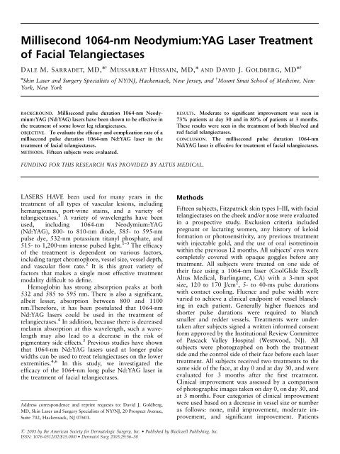
Dermatol Surg 29:1:January 2003 SARRADET ET AL.: FACIAL TELANGIECTASES AND ND: YAG LASER 57were also evaluated for side effects, including edema,erythema, purpura, and blister formation at day 0,day 30, and 3 months.ResultsModerate to significant improvement was seen in 73%of the patients at day 30 and in 80% of the patients atFigure 1. Clinical improvement at Day 30 and at 3 months.Figure 3. (A) Pretreatment. (B) Posttreatment at 3 months.3 months (Figure 1). This improvement was seen inboth larger red/blue facial telangiectases (Figure 2), aswell as in smaller red facial telangiectases (Figure 3).Adverse events were minimal in this study. Twosubjects had mild blistering at their treatment sites.These were treated with topical bacitracin ointmentand were resolved in 2 to 3 days. There was noincidence of infection, scarring, residual erythema, orpostinflammatory pigmentary change in any of thesubjects.Figure 2. (A) Pretreatment. (B) Posttreatment at 3 months.DiscussionRecent studies by our group and others have demonstratedthat millisecond pulse duration Nd:YAG lasersare effective in the treatment of lower extremityvessels. 4,6 This study demonstrated that such lasersare also effective in the treatment of facial telangiectases.Moderate to significant clinical improvementwas seen in both larger blue/red and smaller red facialvessels. Use of a millisecond Nd:YAG laser, such as theone used in this study, allowed for great variations inthe chosen fluence and pulse duration. Such choicesallow adjustment for individual variations in vesselsize, depth, and color.
58 SARRADET ET AL.: FACIAL TELANGIECTASES AND ND: YAG LASER Dermatol Surg 29:1:January 2003This ability for adjustment is a double-edged sword.Treatment of smaller, redder vessels generally requiresthe use of higher energy and shorter pulse widths. Atthese treatment parameters, the risk for blistering andscarring is theoretically increased. We were able tocompensate for this risk with ample pretreatment andposttreatment cooling of each individual treatmentsite. Using the clinical endpoint of vascular blanchingwith adequate precooling and postcooling of eachtreatment site, we were able to obtain consistentclinical outcomes with minimal adverse effects.Clearly, however, the lower the delivered fluence andthe longer the pulse duration used, the less likely one isto see scarring. Further studies are required to comparethe vascular clearing efficacy of millisecond Nd:YAGlasers with that of other vascular-specific lasers.References1. Alster TS. Laser treatment of vascular lesions: recent trends.Cosmetic Derm 2000;47:51–3.2. Dover JS, Sadick NS, Goldman MP. The role of lasers and lightsources in the treatment of leg veins. Dermatol Surg 1999;25:328–36.3. West TB, Alster TS. Comparison of the long-pulsed dye and KTPlasers in the treatment of facial telangiectasia. Dermatol Surg1998;24:221–6.4. Wiess RA, Weiss MA. Early clinical results with a multiplesynchronized pulse 1064 nm laser for leg telangiectasias andreticular veins. Dermatol Surg 1999;25:399–402.5. Sherwood KA, Murray S, Kurban AK, Tan OT. Effect ofwavelength on cutaneous pigment using pulsed irradiation. J InvestDermatol 1989;92:717–20.6. Rogachefsky AS, Silapunt S, Goldberg DJ. 1064 nm Nd: YAG laserirradiation for lower extremity telangiectases and small reticularveins: efficacy as measured by vessel color and size. Dermatol Surg2002;28:220–3.