Toward robot-assisted vascular microsurgery in the retina - Colgate
Toward robot-assisted vascular microsurgery in the retina - Colgate
Toward robot-assisted vascular microsurgery in the retina - Colgate

You also want an ePaper? Increase the reach of your titles
YUMPU automatically turns print PDFs into web optimized ePapers that Google loves.
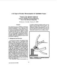
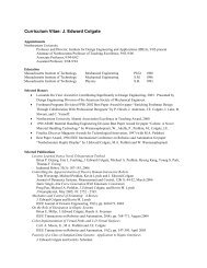
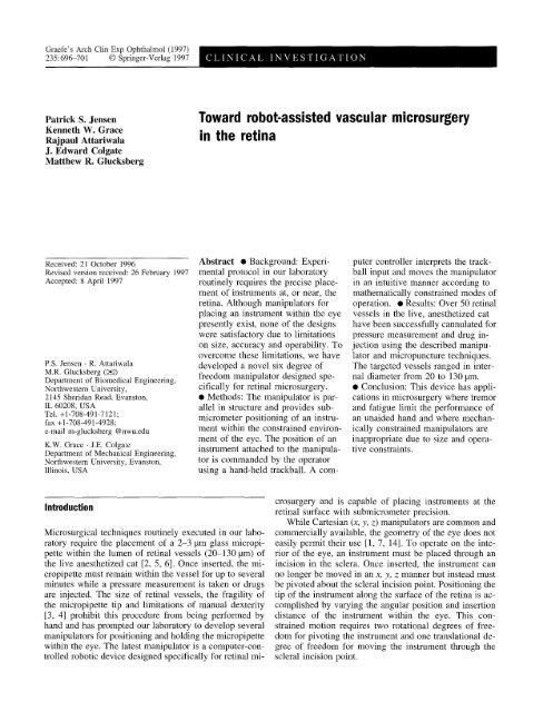
699Center of Rotation Translates With Respect to Base/YFixed H"~r idEn tffal ic~° r ------%~]_ "" ~'- Translate ~ "(f)~ y eaBase I [ Base JCenter of Rotation Fixed With Respect to Base(e)~~ L~~///// ~'/ / ~MMicrornanipulator J///\\\Base]vBase ]Fig. 4 Schematic view of <strong>the</strong> end effector used for ret<strong>in</strong>al <strong>vascular</strong>micropuncture. A micropipette holder (a) provides a pressure-tight<strong>in</strong>terface to <strong>the</strong> glass micropipette. The micropipette assembly is attachedto a Narishige hydraulic actuator (b) which advances and retracts<strong>the</strong> micropipette relative to <strong>the</strong> hypodermic needle (c). A siliconrubber boot (d) provides a seal between <strong>the</strong> micropipette andhypodermic needle to prevent <strong>the</strong> leakage of vitreous. A dovetailslide (e) guides <strong>the</strong> micropipette dur<strong>in</strong>g <strong>in</strong>itially <strong>in</strong>sertion through<strong>the</strong> hypodermic needle. The hypodermic needle is held <strong>in</strong> place witha lock<strong>in</strong>g cam mechanism Q)Fig. 3a, b Splitt<strong>in</strong>g <strong>the</strong> pose variables <strong>in</strong>to functional groups allowsan <strong>in</strong>put device with fewer degrees of freedom than <strong>the</strong> manipulatorto be used <strong>in</strong>tuitively for micropuncture, a The translational mode ofoperation translates <strong>the</strong> enter of rotation us<strong>in</strong>g cartesian coord<strong>in</strong>ateswhile ma<strong>in</strong>ta<strong>in</strong><strong>in</strong>g a fixed tool orientation. This is used to place <strong>the</strong>center of rotation of <strong>the</strong> tool co<strong>in</strong>cident with <strong>the</strong> scleral <strong>in</strong>cisionpo<strong>in</strong>t prior to tool <strong>in</strong>sertion, b The rotational mode of operation fixes<strong>the</strong> center of rotation at <strong>the</strong> scleral <strong>in</strong>cision po<strong>in</strong>t and pivots <strong>the</strong> toolabout that po<strong>in</strong>t or moves <strong>the</strong> tool through that po<strong>in</strong>tto move <strong>the</strong> micropipette through <strong>the</strong> needle. The hypodermic needleprovides a passageway for <strong>the</strong> micropipette through <strong>the</strong> scleraand <strong>in</strong>to <strong>the</strong> eye. A silicon rubber boot placed over <strong>the</strong> micropipetteand hypodermic needle prevents <strong>the</strong> leakage of vitreous.Experimental useThe manipulator has been used extensively <strong>in</strong> our laboratory tomake physiolocial measurements <strong>in</strong> <strong>the</strong> ret<strong>in</strong>al circulation of <strong>the</strong>live, anes<strong>the</strong>tized cat. All animal experiments were performed <strong>in</strong> accordancewith <strong>the</strong> Pr<strong>in</strong>ciples of laboratory animal care (NIH publicationNo. 86-23, revised 1985). Details on our experimental setupand micropuncture can be found elsewhere [2, 5, 6].To prepare <strong>the</strong> manipulator for use, a hypodermic needle wasloaded <strong>in</strong>to <strong>the</strong> end effector such that <strong>the</strong> tip of <strong>the</strong> needle was at<strong>the</strong> software-generated center of rotation. A microscope stand hold<strong>in</strong>g<strong>the</strong> manipulator was used to position <strong>the</strong> tip of <strong>the</strong> needle near <strong>the</strong>sclera and aim <strong>the</strong> needle <strong>in</strong> <strong>the</strong> general direction of <strong>the</strong> targeted vessels.Once approximate position<strong>in</strong>g was complete, <strong>the</strong> "translational"mode of operation was used to place <strong>the</strong> tip of <strong>the</strong> needle (and virtualcenter of rotation) at <strong>the</strong> desired scleral <strong>in</strong>cision po<strong>in</strong>t. The center ofrotation was <strong>the</strong>n fixed at <strong>the</strong> scleral <strong>in</strong>cision po<strong>in</strong>t by chang<strong>in</strong>g <strong>the</strong>mode of operation to "rotational," guarantee<strong>in</strong>g that <strong>the</strong> micropipeneassembly would always pass through this selected po<strong>in</strong>t.The needle was filled with normal sal<strong>in</strong>e to prevent <strong>the</strong> <strong>in</strong>troductionof air <strong>in</strong>to <strong>the</strong> vitreal chamber, rotated us<strong>in</strong>g <strong>the</strong> manipulator untilit was normal to <strong>the</strong> surface of <strong>the</strong> eye, and pushed through <strong>the</strong>sclera. With <strong>the</strong> hypodermic needle successfully <strong>in</strong>serted <strong>in</strong>to <strong>the</strong>eye, a micropipette was filled with ei<strong>the</strong>r a solution to be <strong>in</strong>jectedor 2M sal<strong>in</strong>e for mak<strong>in</strong>g pressure measurements. The micropipettewas placed <strong>in</strong> <strong>the</strong> holder and attached to <strong>the</strong> Narishige hydraulic cyl<strong>in</strong>der.Us<strong>in</strong>g <strong>the</strong> dovetail slide as a guide while view<strong>in</strong>g <strong>the</strong> back of, <strong>the</strong> hypodermic needle with an operat<strong>in</strong>g microscope (Zeiss OPMI1;Carl Zeiss, Thornwood, N.Y.), <strong>the</strong> micropipene was carefully <strong>in</strong>serted<strong>in</strong>to <strong>the</strong> needle so as not to break <strong>the</strong> tip. After <strong>the</strong> <strong>in</strong>itial <strong>in</strong>sertion,<strong>the</strong> micropipette was advanced fur<strong>the</strong>r while <strong>the</strong> operator observed<strong>the</strong> tip of <strong>the</strong> hypodermic needle through <strong>the</strong> pupil us<strong>in</strong>g<strong>the</strong> operat<strong>in</strong>g microscope and a piano-concave lens over <strong>the</strong> cornea.The <strong>in</strong>terior of <strong>the</strong> eye was illum<strong>in</strong>ated through <strong>the</strong> pupil us<strong>in</strong>g awhite light source from <strong>the</strong> operat<strong>in</strong>g microscope. When <strong>the</strong> micropipettetip emerged from <strong>the</strong> tip of <strong>the</strong> hypodermic needle, <strong>the</strong> dovetailslide was locked which rigidly fixed <strong>the</strong> micropipette to <strong>the</strong> manipulator.While cont<strong>in</strong>uously observ<strong>in</strong>g <strong>the</strong> tip of <strong>the</strong> micropipette through<strong>the</strong> operat<strong>in</strong>g microscope, <strong>the</strong> operator directed <strong>the</strong> micropipette towarda target vessel us<strong>in</strong>g <strong>the</strong> hand-held trackball. Once <strong>the</strong> tip of<strong>the</strong> micropipette was centered over a ret<strong>in</strong>al vessel and just touch<strong>in</strong>g<strong>the</strong> surface of <strong>the</strong> ret<strong>in</strong>a, <strong>the</strong> enable button was released, fix<strong>in</strong>g <strong>the</strong>manipulator at that location. Us<strong>in</strong>g <strong>the</strong> Narishige hydraulic actuator,<strong>the</strong> micropipette was carefully advanced until it just penetrated <strong>the</strong>vessel wall [6], after which pressure measurements were taken ordrugs <strong>in</strong>jected.ResultsA pressure measurement obta<strong>in</strong>ed from a 60-tam ret<strong>in</strong>alve<strong>in</strong> us<strong>in</strong>g <strong>the</strong> manipulator, a micropipette and a servonulldevice (model 5A; IPM, LaMesa, Calif.) is shown<strong>in</strong> Fig. 5. The trace shows <strong>the</strong> pressure read<strong>in</strong>g as <strong>the</strong>micropipette was <strong>in</strong>serted, held steady and <strong>the</strong>n retractedfrom <strong>the</strong> lumen of <strong>the</strong> vessel. Over 50 ret<strong>in</strong>alvessels <strong>in</strong> <strong>the</strong> live, anes<strong>the</strong>tized cat rang<strong>in</strong>g <strong>in</strong> diameter
700Fig. 5 Pressure measurement <strong>in</strong>a ret<strong>in</strong>al ve<strong>in</strong> of a live anes<strong>the</strong>tizedcat acquired us<strong>in</strong>g <strong>the</strong> micromanipulatorand <strong>the</strong> micropuncturetechnique140120-I- 100EE80Micropipette placed with<strong>in</strong>lumen of ret<strong>in</strong>al ve<strong>in</strong>Systemic Blood PressureMicropipette withdrawnfrom ret<strong>in</strong>al ve<strong>in</strong>t~o~20Intraocular Pressure0 I I I10 20 30 40 50Time (s)from 20 to 130 ~m have been successfully cannulatedfor pressure measurement and drug <strong>in</strong>jection us<strong>in</strong>g thismanipulator-based micropuncture technique [6].The compact size of <strong>the</strong> <strong>robot</strong>ic manipulator and <strong>the</strong>virtual center of rotation made sett<strong>in</strong>g up <strong>the</strong> experiment<strong>in</strong> preparation for micropuncture significantly easier thanwith previous manipulators [6]. S<strong>in</strong>ce <strong>the</strong> <strong>robot</strong>ic manipulatortakes up less space around <strong>the</strong> head than similarmechanical manipulators, we were also able to performexperiments requir<strong>in</strong>g two <strong>in</strong>struments <strong>in</strong> <strong>the</strong> eye by hold<strong>in</strong>gone <strong>in</strong>strument with <strong>the</strong> <strong>robot</strong>ic manipulator and ano<strong>the</strong>rwith an older mechanical manipulator.DiscussionWe have found <strong>the</strong> <strong>robot</strong>ic manipulator described <strong>in</strong> thispaper to be easy to use and versatile while provid<strong>in</strong>g precise,steady placement of an <strong>in</strong>strument over large areasof <strong>the</strong> ret<strong>in</strong>a. One goal of this research was to develop amanipulator which is easy to set up for ret<strong>in</strong>al <strong>microsurgery</strong>and essentially transparent to an operator us<strong>in</strong>g it.The setup procedure has been simplified <strong>in</strong> that a virtualcenter of rotation at <strong>the</strong> scleral <strong>in</strong>cision po<strong>in</strong>t has been establishedby touch<strong>in</strong>g <strong>the</strong> sclera with <strong>the</strong> tip of <strong>the</strong> <strong>in</strong>strument.Once <strong>the</strong> scleral <strong>in</strong>cision po<strong>in</strong>t has been selected,<strong>the</strong> motion of <strong>the</strong> tool is constra<strong>in</strong>ed to always passthrough that po<strong>in</strong>t. S<strong>in</strong>ce <strong>the</strong> motion of <strong>the</strong> tool is constra<strong>in</strong>ed,<strong>the</strong> operator does not need to worry about damageto <strong>the</strong> <strong>in</strong>strument or <strong>the</strong> eye at <strong>the</strong> scleral <strong>in</strong>cisionpo<strong>in</strong>t due to an erroneous motion command.To move <strong>the</strong> micropipette tip with<strong>in</strong> <strong>the</strong> eye, an operatorobserves <strong>the</strong> tip of <strong>the</strong> micropipette through an operat<strong>in</strong>gmicroscope and guides it based on visual <strong>in</strong>formation.The operator is thus us<strong>in</strong>g <strong>the</strong> manipulator as a tooland always has direct control over how <strong>the</strong> tool is mov<strong>in</strong>g.To make <strong>the</strong> manipulator as <strong>in</strong>tuitive to use as possible,<strong>the</strong> computer and <strong>robot</strong> resid<strong>in</strong>g between <strong>the</strong> operatorand <strong>the</strong> tool should be essentially transparent. This <strong>in</strong>fersthat <strong>the</strong> operator should feel like <strong>the</strong>y are hold<strong>in</strong>g <strong>the</strong> tooland that <strong>the</strong> tool moves <strong>in</strong> a manner which <strong>the</strong> operatorexpects. Transparent operation was addressed by carefullyselect<strong>in</strong>g <strong>the</strong> mapp<strong>in</strong>g schemes which relate a roll<strong>in</strong>g of<strong>the</strong> trackball to a correspond<strong>in</strong>g notion of <strong>the</strong> manipulator.When <strong>the</strong> trackball is rolled <strong>in</strong> one direction, <strong>the</strong> back of<strong>the</strong> <strong>in</strong>strument follows with a reduction <strong>in</strong> scale mak<strong>in</strong>g<strong>the</strong> operator feel as if this thumb is attached directly to<strong>the</strong> back of <strong>the</strong> <strong>in</strong>strument.Us<strong>in</strong>g <strong>the</strong> manipulator <strong>in</strong> a teleoperated manner is onlyone of many possible applications. The computer controlleris also capable of follow<strong>in</strong>g predef<strong>in</strong>ed motion profilesand plac<strong>in</strong>g <strong>the</strong> <strong>in</strong>strument at a specific location with<strong>in</strong><strong>the</strong> eye. This capability could be used to microscopicallyscan surfaces, move <strong>the</strong> <strong>in</strong>strument <strong>in</strong> a complicated preprogrammedmanner or repeatedly visit <strong>the</strong> same ret<strong>in</strong>allocation. By register<strong>in</strong>g <strong>the</strong> <strong>in</strong>strument to <strong>the</strong> eye, <strong>the</strong>computer controller could also be used to set up additionalmotion constra<strong>in</strong>ts based on eye geometry. An examplewould be to keep <strong>the</strong> <strong>in</strong>strument at least 10 pm from <strong>the</strong>ret<strong>in</strong>a or to allow controlled penetration of <strong>the</strong> <strong>in</strong>strument<strong>in</strong>to <strong>the</strong> subret<strong>in</strong>al space for <strong>the</strong> <strong>in</strong>jection of various substances.Acknowledgements The authors would like to thank Dr. SheldonBuzney and Dr. Charles Garcia for <strong>the</strong>ir discussions, suggestionsand many useful <strong>in</strong>sights and also Daniel Laser, Mike Lohse, andPeter Ehman for <strong>the</strong>ir technical assistance and eng<strong>in</strong>eer<strong>in</strong>g expertise.This work was supported by The Margaret W. and Herbert HooverJr. Foundation and by NIH Grant EY09714.
701References1. Allf BE, Juan E de Jr (1987) In vivocannulation of ret<strong>in</strong>al vessels. Graefe'sArch Cl<strong>in</strong> Exp Ophthalmol 225:221-2252. Attariwala R, Giebs CP. Glucksberg MR(1994) The <strong>in</strong>fluence of elevated <strong>in</strong>traocularpressure on <strong>vascular</strong> pressures <strong>in</strong><strong>the</strong> cat ret<strong>in</strong>a. Invest Ophthalmol Vis Sci35:1019-10253. Bose B, Kalra AK, Thurkral S, Sood A,Guha SK, Anand S (1992) Tremorcompensation for <strong>robot</strong>ics <strong>assisted</strong> <strong>microsurgery</strong>.Proc Ann Int'l Conf IEEEEng Med Biol Soc 14:1067-10684. Charles S (1994) Dexterity enhancementfor surgery. Proc First Int'l Symp MedRobotics Comput Assist Surg 2:145-1605. Glucksberg MR, Dunn R (1993) Directmeasurement of ret<strong>in</strong>al micro<strong>vascular</strong>pressure <strong>in</strong> <strong>the</strong> live anes<strong>the</strong>tized cat.Microvasc Res 45:158-1656. Glucksberg MR, Dunn R, Giebs CP(1993) In vivo micropuncture of ret<strong>in</strong>alvessels. Graefe's Arch Cl<strong>in</strong> ExpOphthalmol 231:405-4077. Grace KW (1995) K<strong>in</strong>ematic design ofan ophthalmic surgery <strong>robot</strong> and featureextract<strong>in</strong>g bilateral manipulation. Ph D<strong>the</strong>sis, Northwestern University,Evanston, Ill8. Grace KW, <strong>Colgate</strong> JE, Glucksberg MR,Chun JH (1993) A six degree of freedommicromanipulator for ophthalmic surgery.Proc IEEE Int'l Conf RoboticsAutomation 1:630-6359. Deleted <strong>in</strong> production10. Hunter IW, Doukoglou D, Lafonta<strong>in</strong>eSR, Charette PG, Jones LA, Sagar MA,Mall<strong>in</strong>son GD, Hunter PJ (t993) Ateleoperated microsurgical <strong>robot</strong> andassociated virtual environment for eyesurgery. Presence 2:265-28011. Jensen PS, Glucksberg MR, <strong>Colgate</strong> JE,Grace KW, Attariwala R (1994) Roboticmicromanipulator for ophthalmic surgery.Proc First Int'l Symp Med RoboticsComput Assist Surg 2:204-21012. Merlet J-P (1991) Articulated device,for use <strong>in</strong> particular <strong>in</strong> <strong>robot</strong>ics. US patentno. 5,053,68713. Merlet J-P (1992) Direct k<strong>in</strong>ematics andassembly modes of parallel manipulators.Int J Robotics Res 11 : 150-16214. Pournaras CJ, Shonat RD, Munoz J-L,Petrig BL (1991) New ocular micromanipulatorfor measurements of ret<strong>in</strong>aland vitreous physiologic parameters <strong>in</strong><strong>the</strong> mammalian eye. Exp Eye Res53:723-72715. Steward D (1965) A platform with 6degrees of freedom. Proc Inst Mech Eng180:371-386