Fibrillogenic and Non-fibrillogenic Ensembles of SDS ... - CCMB
Fibrillogenic and Non-fibrillogenic Ensembles of SDS ... - CCMB
Fibrillogenic and Non-fibrillogenic Ensembles of SDS ... - CCMB

Create successful ePaper yourself
Turn your PDF publications into a flip-book with our unique Google optimized e-Paper software.
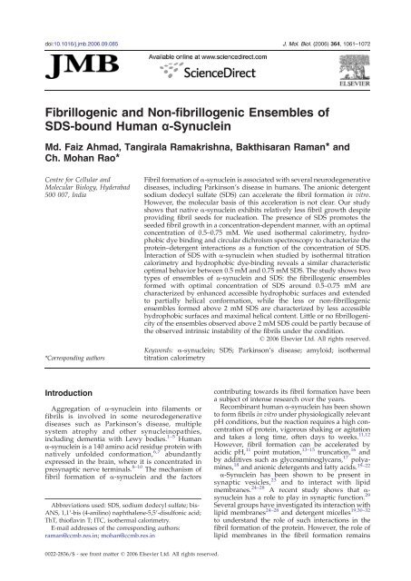
doi:10.1016/j.jmb.2006.09.085 J. Mol. Biol. (2006) 364, 1061–1072<strong>Fibrillogenic</strong> <strong>and</strong> <strong>Non</strong>-<strong>fibrillogenic</strong> <strong>Ensembles</strong> <strong>of</strong><strong>SDS</strong>-bound Human α-SynucleinMd. Faiz Ahmad, Tangirala Ramakrishna, Bakthisaran Raman⁎ <strong>and</strong>Ch. Mohan Rao⁎Centre for Cellular <strong>and</strong>Molecular Biology, Hyderabad500 007, India*Corresponding authorsFibril formation <strong>of</strong> α-synuclein is associated with several neurodegenerativediseases, including Parkinson's disease in humans. The anionic detergentsodium dodecyl sulfate (<strong>SDS</strong>) can accelerate the fibril formation in vitro.However, the molecular basis <strong>of</strong> this acceleration is not clear. Our studyshows that native α-synuclein exhibits relatively less fibril growth despiteproviding fibril seeds for nucleation. The presence <strong>of</strong> <strong>SDS</strong> promotes theseeded fibril growth in a concentration-dependent manner, with an optimalconcentration <strong>of</strong> 0.5–0.75 mM. We used isothermal calorimetry, hydrophobicdye binding <strong>and</strong> circular dichroism spectroscopy to characterize theprotein–detergent interactions as a function <strong>of</strong> the concentration <strong>of</strong> <strong>SDS</strong>.Interaction <strong>of</strong> <strong>SDS</strong> with α-synuclein when studied by isothermal titrationcalorimetry <strong>and</strong> hydrophobic dye-binding reveals a similar characteristicoptimal behavior between 0.5 mM <strong>and</strong> 0.75 mM <strong>SDS</strong>. The study shows twotypes <strong>of</strong> ensembles <strong>of</strong> α-synuclein <strong>and</strong> <strong>SDS</strong>: the <strong>fibrillogenic</strong> ensemblesformed with optimal concentration <strong>of</strong> <strong>SDS</strong> around 0.5–0.75 mM arecharacterized by enhanced accessible hydrophobic surfaces <strong>and</strong> extendedto partially helical conformation, while the less or non-<strong>fibrillogenic</strong>ensembles formed above 2 mM <strong>SDS</strong> are characterized by less accessiblehydrophobic surfaces <strong>and</strong> maximal helical content. Little or no <strong>fibrillogenic</strong>ity<strong>of</strong> the ensembles observed above 2 mM <strong>SDS</strong> could be partly because <strong>of</strong>the observed intrinsic instability <strong>of</strong> the fibrils under the condition.© 2006 Elsevier Ltd. All rights reserved.Keywords: α-synuclein; <strong>SDS</strong>; Parkinson's disease; amyloid; isothermaltitration calorimetryIntroductionAggregation <strong>of</strong> α-synuclein into filaments orfibrils is involved in some neurodegenerativediseases such as Parkinson's disease, multiplesystem atrophy <strong>and</strong> other synucleinopathies,including dementia with Lewy bodies. 1–5 Humanα-synuclein is a 140 amino acid residue protein withnatively unfolded conformation, 6,7 abundantlyexpressed in the brain, where it is concentrated inpresynaptic nerve terminals. 8–10 The mechanism <strong>of</strong>fibril formation <strong>of</strong> α-synuclein <strong>and</strong> the factorsAbbreviations used: <strong>SDS</strong>, sodium dodecyl sulfate; bis-ANS, 1,1′-bis (4-anilino) naphthalene-5,5′-disulfonic acid;ThT, thi<strong>of</strong>lavin T; ITC, isothermal calorimetry.E-mail addresses <strong>of</strong> the corresponding authors:raman@ccmb.res.in; mohan@ccmb.res.incontributing towards its fibril formation have beena subject <strong>of</strong> intense research over the years.Recombinant human α-synuclein has been shownto form fibrils in vitro under physiologically relevantpH conditions, but the reaction requires a high concentration<strong>of</strong> protein, vigorous shaking or agitation<strong>and</strong> takes a long time, <strong>of</strong>ten days to weeks. 11,12However, fibril formation can be accelerated byacidic pH, 11 point mutation, 13–15 truncation, 16 <strong>and</strong>by additives such as glycosaminoglycans, 17 polyamines,18 <strong>and</strong> anionic detergents <strong>and</strong> fatty acids. 19–22α-Synuclein has been shown to be present insynaptic vesicles, 23 <strong>and</strong> to interact with lipidmembranes. 24–28 A recent study shows that α-synuclein has a role to play in synaptic function. 29Several groups have investigated its interaction withlipid membranes 24–28 <strong>and</strong> detergent micelles 19,30–32to underst<strong>and</strong> the role <strong>of</strong> such interactions in thefibril formation <strong>of</strong> the protein. However, the role <strong>of</strong>lipid membranes in the fibril formation remains0022-2836/$ - see front matter © 2006 Elsevier Ltd. All rights reserved.
1062 <strong>SDS</strong>-induced Fibril Growth <strong>of</strong> a-Synucleincomplex <strong>and</strong> debatable. Two schools <strong>of</strong> thoughtsexist: one contends that membrane favors aggregation<strong>and</strong> fibril formation, 20,33,34 while the othercontends that membrane disaggregates <strong>and</strong> inhibitsfibril formation. 28,35,36 Zhu <strong>and</strong> Fink have reportedthat the α-helical conformation induced by theanionic lipid vesicles is not <strong>fibrillogenic</strong>. 28 Fromthe forgoing, it is clear that the role <strong>of</strong> membranes ormembrane mimetic conditions in α-synuclein fibrilformation is not yet clearly understood. Unequivocalunderst<strong>and</strong>ing <strong>of</strong> the role <strong>of</strong> membraneenvironment in the fibril formation <strong>of</strong> α-synucleinis important in underst<strong>and</strong>ing synucleinopathies.The anionic detergent, sodium dodecyl sulfate(<strong>SDS</strong>) has been used in some studies to underst<strong>and</strong>the role <strong>of</strong> a membrane-mimetic environment in thestructure <strong>and</strong> <strong>fibrillogenic</strong> properties <strong>of</strong> α-synuclein.19,30,31 The presence <strong>of</strong> <strong>SDS</strong> has been shown toaccelerate fibril formation. 19,37 Various structuralstudies have used very high concentrations <strong>of</strong> <strong>SDS</strong>(at which it forms micelles) to determine the <strong>SDS</strong>inducedfolded conformation <strong>of</strong> α-synuclein. The<strong>fibrillogenic</strong>ity <strong>of</strong> such a folded conformation is notclear. We set out to systematically compare theseeded fibril growth <strong>of</strong> α-synuclein as well as <strong>SDS</strong>–αsynucleininteractions as a function <strong>of</strong> the concentration<strong>of</strong> <strong>SDS</strong>. In contrast to the rapid fibril growthobserved earlier with other <strong>fibrillogenic</strong> polypeptidesor proteins such as amyloid β peptides 38 or β2-microglobulin 39 upon seeding, our present studyshows that α-synuclein, though natively unfolded orintrinsically unstructured, 6,7 exhibits subtle differencesin the seeded fibril growth. Our isothermaltitration calorimetry (ITC), circular dichroism (CD)spectroscopy <strong>and</strong> 1,1′-bis (4-anilino) naphthalene-5,5′-disulfonic acid (bis-ANS) binding studies alongwith studies on the seeded fibril growth show thepresence <strong>of</strong> two types <strong>of</strong> ensembles <strong>of</strong> <strong>SDS</strong>–αsynuclein.These two ensembles show interestingdifferences, including <strong>fibrillogenic</strong>ity.ResultsDual behavior <strong>of</strong> <strong>SDS</strong> in the fibril growth <strong>of</strong>α-synucleinWhen native α-synuclein in buffer alone wasincubated (with no stirring or agitation) with fibrilseeds, no increase in thi<strong>of</strong>lavin T (ThT) fluorescencewas observed (Figure 1(a)). Prolonged incubation(34 h) did not yield a significant increase in ThTfluorescence (data not shown). Wood et al. havereported that α-synuclein fibrillogenesis is nucleation-dependent.40 Their results show that the fibrillationoccurs only after prolonged incubation (a fewdays) even in the presence <strong>of</strong> seeds. In comparison,other nucleation-dependent, fibril-forming systems,such as amyloid β peptides 38 or β 2 -microglobulinunder acidic conditions, 39 exhibit rapid fibril growth(in minutes) once seeded. This implies that α-synuclein exhibits subtle differences in its nucleationFigure 1. <strong>SDS</strong>-perturbed state, but not native humanα-synuclein, is compatible with fibril growth. (a) <strong>SDS</strong>inducedfibril growth <strong>of</strong> α-synuclein monitored by ThTfluorescence. α-Synuclein (1 mg/ml) in 20 mM Hepesbuffer (pH 7.0) was incubated at 37 °C with 0.1 mg/ml <strong>of</strong>fibril seed in the absence (open circles) <strong>and</strong> in the presence(filled circles) <strong>of</strong> 0.15 mM <strong>SDS</strong> <strong>and</strong> the sample <strong>of</strong> α-synuclein (1 mg/ml) in the presence <strong>of</strong> 0.15 mM <strong>SDS</strong>incubated without fibril seeds (open triangles). The insetshows transmission electron microscopic image <strong>of</strong> fibrils<strong>of</strong> α-synuclein formed in the presence <strong>of</strong> both the fibrilseed <strong>and</strong> <strong>SDS</strong>. (b) Association-induced β-sheet structureupon fibril growth <strong>of</strong> α-synuclein studied by far-UV CDspectroscopy. Far-UV CD spectra <strong>of</strong> the samples <strong>of</strong> α-synuclein (1 mg/ml) in the presence <strong>of</strong> 0.15 mM <strong>SDS</strong>incubated at 37 °C for 4 h in the absence (curve 1) <strong>and</strong> inthe presence <strong>of</strong> 0.1 mg/ml fibril <strong>of</strong> seed (curve 2). The far-UV CD spectrum (curve 3) <strong>of</strong> the sample <strong>of</strong> α-synucleinincubated with only fibril seed (without <strong>SDS</strong>) is shown.[θ] MRM is the mean residue mass ellipticity.dependency; nucleation alone is not sufficient for therapid formation <strong>of</strong> α-synuclein fibrils.Anionic amphiphilic molecules such as <strong>SDS</strong> <strong>and</strong>fatty acids are known to induce fibril formation <strong>of</strong> α-synuclein. 19–22 We have studied the seeded fibrilgrowth <strong>of</strong> α-synuclein in the presence <strong>of</strong> 0.15 mM<strong>SDS</strong> <strong>and</strong> found that ThT fluorescence intensityincreased as a function <strong>of</strong> time <strong>and</strong> was saturatedby 4 h (Figure 1(a)), indicating that fibril growthoccurs rapidly. Electron microscopic examination <strong>of</strong>the sample showed fibrillar morphology (inset inFigure 1(a)). Incubation <strong>of</strong> α-synuclein in thepresence <strong>of</strong> <strong>SDS</strong> without the seed did not result inincrease in ThT fluorescence over the observedperiod <strong>of</strong> time (Figure 1(a)).
<strong>SDS</strong>-induced Fibril Growth <strong>of</strong> a-Synuclein1063Ordered aggregation <strong>of</strong> proteins to amyloid fibrilsis known to be associated with cross β-sheetstructure. 41 We have investigated the generation <strong>of</strong>β-sheet structure upon fibril growth <strong>of</strong> α-synucleinby far-UV CD spectroscopy. Consistent with theresults <strong>of</strong> ThT-binding, the association-inducedβ-sheet generation is observed only with samples<strong>of</strong> α-synuclein incubated with both <strong>SDS</strong> <strong>and</strong> thefibril seed (Figure 1(b)). The small extent <strong>of</strong> β-sheetstructure indicated by the spectrum <strong>of</strong> the sample <strong>of</strong>native α-synuclein incubated with seed was contributedby the seed <strong>and</strong> was not due to the fibrilgrowth, as judged by the far-UV CD spectrum <strong>of</strong>seed alone (spectrum not shown).Figure 2 shows the extent <strong>of</strong> fibril growth <strong>of</strong> theprotein as a function <strong>of</strong> the concentration <strong>of</strong> <strong>SDS</strong> asmeasured by ThT fluorescence. The extent <strong>of</strong> fibrilgrowth increased dramatically <strong>and</strong> attained amaximum at a concentration <strong>of</strong> 0.5 mM <strong>SDS</strong>; itdecreased progressively with further increase in theconcentration <strong>of</strong> <strong>SDS</strong>. Fibril growth did not occur atconcentrations <strong>of</strong> <strong>SDS</strong> above 2 mM. One possibilityfor this biphasic nature <strong>of</strong> the pr<strong>of</strong>ile is that theprotein to detergent ratio with some stoichiometry isimportant in the fibril formation <strong>of</strong> α-synuclein.Even when two different concentrations <strong>of</strong> α-synuclein(1 mg/ml or 0.22 mg/ml) were used, theoptimal concentration <strong>of</strong> <strong>SDS</strong> for fibril growthremained around 0.5 mM (see Figure 2 (a) <strong>and</strong>(b)). However, the width <strong>of</strong> the pr<strong>of</strong>ile is relativelysharper at the lower concentration <strong>of</strong> proteincompared to that at the higher concentration.Thus, all our results show that <strong>SDS</strong> exhibits dualbehavior in the fibril formation <strong>of</strong> α-synuclein. Inorder to underst<strong>and</strong> the dual behavior <strong>of</strong> <strong>SDS</strong>induced<strong>fibrillogenic</strong>ity, we have further studied theprotein–<strong>SDS</strong> interactions by ITC, hydrophobic dye,bis-ANS binding, time-resolved fluorescence spectroscopy<strong>and</strong> CD.Interaction <strong>of</strong> <strong>SDS</strong> with α-synuclein studied byisothermal calorimetryFigure 3(a) illustrates an ITC experiment wherewe have injected 10 μl <strong>of</strong> 7 mM <strong>SDS</strong> repeatedly intothe reaction cell containing 15 μM α-synuclein.Relatively low <strong>and</strong> constant exothermic heat flowwith the enthalpy change, comparable to theenthalpy change <strong>of</strong> dilution <strong>of</strong> the detergent(blank), was observed in the initial few additions<strong>of</strong> the injectant (up to a certain concentration <strong>of</strong><strong>SDS</strong>). Further increase in the concentration <strong>of</strong> <strong>SDS</strong>led to a large exothermic enthalpy change, reachinga maximum at 0.5 mM <strong>SDS</strong>, which decreased uponfurther increase in the concentration <strong>of</strong> <strong>SDS</strong>. Thisinteresting ITC pr<strong>of</strong>ile <strong>of</strong> <strong>SDS</strong>–synuclein interactionmight be described in two parts: the first part mightbe largely driven by entropy, with little or no changein enthalpy, thus showing no observable heattransfer in the first few injections; after saturatingthis interaction, further injections might lead to asecond part <strong>of</strong> the interaction that is driven byenthalpy, as seen by significant exothermic heattransfer. If such a description was true, altering theconcentration <strong>of</strong> synuclein should shift the pr<strong>of</strong>ilesignificantly. Figure 3(b) shows the ITC pr<strong>of</strong>ile withonly 5 μM α-synuclein. Despite such a large decreasein the concentration <strong>of</strong> α-synuclein, the ITC pr<strong>of</strong>iledid not show a shift to lower concentrations <strong>of</strong> <strong>SDS</strong>(maximum is ∼0.65 mM <strong>SDS</strong>). Moreover, when weincreased the concentration <strong>of</strong> α-synuclein to 70 μM,the ITC pr<strong>of</strong>ile still exhibited a peak around 0.5 mM<strong>SDS</strong>. The peak, however, was broad, taperinggradually beyond 2 mM <strong>SDS</strong> (data not shown).Thus, the ITC pr<strong>of</strong>ile cannot be used to derivestoichiometric thermodynamic parameters. However,the study shows a characteristic optimumaround 0.5 mM <strong>SDS</strong> (which parallels the <strong>fibrillogenic</strong>ity),suggesting formation <strong>of</strong> some non-stoichiometricensembles <strong>of</strong> the protein with thedetergent.<strong>Fibrillogenic</strong> ensembles <strong>of</strong> α-synuclein with <strong>SDS</strong>exhibit enhanced hydrophobic surfacesFigure 2. <strong>SDS</strong>-induced fibril growth <strong>of</strong> human α-synuclein as a function <strong>of</strong> <strong>SDS</strong> concentration monitored byThT fluorescence. α-Synuclein at (a) 1 mg/ml or (b)0.22 mg/ml in Hepes–NaOH buffer (pH 7.0), wasincubated with 10% fibril seed at 37 °C for 4 h at variousconcentrations <strong>of</strong> <strong>SDS</strong>.Accessible hydrophobic surfaces on proteins canbe studied using the fluorescent hydrophobic probebis-ANS. 42 The probe is non-fluorescent in a polarenvironment. Upon binding to hydrophobic surfaces,its fluorescence increases accompanied by ablue shift in its emission maximum. 42 bis-ANS bindsto native α-synuclein <strong>and</strong> exhibits enhanced fluorescence(Figure 4(a)). The fluorescence intensity
1064 <strong>SDS</strong>-induced Fibril Growth <strong>of</strong> a-SynucleinFigure 3. Isothermal titration calorimetry showing a cooperative exothermic reaction between human α-synuclein<strong>and</strong> <strong>SDS</strong> at 30 °C. (a) Titration <strong>of</strong> 15 μM (0.22 mg/ml) α-synuclein with 7 mM monomeric <strong>SDS</strong>. (b) Titration <strong>of</strong> 5 μM(0.072 mg/ml) α-synuclein with 7 mM monomeric <strong>SDS</strong>. Titrations were performed by adding aliquots <strong>of</strong> 10 μl each time.Both the protein <strong>and</strong> the detergent were buffered at pH 7.0 with 20 mM Hepes–NaOH. The thick line in the dQ/dt (rate <strong>of</strong>enthalpy change) window <strong>and</strong> filled circles in Q (enthalpy change) window represent the observed heat flow upontitration <strong>of</strong> the protein with the detergent. Thin line in the dQ/dt window <strong>and</strong> open circle in the Q window represent theblank titration (the heat flow <strong>of</strong> the dilution <strong>of</strong> the detergent to buffer alone).increases further upon addition <strong>of</strong> 0.25 mM, 0.5 mM<strong>and</strong> 1.0 mM <strong>SDS</strong>, <strong>and</strong> then decreases at 2 mM <strong>SDS</strong>.bis-ANS in 10 mM <strong>SDS</strong> without the protein shows anegligibly low level <strong>of</strong> fluorescence. Figure 4(b)further explores this biphasic behavior; the plot <strong>of</strong>fluorescence intensity versus the concentration <strong>of</strong><strong>SDS</strong> peaks at 0.75 mM <strong>SDS</strong> <strong>and</strong> decreases graduallywith increasing concentration <strong>of</strong> <strong>SDS</strong>. This resultshows an increase in the accessible hydrophobicsurfaces <strong>of</strong> the protein or the protein–detergentensembles up to 0.75 mM <strong>SDS</strong>. The emissionmaximum red shifts slightly below 2 mM <strong>SDS</strong> <strong>and</strong>in a more pronounced manner above 2 mM <strong>SDS</strong>(Figure 4(b) inset). The minor red shift (below 2 mM<strong>SDS</strong>), despite an increase in the fluorescenceintensity, indicates that the intrinsic hydrophobicity<strong>of</strong> at least some sites decreases or some relativelymoderate hydrophobic sites become accessible. Thedecrease in the fluorescence intensity accompanyingthe pronounced red shift above 2 mM <strong>SDS</strong> reflects adecrease in the accessible hydrophobic surfaces.Similar results were obtained with 0.22 mg/ml <strong>of</strong> α-synuclein (Figure 4(c)). Thus, our results demonstratesubstantial rearrangement <strong>of</strong> hydrophobicsurfaces <strong>of</strong> the protein–detergent ensembles, whichmay be important for <strong>fibrillogenic</strong>ity.The fluorescence life-time <strong>of</strong> the polarity-sensitivefluorophore ANS has been used to study conformationalchanges <strong>and</strong> partially unfolded states <strong>of</strong>proteins; 43,44 it exhibits longer lifetimes upon bindingto protein hydrophobic surfaces. We have usedtime-resolved fluorescence spectroscopy to study thebis-ANS binding to α-synuclein. The fluorescencedecay pr<strong>of</strong>ile <strong>of</strong> the sample <strong>of</strong> bis-ANS bound tonative α-synuclein (Figure 5) can be fit with a threeexponentialdecay model, with lifetimes τ1, τ2 <strong>and</strong> τ3<strong>of</strong> around 0.15 ns, 2.6 ns <strong>and</strong> 7.6 ns, respectively. Thefast decay component (τ1, 0.15 ns) could representunbound bis-ANS, while τ2 <strong>and</strong> τ3 represent thepopulations <strong>of</strong> bis-ANS bound to at least two types <strong>of</strong>hydrophobic surfaces <strong>of</strong> α-synuclein.The fluorescence decay pr<strong>of</strong>iles <strong>of</strong> bis-ANS in thesample <strong>of</strong> α-synuclein with increasing concentrations<strong>of</strong> <strong>SDS</strong> could also be fit with a threeexponentialdecay model. The fast component, τ1,remains unchanged. Remarkable changes could beobserved in the lifetimes <strong>of</strong> τ2 <strong>and</strong> τ3, as well as intheir relative amplitudes as a function <strong>of</strong> theconcentration <strong>of</strong> <strong>SDS</strong>. τ2 increases from 2.6 ns inthe absence <strong>of</strong> <strong>SDS</strong> to almost 3.5 ns in the presence <strong>of</strong>0.5 mM <strong>SDS</strong> (Figure 6(a)). With further increase inthe concentration <strong>of</strong> <strong>SDS</strong>, τ2 decreases progressively.On the other h<strong>and</strong>, τ3 drops sharply from7.6 ns in the absence <strong>of</strong> <strong>SDS</strong> to
<strong>SDS</strong>-induced Fibril Growth <strong>of</strong> a-Synuclein1065predominantly binding to <strong>and</strong> reporting the hydrophobicsurface <strong>of</strong> the protein component. (1) Thefluorescence intensity <strong>of</strong> bis-ANS is not enhancedsignificantly by <strong>SDS</strong> alone. (2) The emission maximum,even in the presence <strong>of</strong> 20 mM <strong>SDS</strong> (in micelleform), was found to be 510 nm, which is significantlydifferent compared to the probe bound to α-synuclein alone (486 nm) or to the protein–detergentcomplex (486–496 nm, depending on the concentration<strong>of</strong> <strong>SDS</strong> as shown in the inset in Figure 4(b)). Evenin the presence <strong>of</strong> 20 mM <strong>SDS</strong>, the fluorescenceintensity <strong>of</strong> bis-ANS was found to be less than sevenarbitrary units compared to a high value rangingfrom 20–75 arbitrary units observed in the presence<strong>of</strong> α-synuclein alone or both the protein <strong>and</strong> <strong>SDS</strong>(Figure 4(b)). (3) The decay pr<strong>of</strong>ile <strong>of</strong> the probe in thepresence <strong>of</strong> the <strong>SDS</strong> micelle could be fit with a twoexponentialdecay model, with the population havingan average lifetime <strong>of</strong> 1.97 (with st<strong>and</strong>ard error(S.E.) <strong>of</strong> 0.005, n=4) contributing 90% (S.E.=1.07) <strong>of</strong>the fluorescence intensity representing the micelleboundbis-ANS molecules. The population having alifetime <strong>of</strong> 0.14 ns (S.E.=0.04) contributing 9.4%(S.E.=1.07) fluorescence intensity may represent theFigure 4. Binding <strong>of</strong> the hydrophobic fluorescentprobe, bis-ANS to the ensembles <strong>of</strong> α-synuclein <strong>and</strong><strong>SDS</strong>. (a) Fluorescence spectra <strong>of</strong> 5 μM bis-ANS bound to1 mg/ml <strong>of</strong> α-synuclein in the absence <strong>and</strong> in the presence<strong>of</strong> <strong>SDS</strong> (concentrations as indicated). The fluorescencespectrum <strong>of</strong> bis-ANS in the presence <strong>of</strong> 10 mM <strong>SDS</strong> aloneis also shown (curve a) (b) Variation <strong>of</strong> fluorescenceintensity at the emission maximum <strong>of</strong> bis-ANS when1 mg/ml <strong>of</strong> α-synuclein was titrated with increasingconcentrations <strong>of</strong> <strong>SDS</strong>. The inset shows the change in thewavelength <strong>of</strong> emission maximum as a function <strong>of</strong> theconcentration <strong>of</strong> <strong>SDS</strong>. (c) Variation <strong>of</strong> fluorescenceintensity at the emission maximum <strong>of</strong> bis-ANS when0.22 mg/ml <strong>of</strong> α-synuclein was titrated with increasingconcentrations <strong>of</strong> <strong>SDS</strong>. The samples were excited at390 nm with excitation <strong>and</strong> emission b<strong>and</strong> passes set at5 nm; the buffer was 20 mM Hepes–NaOH (pH 7.0).Thus, these results show a significant reorganization<strong>of</strong> hydrophobic sites, which may represent eitherchange in the conformation <strong>and</strong> polarity <strong>of</strong> theexisting sites or generation <strong>of</strong> new surfaces upon α-synuclein–detergent interactions.It is possible that bis-ANS binds to the protein or tothe detergent component <strong>of</strong> the ensembles. Thefollowing observations suggest that bis-ANS isFigure 5. Time-resolved fluorescence measurement <strong>of</strong>bis-ANS bound to α-synuclein. (a) Fluorescence decay <strong>of</strong>bis-ANS bound to α-synuclein in 20 mM Hepes–NaOHbuffer (pH 7.0), could be fit with a three-exponential decaymodel. The goodness <strong>of</strong> the fit was assessed by the χ 2 test,which gave a value close to 1, <strong>and</strong> the weighted residualsare shown in the lower panels <strong>of</strong> the Figure. Arepresentative decay pr<strong>of</strong>ile out <strong>of</strong> four experimentaldata is shown in each case. The insets show mean lifetimes(τ) in nanoseconds <strong>and</strong> pre-exponential amplitude (α) inpercentage with the st<strong>and</strong>ard errors indicated within theparentheses. bis-ANS was excited at 337 nm using apicosecond LED light source.
1066 <strong>SDS</strong>-induced Fibril Growth <strong>of</strong> a-Synuclein<strong>SDS</strong>-induced secondary structural generation<strong>and</strong> <strong>fibrillogenic</strong>ity <strong>of</strong> α-synucleinFigure 7 shows the change in the secondarystructure <strong>of</strong> α-synuclein from an unstructuredconformation to an α-helical conformation uponinteracting with <strong>SDS</strong>. The negative ellipticity at222 nm, <strong>and</strong> hence the helical content, increasesprogressively as the concentration <strong>of</strong> <strong>SDS</strong> increasesto 2 mM, <strong>and</strong> becomes constant with furtherincrease in the concentration <strong>of</strong> <strong>SDS</strong>. The <strong>SDS</strong>inducedfolding <strong>of</strong> α-synuclein to an α-helicalstructure is known. 30,45,46 However, the <strong>fibrillogenic</strong>nature <strong>of</strong> such an <strong>SDS</strong>-induced folded structure wasnot investigated. We have compared the extent <strong>of</strong><strong>SDS</strong>-induced folding with <strong>SDS</strong>-induced exposure <strong>of</strong>hydrophobic surfaces <strong>and</strong> fibril growth. Maximallyα-helically folded α-synuclein in the protein–detergentensembles above 2 mM <strong>SDS</strong>, as judged byellipticity at 222 nm (Figure 7), exhibit relatively lessexposed hydrophobic surfaces (Figure 4) <strong>and</strong> do notexhibit <strong>fibrillogenic</strong>ity (Figure 2). However, partiallyfolded species <strong>of</strong> α-synuclein in protein–detergentensembles below 2 mM <strong>SDS</strong> exhibit highly exposedhydrophobic surfaces <strong>and</strong> are <strong>fibrillogenic</strong>.Dual behavior <strong>of</strong> <strong>SDS</strong> in fibril formation <strong>and</strong> fibrildepolymerization <strong>of</strong> α-synucleinThe reason for poor fibril growth <strong>of</strong> α-synucleinabove 2 mM <strong>SDS</strong> could either be the conformationalconstraint in the protein–detergent ensembles, suchthat it is not competent for fibril growth, or therelative fibril instability under such conditions, orboth. We have measured the fibrils using ThTbindingafter incubating fibrils <strong>of</strong> α-synuclein(0.2 mg/ml with respect to α-synuclein concentration)at different concentrations <strong>of</strong> <strong>SDS</strong> for 1 h at37 °C (Figure 8(a)). Diluting the <strong>SDS</strong> (to approxi-Figure 6. Variation <strong>of</strong> fluorescence lifetimes <strong>of</strong> bis-ANS bound to α-synuclein as a function <strong>of</strong> the concentration<strong>of</strong> <strong>SDS</strong>. Dotted vertical lines approximately divide thepopulations <strong>of</strong> <strong>fibrillogenic</strong> <strong>and</strong> less or non-<strong>fibrillogenic</strong>ensembles <strong>of</strong> α-synuclein <strong>and</strong> <strong>SDS</strong>. Traces 1 <strong>and</strong> 2 in therelative amplitude panel correspond to the lifetimes τ2<strong>and</strong> τ3, respectively.unbound probe. Thus, fluorescence lifetimes <strong>of</strong> bis-ANS bound to α-synuclein <strong>and</strong> the <strong>SDS</strong> micelle aresignificantly different. Since we could not observe afluorescence lifetime component corresponding tothe lifetime <strong>of</strong> the probe in the micellar environment(around 1.9 ns) at any concentration <strong>of</strong> <strong>SDS</strong> in thepresence <strong>of</strong> protein (Figure 6), the changes in thefluorescence lifetimes <strong>of</strong> bis-ANS are likely to reportconformational changes associated with hydrophobicsurface alteration in the protein component <strong>of</strong> theprotein–detergent ensembles.Figure 7. <strong>SDS</strong>-induced folding <strong>of</strong> α-synuclein monitoredby far-UV CD spectroscopy. Variation <strong>of</strong> the meanresidue mass ellipticity ([θ] MRM ) at 222 nm <strong>of</strong> α-synuclein(1 mg/ml) as a function <strong>of</strong> the concentration <strong>of</strong> <strong>SDS</strong>. Inset:far-UV CD spectra <strong>of</strong> α-synuclein in 20 mM Hepes–NaOHbuffer (pH 7.0) in the absence (curve 1) <strong>and</strong> in the presence<strong>of</strong> 0.15 mM <strong>SDS</strong> (curve 2), 0.5 mM <strong>SDS</strong> (curve 3), 1 mM<strong>SDS</strong> (curve 4) <strong>and</strong> 2 mM <strong>SDS</strong> (curve 5).
<strong>SDS</strong>-induced Fibril Growth <strong>of</strong> a-Synuclein1067from β2-microglobulin (referred to as K3) underacidic conditions. 47 We have tested if amorphousaggregation occurs with α-synuclein under ourexperimental conditions. Light-scattering <strong>of</strong> α-synucleindid not change significantly with increasingconcentration <strong>of</strong> <strong>SDS</strong> (Figure 9(a)). Since the multimericassembly <strong>of</strong> αB-crystallin (∼550 kDa) isknown to be sensitive to <strong>SDS</strong>, 48 we have monitoredthe <strong>SDS</strong>-induced dissociation <strong>of</strong> the protein forcomparison (Figure 9(a)). The scattering value <strong>of</strong>α-synuclein in the absence or in the presence <strong>of</strong> <strong>SDS</strong>is far lower than that <strong>of</strong> αB-crystallin, indicating theabsence <strong>of</strong> even soluble large oligomers <strong>of</strong> α-synuclein; if present, their population is not highenough to be detected. Under similar conditions, theDTT-induced aggregation <strong>of</strong> insulin gives a verylarge scatter value (approximately 250 arbitraryunits, data not shown). All these results show that<strong>SDS</strong> does not induce amorphous aggregation <strong>of</strong> α-synuclein significantly.Figure 8. <strong>SDS</strong> concentration-dependent fibril dissociation<strong>of</strong> α-synuclein. (a) Fibrils <strong>of</strong> α-synuclein present afterincubation <strong>of</strong> 0.2 mg/ml <strong>of</strong> fibrils in 20 mM Hepes–NaOHbuffer (pH 7.0) at 37 °C for 1 h as a function <strong>of</strong> theconcentration <strong>of</strong> <strong>SDS</strong> determined by ThT binding. Theinset shows the time-dependent dissociation <strong>of</strong> the fibrilswhen incubated in the presence <strong>of</strong> 1 mM (open circles),2 mM <strong>SDS</strong> (filled circles) <strong>and</strong> 5 mM <strong>SDS</strong> (filled triangles).(b) β-Sheet to α-helical conversion <strong>of</strong> the fibrils <strong>of</strong> α-synuclein upon its dissociation in the presence <strong>of</strong> highconcentrations <strong>of</strong> <strong>SDS</strong>. Far-UV CD spectra <strong>of</strong> the fibrils <strong>of</strong>α-synuclein incubated at 37 °C in the presence <strong>of</strong> 0.5 mM<strong>SDS</strong> (curve 1) <strong>and</strong> in the presence <strong>of</strong> 4 mM <strong>SDS</strong> at 30 min(curve 2) <strong>and</strong> at 60 min (curve 3) <strong>of</strong> incubation.mately 15 μM) due to the dilution <strong>of</strong> the fibrils stockresults in significant amount <strong>of</strong> dissociation <strong>and</strong> loss<strong>of</strong> the fibrils. The fibrils are significantly stable up toa concentration <strong>of</strong> <strong>SDS</strong> <strong>of</strong> 1 mM. However, increasingthe concentration <strong>of</strong> <strong>SDS</strong> further leads to drasticloss <strong>of</strong> fibrils due to dissociation. The dissociation isassociated with conversion <strong>of</strong> β-sheet structure to α-helical structure, as revealed by far-UV CD spectroscopy(Figure 8(b)). This result shows that fibrilinstability is one <strong>of</strong> the factors contributing to theobserved lack <strong>of</strong> fibril growth above 2 mM <strong>SDS</strong>.Treatment with <strong>SDS</strong> does not lead to amorphousaggregation <strong>of</strong> α-synucleinA recent study by Yamaguchi et al. has shown thatamorphous aggregation competes with <strong>SDS</strong>inducedfibril formation <strong>of</strong> a small peptide derivedFigure 9. Measurements <strong>of</strong> light-scattering <strong>and</strong> criticalmicellar concentration. (a) Light-scattering (at 465 nm) <strong>of</strong>samples <strong>of</strong> 0.3 mg/ml <strong>of</strong> α-synuclein (filled circles) <strong>and</strong>0.3 mg/ml αB-crystallin (filled triangles) upon treatingwith increasing concentrations <strong>of</strong> <strong>SDS</strong>; the buffer was20 mM Hepes–NaOH (pH 7.0). The light scattering <strong>of</strong> thebuffer alone with increasing concentrations <strong>of</strong> <strong>SDS</strong> (opencircles) is also shown. (b) The critical micellar concentration<strong>of</strong> <strong>SDS</strong> in 20 mM Hepes–NaOH buffer (pH 7.0)measured using 10 μM diphenylhexatriene as the probe.The fluorescence intensity <strong>of</strong> the probe at 430 nm withincreasing concentrations <strong>of</strong> <strong>SDS</strong> in the absence (opencircles) <strong>and</strong> in the presence (filled circles) <strong>of</strong> 0.3 mg/ml <strong>of</strong>α-synuclein is shown. The probe was excited at 358 nm.The broken line indicates a slope change in the pr<strong>of</strong>ile <strong>of</strong>fluorescence intensity versus concentration <strong>of</strong> <strong>SDS</strong> correspondingto the α-synuclein sample.
1068 <strong>SDS</strong>-induced Fibril Growth <strong>of</strong> a-SynucleinCritical micellar concentration <strong>of</strong> <strong>SDS</strong> in theabsence <strong>and</strong> in the presence <strong>of</strong> α-synucleinAn earlier study by Yamamoto et al. showedthatβ2-microglobulin exhibits optimal concentrations <strong>of</strong><strong>SDS</strong> around 0.5 mM in its <strong>SDS</strong>-induced fibrilformation <strong>and</strong> proposed that fibril formation occursnear the critical micellar concentration (cmc) <strong>of</strong>thedetergent in the presence <strong>of</strong> the protein. 49 Wemeasured the cmc <strong>of</strong> <strong>SDS</strong> in our buffer system usingdiphenylhexatriene as a probe. The fluorescence <strong>of</strong> theprobe is low below the cmc <strong>and</strong> increases dramaticallyabove the cmc. 50 As this probe is uncharged, thismethod avoids interference from detergent charge indetermining cmc. 50 Figure 9(b) shows that the cmc <strong>of</strong><strong>SDS</strong> is around 7.5 mM in the buffer. In the presence <strong>of</strong>α-synuclein, the fluorescence intensity increases as afunction <strong>of</strong> the concentration <strong>of</strong> <strong>SDS</strong> without a distinctinflection point, in contrast to that observed in theabsence <strong>of</strong> the protein. However, there is a slopechange around 0.5 mM <strong>SDS</strong>. Considering the increasein the fluorescence intensity even before 0.5 mM <strong>SDS</strong>,it is not clear whether this point represents the cmc.However, it is evident that the presence <strong>of</strong> the proteinalters the property <strong>of</strong> the detergent. Thus, our studyshows that the protein–detergent interactions affectthe properties <strong>of</strong> each other.DiscussionInvestigating the factors that are involved in the<strong>fibrillogenic</strong>ity <strong>of</strong> α-synuclein is important to underst<strong>and</strong>the synucleinopathies, including Parkinson'sdiseases. Amphiphiles such as lipids, fatty acids <strong>and</strong>detergents like <strong>SDS</strong> are known to affect the<strong>fibrillogenic</strong>ity <strong>of</strong> the protein. Our present studyshows two important aspects in the <strong>SDS</strong>-inducedfibril formation <strong>of</strong> α-synuclein. (i) Dual behavior <strong>of</strong><strong>SDS</strong> in promoting fibril formation <strong>and</strong> fibril depolymerization<strong>of</strong> α-synuclein. (ii) Our study characterizesthe <strong>fibrillogenic</strong> <strong>and</strong> non-<strong>fibrillogenic</strong>ensembles <strong>of</strong> <strong>SDS</strong>–α-synuclein <strong>and</strong> shows that the<strong>fibrillogenic</strong> ensembles have highly exposed hydrophobicsurfaces <strong>and</strong> are partially structured.Figure 10 describes the dual nature <strong>of</strong> <strong>SDS</strong>inducedfibril formation <strong>and</strong> depolymerization <strong>of</strong>α-synuclein observed in our study. Native α-synuclein (NU), though intrinsically unstructuredor natively unfolded, exhibits relatively less <strong>fibrillogenic</strong>ityeven in the presence <strong>of</strong> fibril seed fornucleation. ITC, CD spectroscopy <strong>and</strong> hydrophobicdye binding along with fluorescence lifetime studiesshow that the protein–detergent ensembles (I) below2 mM <strong>SDS</strong> have extended to a partially helicalconformation with highly exposed hydrophobicsurfaces <strong>and</strong> are <strong>fibrillogenic</strong>. A recent study hasshown that, although α-synuclein lacks propersecondary structure, dynamic long-range tertiaryinteractions are present. 51 Perturbations <strong>of</strong> suchlong-range interactions by <strong>SDS</strong> may be involved inthe <strong>SDS</strong>-promoted fibril growth <strong>of</strong> α-synuclein.Our study shows that <strong>SDS</strong> inhibits fibril formation<strong>of</strong> α-synuclein at higher concentrations. The protein–detergentensembles (F) formed above 2 mM<strong>SDS</strong>, which represent fully folded α-synuclein withFigure 10. A model describing the <strong>fibrillogenic</strong> <strong>and</strong> non-<strong>fibrillogenic</strong> ensembles <strong>of</strong> α-synuclein <strong>and</strong> <strong>SDS</strong>, <strong>and</strong> theirproperties. NU represents the natively unfolded α-synuclein. I represents partially folded α-synuclein with increasedaccessible hydrophobic surfaces <strong>of</strong> the <strong>fibrillogenic</strong> ensembles. F represents the fully folded α-synuclein with lessaccessible hydrophobic surfaces <strong>of</strong> the non-<strong>fibrillogenic</strong> ensembles.
<strong>SDS</strong>-induced Fibril Growth <strong>of</strong> a-Synuclein1069less accessible hydrophobic surfaces, are less or non<strong>fibrillogenic</strong>in nature. Such lack <strong>of</strong> <strong>fibrillogenic</strong>itymay stem either from the incompetence <strong>of</strong> theconformation for fibril growth or from the instability<strong>of</strong> the fibril under these conditions. Our results showthat fibril instability, which leads to dissociation <strong>of</strong>the fibril accompanied by conversion <strong>of</strong> a β-sheet toan α-helical conformation, is one <strong>of</strong> the factorscontributing to the non-<strong>fibrillogenic</strong> nature <strong>of</strong> α-synuclein observed above 2 mM <strong>SDS</strong>. It is importantto note that earlier studies have addressed thestructure <strong>of</strong> the protein in <strong>SDS</strong> micelles (at highconcentrations <strong>of</strong> <strong>SDS</strong>) in which α-synuclein isshown to adopt an α-helical structure. 30,45,46 Thus,these fully structured, micelle-bound species <strong>of</strong> α-synuclein are less or non-<strong>fibrillogenic</strong> in nature.α-Synuclein is known to interact with lipidmembrane. 28 The membrane environment has beenthought to be one <strong>of</strong> the factors that are involved inthe fibril formation <strong>of</strong> α-synuclein, although its exactrole remains controversial. 20,28,33–36 Our presentstudy shows a biphasic behavior in the <strong>SDS</strong>-inducedfibril growth, which parallels distinct changes in thefolded state <strong>and</strong> hydrophobic surfaces <strong>of</strong> α-synuclein.This suggests that the contradictory resultsreported with regard to the lipid vesicles could bedue to the different types <strong>of</strong> interactions <strong>of</strong> theprotein molecule with lipid vesicles under givenconditions. Such interactions can be modulated bymany factors in vivo, such as lipid composition <strong>and</strong>membrane heterogeneity, interactions with fattyacids <strong>and</strong> cholesterol, <strong>and</strong> their role in membranepacking <strong>and</strong> fluidity alterations. Further systematicstudies are required to underst<strong>and</strong> their individualroles in modulating the <strong>fibrillogenic</strong>ity <strong>of</strong> α-synuclein.In this context, our present study with themembrane-mimicking agent <strong>SDS</strong> should proveuseful in underst<strong>and</strong>ing the role <strong>of</strong> amphiphilicmolecules in the <strong>fibrillogenic</strong>ity <strong>of</strong> α-synuclein.Materials <strong>and</strong> MethodsMaterialsThi<strong>of</strong>lavin T (ThT), <strong>SDS</strong> <strong>and</strong> 1,1′-bis (4-anilino) naphthalene-5,5′-disulfonicacid (bis-ANS) were purchased fromthe Sigma Chemical Company, USA. The expressionvector bearing the coding sequence <strong>of</strong> wild-type humanα-synuclein (pRK172/α-synuclein) was a kind gift fromPr<strong>of</strong>essor Anthony L. Fink (University <strong>of</strong> California, SantaCruz, California).Preparation <strong>of</strong> α-synucleinWild-type α-synuclein was expressed in Escherichia coliBL21 (DE3) cells transformed with the pRK172/α-synucleinplasmid. Expression <strong>of</strong> the protein was induced bythe addition <strong>of</strong> IPTG (to 1 mM). Cells were harvested 4 hafter induction. The cell pellet was suspended in 50 mMTris–HCl (pH 7.4), 1 mM EDTA, 100 mM NaCl, 0.1 mg/ml<strong>of</strong> lysozyme <strong>and</strong> 20 μM PMSF. The suspended cells werelysed by sonication on ice <strong>and</strong> centrifuged at 5000g toremove the cell debris. The supernatant containing theexpressed α-synuclein was subjected to ammoniumsulfate fractionation. The protein that precipitated in the15%–45% saturated ammonium sulfate fraction wasresolubilized in the above-mentioned buffer, but lackinglysozyme <strong>and</strong> PMSF, <strong>and</strong> loaded onto a Sephadex G75 gelfiltrationchromatography column (150 cm×1.8 cm diameter)previously equilibrated with the same buffer. Thecolumn was eluted at a flow-rate <strong>of</strong> 20 ml/h <strong>and</strong> 2 mlfractions were collected. The fractions corresponding to α-synuclein were pooled, exchanged in 20 mM Hepes–NaOH buffer (pH 7.0), <strong>and</strong> concentrated at 4 °C using anAmicon ultrafiltration unit with a 5 kDa cut<strong>of</strong>f filter. Theprotein preparation was found to be highly homogeneousas assessed by <strong>SDS</strong>-PAGE (15% polyacrylamide gel). Theextinction coefficient <strong>of</strong> α-synuclein, determined asdescribed, 52 was 0.354 at 280 nm for a 1 mg/ml solution.The concentration <strong>of</strong> α-synuclein was determined usingthis extinction coefficient.Fibril growth <strong>of</strong> α-synucleinPurified α-synuclein was filtered through 0.22 μm poresize filters to remove any particulate matter or biggeraggregates. The fibrils <strong>of</strong> α-synuclein were obtainedinitially by incubating α-synuclein (1 mg/ml) in 20 mMHepes–NaOH buffer (pH 7.0), containing 0.15 mM <strong>SDS</strong> at37 °C overnight with vigorous stirring. Formation <strong>of</strong> thefibrils was measured by ThT binding: 53 at various timepoints,a small portion <strong>of</strong> the sample was added to 1 ml<strong>of</strong> 10 μM ThT in 50 mM glycine–NaOH buffer (pH 8.5),<strong>and</strong> the fluorescence intensity at 485 nm upon excitingthe sample at 445 nm was measured using a Hitachi F-4000 fluorescence spectrophotometer. The fluorescenceintensity is proportional to the extent <strong>of</strong> fibril formation.The fibril sample was used for the preparation <strong>of</strong>fragmented fibril seeds by sonication. In all furtherexperiments, seeded fibril growth <strong>of</strong> α-synuclein wasstudied using these fibril seeds. No stirring was used inthese experiments.Seeded fibril growth <strong>of</strong> α-synuclein was studied byincubating the sample <strong>of</strong> α-synuclein (at the indicatedconcentrations) in 20 mM Hepes buffer (pH 7.0), in theabsence or in the presence <strong>of</strong> indicated concentrations <strong>of</strong><strong>SDS</strong> with 10% fibril seeds (with respect to the monomericα-synuclein concentration) at 37 °C. A 10 μl portion <strong>of</strong> thesample was withdrawn at different time-points <strong>and</strong>added to 1 ml <strong>of</strong> 10 μM ThT in 50 mM glycine–NaOHbuffer (pH 8.5), <strong>and</strong> the ThT fluorescence was measuredas described above.Electron microscopyA portion <strong>of</strong> the sample <strong>of</strong> α-synuclein fibrils was placedon Formavar/carbon-coated grids (300-mesh). Excesssample was removed <strong>and</strong> the grid was air-dried. Thefibril-bearing grid was stained with 2% (w/v) uranyl acetatefor 2 min. Images from r<strong>and</strong>omly selected areas werecaptured on a film at 33,000–50,000× magnification. Imageswere processed <strong>and</strong> printed using Adobe Photoshop.Isothermal titration calorimetryITC <strong>of</strong> α-synuclein in the presence <strong>of</strong> <strong>SDS</strong> wasperformed using a VP-ITC instrument (Microcal, Northampton,MA 01060, USA). In all titration experiments,7 mM <strong>SDS</strong> in 20 mM Hepes–NaOH buffer (pH 7.0) wasinjected (10 μl per addition) into the cell containing the
1070 <strong>SDS</strong>-induced Fibril Growth <strong>of</strong> a-Synucleingiven concentration <strong>of</strong> α-synuclein in the same buffer at30 °C. In a control experiment (blank titration), thecorresponding <strong>SDS</strong> solution was injected into the bufferwithout the protein.bis-ANS bindingThe sample <strong>of</strong> α-synuclein (1 mg/ml or 0.22 mg/ml) in20 mM Hepes–NaOH buffer (pH 7.0) containing 5 μM bis-ANS was titrated with increasing concentrations <strong>of</strong> <strong>SDS</strong>.The fluorescence spectrum <strong>of</strong> bis-ANS was recorded usinga Hitachi F-4000 fluorescence spectrophotometer with theexcitation wavelength set at 390 nm. The excitation <strong>and</strong>emission b<strong>and</strong> passes were set at 5 nm. All the spectrawere recorded in the corrected spectrum mode.Time-resolved fluorescence measurementsFluorescence lifetime measurements were performedusing the TCSPC Picosecond Lifetime measurementsystem, model 5000 U (Horiba Jobin Yvon, France) intime-correlated single photon counting (TCSPC) mode.The excitation source was a 337 nm wavelength NanoLEDpulsed laser with a repetition rate <strong>of</strong> 250 kHz. Theinstrument response function (prompt) was obtained at337 nm using Ludox suspension. The sample <strong>of</strong> α-synuclein (0.22 mg/ml) in 20 mM Hepes–NaOH buffer(pH 7.0) containing 5 μM bis-ANS was titrated withincreasing concentrations <strong>of</strong> <strong>SDS</strong>. The emission decay (at485 nm) data were analyzed using the DAS6s<strong>of</strong>twareprovided with the instrument. The analysis used astatistical method <strong>of</strong> iterative non-linear, least-squaresreconvolution, assuming the three-exponential decayfunction:ðA þ B 1 exp½ i=s 1 ŠþB 2 exp½ i=s 2 ŠþB 3 exp½ i=s 3 ŠÞThe best fit was assessed based on χ2, which was closeto 1 for all the samples. The goodness <strong>of</strong> the fit is evidentalso from the distribution <strong>of</strong> weighted residuals along thezero line. In another experiment, a sample <strong>of</strong> <strong>SDS</strong> micelle(20 mM) in the same buffer containing 5 μM bis-ANS wasused. The decay data could be fit best with a twoexponentialdecay model.Circular dichroism spectroscopyFar-UV CD spectra (200–250 nm) <strong>of</strong> α-synuclein (eitherat 1 mg/ml or 0.22 mg/ml) in 20 mM Hepes–NaOH buffer(pH 7.0) in the absence or in the presence <strong>of</strong> variousconcentrations <strong>of</strong> <strong>SDS</strong> were recorded at room temperatureusing a Jasco J-715 spectropolarimeter. The spectra shownare the average <strong>of</strong> four scans.<strong>SDS</strong> concentration-dependent dissociation <strong>of</strong>α-synuclein fibrilsFibrils <strong>of</strong> α-synuclein (1 mg/ml with respect to theconcentration <strong>of</strong> monomeric protein) were prepared asdescribed earlier. The fibrils were resuspended at a finalconcentration <strong>of</strong> 0.2 mg/ml in 20 mM Hepes buffer (pH7.0) in the presence <strong>of</strong> various concentrations <strong>of</strong> <strong>SDS</strong> <strong>and</strong>the samples were incubated at 37 °C without agitation. A(5 μl) portion was withdrawn from the samples atdifferent time-points <strong>and</strong> added to 1 ml <strong>of</strong> 10 μM ThT in50 mM glycine–NaOH buffer (pH 8). The fluorescence <strong>of</strong>fibril-bound ThT, which is a measure <strong>of</strong> amount <strong>of</strong> fibrilspresent in the sample at the indicated time-point, wasmeasured as described earlier.Far-UV CD spectral changes upon fibril dissociationwere studied by incubating the α-synuclein fibrils (1 mg/ml) in 20 mM Hepes (pH 7.0) in the presence <strong>of</strong> either0.5 mM or 4 mM <strong>SDS</strong> at 37 °C. The CD spectra wererecorded at the indicated incubation time-point using aJasco J-715 spectropolarimeter. Spectra shown are theaverage <strong>of</strong> four scans.Light-scattering <strong>of</strong> α-synuclein upon treating with<strong>SDS</strong>To test whether treating α-synuclein with <strong>SDS</strong> promotesamorphous aggregation, a sample <strong>of</strong> 0.3 mg/ml <strong>of</strong> α-synuclein in 20 mM Hepes–NaOH buffer (pH 7.0) wasequilibrated at 37 °C in the cuvette holder <strong>of</strong> a Hitachi F-4000 fluorescence spectrophotometer <strong>and</strong> the sample wastreated with increasing concentration <strong>of</strong> <strong>SDS</strong>. Lightscatteringwas measured by setting both the excitation<strong>and</strong> emission monochromator at 465 nm. Similarly, asample <strong>of</strong> 0.3 mg/ml <strong>of</strong> human recombinant αB-crystallinin the same buffer was treated with <strong>SDS</strong> <strong>and</strong> the lightscatteringwas measured.Measurement <strong>of</strong> critical micellar concentration (cmc)<strong>of</strong> <strong>SDS</strong>The cmc <strong>of</strong> <strong>SDS</strong> in 20 mM Hepes–NaOH buffer (pH 7.0)was measured using the fluorescent probe diphenylhexatriene.50 The fluorescence intensity <strong>of</strong> the probe(10 μM) at 430 nm was measured in buffer alone or in thepresence <strong>of</strong> 0.3 mg/ml <strong>of</strong> α-synuclein with differentconcentrations <strong>of</strong> <strong>SDS</strong> using a Hitachi F-4000 fluorescencespectrophotometer. The probe was excited at 358 nm. Theexcitation <strong>and</strong> emission b<strong>and</strong> passes were set at 1.5 nm <strong>and</strong>10 nm, respectively.AcknowledgementsWe thank Pr<strong>of</strong>essor Antony L. Fink (University <strong>of</strong>California, California, USA) for the kind gift <strong>of</strong>pRK172-α-synuclein plasmid <strong>and</strong> Dr Shashi Singh(Centre for Cellular <strong>and</strong> Molecular Biology, Hyderabad,India) for electron microscopy. M.F.Aacknowledges the Council <strong>of</strong> Scientific <strong>and</strong> IndustrialResearch, New Delhi, India for the grant <strong>of</strong> aSenior Research Fellowship.References1. Forster, E. & Lewy, F. H. (1912). Paralysis agitans. InPathologische Anatomie. H<strong>and</strong>buch der Neurologie(Lew<strong>and</strong>owski, M., ed), pp. 920–933, Springer Verlag,Berlin.2. Forno, L. S. (1996). Neuropathology <strong>of</strong> Parkinson'sdisease. J. Neuropathol. Expt. Neurol. 55, 259–272.3. Spillantini, M. G., Crowther, R. A., Jakes, R.,Hasegawa, M. & Goedert, M. (1998). Alpha-synucleinin filamentous inclusions <strong>of</strong> Lewy bodies fromParkinson's disease <strong>and</strong> dementia with lewy bodies.Proc. Natl Acad. Sci. USA, 95, 6469–6473.
<strong>SDS</strong>-induced Fibril Growth <strong>of</strong> a-Synuclein10714. Arima, K., Ueda, K., Sunohara, N., Arakawa, K., Hirai,S., Nakamura, M. et al. (1998). NACP/alpha-synucleinimmunoreactivity in fibrillary components <strong>of</strong> neuronal<strong>and</strong> oligodendroglial cytoplasmic inclusions in thepontine nuclei in multiple system atrophy. ActaNeuropathol. (Berl), 96, 439–444.5. Wakabayashi, K., Hayashi, S., Kakita, A., Yamada, M.,Toyoshima, Y., Yoshimoto, M. et al. (1998). Accumulation<strong>of</strong> alpha-synuclein/NACP is a cytopathologicalfeature common to Lewy body disease <strong>and</strong> multiplesystem atrophy. Acta Neuropathol. (Berl), 96, 445–452.6. Weinreb, P. H., Zhen, W., Poon, A. W., Conway, K. A.& Lansbury, P. T., Jr (1996). A protein implicated inAlzheimer's disease <strong>and</strong> learning, is nativelyunfolded. Biochemistry, 35, 13709–13715.7. Uversky, V. N. (2002). What does it mean to benatively unfolded? Eur. J. Biochem. 269, 2–12.8. Jakes, R., Spillantini, M. G. & Goedert, M. (1994).Identification <strong>of</strong> two distinct synucleins from humanbrain. FEBS Letters, 345, 27–32.9. Iwai, A., Masliah, E., Yoshimoto, M., Ge, N., Flanagan,L., de Silva, H. A. et al. (1995). The precursor protein <strong>of</strong>non-A beta component <strong>of</strong> Alzheimer's disease amyloidis a presynaptic protein <strong>of</strong> the central nervoussystem. Neuron, 14, 467–475.10. Irizarry, M. C., Growdon, W., Gomez-Isla, T., Newell,K., George, J. M., Clayton, D. F. et al. (1998). Nigral <strong>and</strong>cortical Lewy bodies <strong>and</strong> dystrophic nigral neurites inParkinson's disease <strong>and</strong> cortical Lewy body diseasecontain alpha-synuclein immunoreactivity. J. Neuropathol.Expt. Neurol. 57, 334–337.11. Uversky, V. N., Cooper, E., Bower, K. S., Li, J. & Fink,A. L. (2002). Accelerated alpha-synuclein fibrillationin crowded milieu. FEBS Letters, 515, 99–103.12. Uversky, V. N., Li, J. & Fink, A. L. (2001). Evidence fora partially folded intermediate in alpha-synucleinfibril formation. J. Biol. Chem. 276, 10737–10744.13. Conway, K. A., Harper, J. D. & Lansbury, P. T., Jr(2000). Fibrils formed in vitro from alpha-synuclein<strong>and</strong> two mutant forms linked to Parkinson's diseaseare typical amyloid. Biochemistry, 39, 2552–2563.14. Conway, K. A., Lee, S. J., Rochet, J. C., Ding, T. T.,Williamson, R. E. & Lansbury, P. T., Jr (2000).Acceleration <strong>of</strong> oligomerization, not fibrillization, isa shared property <strong>of</strong> both alpha-synuclein mutationslinked to early-onset Parkinson's disease: implicationsfor pathogenesis <strong>and</strong> therapy. Proc. Natl Acad. Sci.USA, 97, 571–576.15. Conway, K. A., Harper, J. D. & Lansbury, P. T. (1998).Accelerated in vitro fibril formation by a mutantalpha-synuclein linked to early-onset Parkinson disease.Nature Med. 4, 1318–1320.16. Serpell, L. C., Berriman, J., Jakes, R., Goedert, M. &Crowther, R. A. (2000). Fiber diffraction <strong>of</strong> syntheticalpha-synuclein filaments shows amyloid-like crossbetaconformation. Proc. Natl Acad. Sci. USA, 97,4897–4902.17. Naiki, H., Higuchi, K., Matsushima, K., Shimada, A.,Chen, W. H., Hosokawa, M. et al. (1990). Fluorometricexamination <strong>of</strong> tissue amyloid fibrils in murine senileamyloidosis: use <strong>of</strong> the fluorescent indicator, thi<strong>of</strong>lavineT. Lab. Invest. 62, 768–773.18. Oberg, K., Chrunyk, B. A., Wetzel, R. & Fink, A. L.(1994). Native-like secondary structure in interleukin-1 beta inclusion bodies by attenuated total reflectanceFTIR. Biochemistry, 33, 2628–2634.19. Necula, M., Chirita, C. N. & Kuret, J. (2003). Rapidanionic micelle-mediated alpha-synuclein fibrillizationin vitro. J. Biol. Chem. 278, 46674–46680.20. Sharon, R., Goldberg, M. S., Bar-Josef, I., Betensky,R. A., Shen, J. & Selkoe, D. J. (2001). Alpha-Synuclein occurs in lipid-rich high molecular weightcomplexes, binds fatty acids <strong>and</strong> shows homology tothe fatty acid-binding proteins. Proc. Natl Acad. Sci.USA, 98, 9110–9115.21. Sharon, R., Bar-Joseph, I., Frosch, M. P., Walsh, D. M.,Hamilton, J. A. & Selkoe, D. J. (2003). The formation <strong>of</strong>highly soluble oligomers <strong>of</strong> alpha-synuclein is regulatedby fatty acids <strong>and</strong> enhanced in Parkinson'sdisease. Neuron, 37, 583–595.22. Perrin, R. J., Woods, W. S., Clayton, D. F. & George,J. M. (2001). Exposure to long chain polyunsaturatedfatty acids triggers rapid multimerization <strong>of</strong> synucleins.J. Biol. Chem. 276, 41958–41962.23. Jensen, P. H., Nielsen, M. S., Jakes, R., Dotti, C. G. &Goedert, M. (1998). Binding <strong>of</strong> alpha-synuclein tobrain vesicles is abolished by familial Parkinson'sdisease mutation. J. Biol. Chem. 273, 26292–26294.24. Davidson, W. S., Jonas, A., Clayton, D. F. & George,J. M. (1998). Stabilization <strong>of</strong> alpha-synuclein secondarystructure upon binding to synthetic membranes.J. Biol. Chem. 273, 9443–9449.25. Eliezer, D., Kutluay, E., Bussell, R., Jr & Browne, G.(2001). Conformational properties <strong>of</strong> alpha-synucleinin its free <strong>and</strong> lipid-associated states. J. Mol. Biol. 307,1061–1073.26. Perrin, R. J., Woods, W. S., Clayton, D. F. & George,J. M. (2000). Interaction <strong>of</strong> human alpha-Synuclein<strong>and</strong> Parkinson's disease variants with phospholipids.Structural analysis using site-directed mutagenesis.J. Biol. Chem. 275, 34393–34398.27. Jo, E., McLaurin, J., Yip, C. M., St. George-Hyslop, P. &Fraser, P. E. (2000). alpha-Synuclein membrane interactions<strong>and</strong> lipid specificity. J. Biol. Chem. 275,34328–34334.28. Zhu, M. & Fink, A. L. (2003). Lipid binding inhibitsalpha-synuclein fibril formation. J. Biol. Chem. 278,16873–16877.29. Ch<strong>and</strong>ra, S., Gallardo, G., Fern<strong>and</strong>ez-Chacon, R.,Schluter, O. M. & Sudh<strong>of</strong>, T. C. (2005). Alphasynucleincooperates with CSPalpha in preventingneurodegeneration. Cell, 123, 383–396.30. Bisaglia, M., Tessari, I., Pinato, L., Bell<strong>and</strong>a, M.,Giraudo, S., Fasano, M. et al. (2005). A topologicalmodel <strong>of</strong> the interaction between alpha-synuclein<strong>and</strong> sodium dodecyl sulfate micelles. Biochemistry,44, 329–339.31. Lee, J. C., Langen, R., Hummel, P. A., Gray, H. B. &Winkler, J. R. (2004). Alpha-synuclein structures fromfluorescence energy-transfer kinetics: implications forthe role <strong>of</strong> the protein in Parkinson's disease. Proc. NatlAcad. Sci. USA, 101, 16466–16471.32. Ulmer, T. S., Bax, A., Cole, N. B. & Nussbaum, R. L.(2005). Structure <strong>and</strong> dynamics <strong>of</strong> micelle-boundhuman alpha-synuclein. J. Biol. Chem. 280, 9595–9603.33. Lee, H. J., Choi, C. & Lee, S. J. (2002). Membraneboundalpha-synuclein has a high aggregation propensity<strong>and</strong> the ability to seed the aggregation <strong>of</strong> thecytosolic form. J. Biol. Chem. 277, 671–678.34. Lee, E. N., Lee, S. Y., Lee, D., Kim, J. & Paik, S. R.(2003). Lipid interaction <strong>of</strong> alpha-synuclein during themetal-catalyzed oxidation in the presence <strong>of</strong> Cu 2+ <strong>and</strong>H 2 O 2 . J. Neurochem. 84, 1128–1142.35. Narayanan, V. & Scarlata, S. (2001). Membranebinding <strong>and</strong> self-association <strong>of</strong> alpha-synucleins.Biochemistry, 40, 9927–9934.36. Munishkina, L. A., Phelan, C., Uversky, V. N. &Fink, A. L. (2003). Conformational behavior <strong>and</strong>
1072 <strong>SDS</strong>-induced Fibril Growth <strong>of</strong> a-Synucleinaggregation <strong>of</strong> alpha-synuclein in organic solvents:modeling the effects <strong>of</strong> membranes. Biochemistry, 42,2720–2730.37. Pertinhez, T. A., Bouchard, M., Smith, R. A., Dobson,C. M. & Smith, L. J. (2002). Stimulation <strong>and</strong> inhibition<strong>of</strong> fibril formation by a peptide in the presence <strong>of</strong>different concentrations <strong>of</strong> <strong>SDS</strong>. FEBS Letters, 529,193–197.38. Jarrett, J. T., Berger, E. P. & Lansbury, P. T., Jr (1993).The carboxy terminus <strong>of</strong> the beta amyloid protein iscritical for the seeding <strong>of</strong> amyloid formation: implicationsfor the pathogenesis <strong>of</strong> Alzheimer's disease.Biochemistry, 32, 4693–4697.39. Naiki, H., Hashimoto, N., Suzuki, S., Kimura, H.,Nakakuki, K. & Gejyo, F. (1997). Establishment <strong>of</strong> akinetic model <strong>of</strong> dialysis-related amyloid fibril extensionin vitro. Amyloid, 4, 223–232.40. Wood, S. J., Wypych, J., Steavenson, S., Louis, J.-C.,Citron, M. & Biere, A. L. (1999). α-Synuclein fibrillogenesisis nucleation-dependent. J. Biol. Chem. 274,19509–19512.41. Sunde, M. & Blake, C. C. (1998). From the globular tothe fibrous state: protein structure <strong>and</strong> structuralconversion in amyloid formation. Quart. Rev. Biophys.31, 1–39.42. Musci, G., Metz, G. D., Tsunematsu, H. & Berliner,L. J. (1985). 4,4′-Bis[8-(phenylamino) naphthalene-1-sulfonate] binding to human thrombins: a sensitiveexo site fluorescent affinity probe. Biochemistry, 24,2034–2039.43. Uversky, V. N., Winter, S. & Lober, G. (1996). Use <strong>of</strong>fluorescence decay times <strong>of</strong> 8-ANS-protein complexesto study the conformational transitions in proteinswhich unfold through the molten globule state.Biophys. Chem. 60, 79–88.44. Bushmarina, N. A., Muznetsova, I. M., Biktashev,A. G., Turoverov, K. K. & Uversky, V. N. (2001).Partially folded conformations in the folding pathway<strong>of</strong> bovine carbonic anydrase II: A fluorescencespectroscopic analysis. Chem. Biochem. 2, 813–821.45. Ch<strong>and</strong>ra, S., Chen, X., Rizo, J., Jahn, R. & Sudh<strong>of</strong>, C. T.(2003). A broken α-helix in folded α-synuclein. J. Biol.Chem. 278, 15313–15318.46. Ulmer, S. T. & Bax, A. D. (2005). Comparison <strong>of</strong>structure <strong>and</strong> dynamics <strong>of</strong> micelle-bound human α-synuclein <strong>and</strong> its Parkinson disease variants. J. Biol.Chem. 280, 43179–43187.47. Yamaguchi, K. I., Naiki, H. & Goto, Y. (2006).Mechanism by which the amyloid-like fibrils <strong>of</strong> a β 2 -microglobulin fragment are induced by fluorinesubstitutedalcohols. J Mol. Biol. 363, 279–288.48. Biswas, A. & Das, K. P. (2004). <strong>SDS</strong> induced structuralchanges in alpha-crystallin <strong>and</strong> it's effect on refolding.Protein J. 23, 529–538.49. Yamamoto, S., Hasegawa, K., Yamaguchi, I., Tsutsumi,S., Kardos, J., Goto, Y. et al. (2004). Low concentrations<strong>of</strong> sodium dodecyl sulfate induce the extension <strong>of</strong> β 2 -microglobulin-related amyloid fibrils at a neutral pH.Biochemistry, 43, 11075–11082.50. Chattopadhyay, A. & London, E. (1984). Fluorimetricdetermination <strong>of</strong> critical micelle concentration avoidinginterference from detergent charge. Anal. Biochem.139, 408–412.51. Bertoncini, C. W., Jung, Y. S., Fern<strong>and</strong>ez, C. O., Hoyer,W., Griesinger, C., Jovin, T. M. et al. (2005). Release <strong>of</strong>long-range tertiary interactions potentiates aggregation<strong>of</strong> natively unstructured alpha-synuclein. Proc.Natl Acad. Sci. USA, 102, 1430–1435.52. Pace, C. N., Vajdos, F., Fee, L., Grimsley, G. & Gray, T.(1995). How to measure <strong>and</strong> predict the molarabsorption coefficient <strong>of</strong> a protein. Protein Sci. 4,2411–2423.53. Naiki, H., Higuchi, K., Hosokawa, M. & Takeda, T.(1989). Fluorometric determination <strong>of</strong> amyloid fibrilsin vitro using the fluorescent dye, thi<strong>of</strong>lavin T1. Anal.Biochem. 177, 244–249.Edited by K. Kuwajima(Received 17 August 2006; received in revised form 22 September 2006; accepted 29 September 2006)Available online 3 October 2006