2. Fluid Therapy for Veterinary Technicians
2. Fluid Therapy for Veterinary Technicians
2. Fluid Therapy for Veterinary Technicians

Create successful ePaper yourself
Turn your PDF publications into a flip-book with our unique Google optimized e-Paper software.
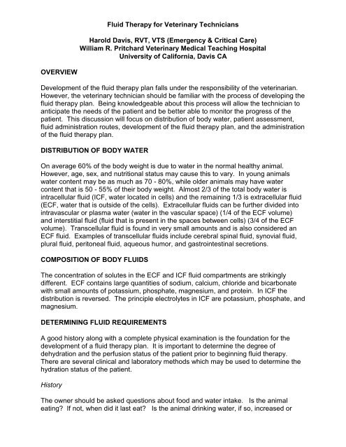
<strong>Fluid</strong> <strong>Therapy</strong> <strong>for</strong> <strong>Veterinary</strong> <strong>Technicians</strong>Harold Davis, RVT, VTS (Emergency & Critical Care)William R. Pritchard <strong>Veterinary</strong> Medical Teaching HospitalUniversity of Cali<strong>for</strong>nia, Davis CAOVERVIEWDevelopment of the fluid therapy plan falls under the responsibility of the veterinarian.However, the veterinary technician should be familiar with the process of developing thefluid therapy plan. Being knowledgeable about this process will allow the technician toanticipate the needs of the patient and be better able to monitor the progress of thepatient. This discussion will focus on distribution of body water, patient assessment,fluid administration routes, development of the fluid therapy plan, and the administrationof the fluid therapy plan.DISTRIBUTION OF BODY WATEROn average 60% of the body weight is due to water in the normal healthy animal.However, age, sex, and nutritional status may cause this to vary. In young animalswater content may be as much as 70 - 80%, while older animals may have watercontent that is 50 - 55% of their body weight. Almost 2/3 of the total body water isintracellular fluid (ICF, water located in cells) and the remaining 1/3 is extracellular fluid(ECF, water that is outside of the cells). Extracellular fluids can be further divided intointravascular or plasma water (water in the vascular space) (1/4 of the ECF volume)and interstitial fluid (fluid that is present in the spaces between cells) (3/4 of the ECFvolume). Transcellular fluid is found in very small amounts and is also considered anECF fluid. Examples of transcellular fluids include cerebral spinal fluid, synovial fluid,plural fluid, peritoneal fluid, aqueous humor, and gastrointestinal secretions.COMPOSITION OF BODY FLUIDSThe concentration of solutes in the ECF and ICF fluid compartments are strikinglydifferent. ECF contains large quantities of sodium, calcium, chloride and bicarbonatewith small amounts of potassium, phosphate, magnesium, and protein. In ICF thedistribution is reversed. The principle electrolytes in ICF are potassium, phosphate, andmagnesium.DETERMINING FLUID REQUIREMENTSA good history along with a complete physical examination is the foundation <strong>for</strong> thedevelopment of a fluid therapy plan. It is important to determine the degree ofdehydration and the perfusion status of the patient prior to beginning fluid therapy.There are several clinical and laboratory methods which may be used to determine thehydration status of the patient.HistoryThe owner should be asked questions about food and water intake. Is the animaleating? If not, when did it last eat? Is the animal drinking water, if so, increased or
decreased amounts? Is the animal suffering any abnormal losses such as vomiting,diarrhea, or polyuria? What is the duration of theses abnormal losses?Physical FindingsSkin turgor or skin elasticity is a crude way of determining the interstitial compartmentvolume status. When assessing skin turgor its best to use the same location <strong>for</strong>consistency in technique. The lateral thorax or between the shoulder blades are goodlocations to assess skin turgor. With 5% dehydration the skin, when lifted, will return toit's normal position fairly quickly but slightly slower than normal. With 8% dehydrationthe skin returns to its normal position slower than 5% dehydration but faster than 12%dehydration. When the patient is 12% dehydrated the skin will remain tented and notreturn to it's normal position. Elasticity of the skin is affected by cachexia and obesity.It is possible to have a normally hydrated patient that has reduced skin elasticity due tocachexia; or a dehydrated patient that has normal skin elasticity as a result of being fat.Other signs consistent with dehydration include dry skin and mucus membranes,oliguria, and signs of compensatory peripheral vasoconstriction.Laboratory AnalysisPacked cell volume (PCV) and total protein (T.P.) are simple tests that can be used toevaluate hydration. PVC and T.P. are often elevated with dehydration. In the case ofanemia, the PCV may appear to be normal but this is due only to hemoconcentration.A urine specific gravity greater than 1.030 usually indicates that the kidneys areresponding to the dehydration in an appropriate manner. Electrolyte and acid basestatus should be determined. Depending on the disease process, It is not uncommonto find electrolyte abnormalities with sodium and / or potassium. Like the electrolytes,the acid base status of a patient may be altered due to a variety of disease processes.The normal pH (hydrogen ion concentration ) is maintained in a narrow range of 7.35 -7.45. If the pH is less than 7.35 or greater than 7.45 the patient is academic oralkalemic respectively.FLUID ADMINISTRATION ROUTESOralThe oral route is the most physiologic. <strong>Fluid</strong>s can be administered rapidly with minimalside effects. This route should not be used in the presence of vomiting. This route isalso inadequate <strong>for</strong> animals that have had acute or extensive fluid losses. <strong>Fluid</strong>absorption is not sufficiently rapid via the oral route, in those cases where the fluid losshas been extensive and blood flow inadequate.Subcutaneous<strong>Fluid</strong>s are usually administered in the subcutaneous tissues over the dorsal neck andcranial trunk. In the absences of vasoconstriction and hypovolemia the rate ofabsorption is approximately six to eight hours. <strong>Fluid</strong>s should be administered at bodytemperature to decrease the discom<strong>for</strong>t to the patient and improve absorption. Onlyisotonic fluids should be administered by this route. Potassium supplementation up to40 mEq/L may be added to the fluids. The rate and volume of administration will varyfrom patient to patient. Skin necrosis and infection are complications associated withthis route of fluid administration.
IntravenousThis is the route of choice when vascular volume restoration is desired. This route issuperior to all others with perhaps the exception of intraosseous. <strong>Fluid</strong> absorption israpid. In addition to isotonic solutions, hyper and hypotonic solutions may beadministered via this route. The rate and volume administered will vary from patient topatient based upon the desired endpoint.Intraosseous<strong>Fluid</strong>s are administered via the bone marrow. Like intravenous administration, fluidabsorption is rapid. This route is indicated when it is difficult to gain venous accessusing standard techniques. This route is best used <strong>for</strong> the short-term administration offluids and or drugs. <strong>Fluid</strong> rates of 11 ml/min with gravity and 24 ml/min under 300 mmHg pressure have been used to deliver the fluids.IntraperitonealIntraperitoneal administration is the administration of fluids into the peritoneal cavity.The rate of absorption from this route is roughly equivalent to the subcutaneous route.Peritonitis and intra-abdominal abscess are potential complications associated with thisroute. Intraperitoneal administration does not offer any advantages over other routes;there<strong>for</strong>e it is reserved as a last resort.DEVELOPMENT OF A FLUID THERAPY PLANThe basic components of a fluid therapy plan include the determination, calculation,and replacement of the volume deficit (percent dehydration); abnormal on going losses;and maintenance needs. The veterinarian will determine the types of fluids used andthe rates. In addition to the previously mentioned components, potassiumsupplementation must be taken into consideration.Volume Deficit RepairTo determine the volumereplacement, multiply the percentdehydration by the patient's bodyweight, this will equal the amount offluids in liters estimated to correctdehydration (Figure 1). When thenature of fluid losses are not known,are due to trauma, and or electrolytemeasurements are not available anexcellent replacement solution is Lactated Ringer's or it's equivalent. The electrolyteconcentration of Lactated Ringer's is similar to that of the ECF.Abnormal Ongoing LossesCalculation of volume deficit 20 kg patient is 10%dehydrated.20 kg x.10 = <strong>2.</strong>0 liters (2000 ml)Figure 1 The calculation <strong>for</strong> determining the volume of fluids tocorrect the dehydration.Losses through vomiting, diarrhea, excessive urination, burns and transudation intobody cavities are considered to be abnormal ongoing losses. These losses should be
calculated into the fluid therapy plan. Typically, abnormal losses should be replacedmL <strong>for</strong> mL. In the event that the volume of abnormal losses is unknown, initially,administer a volume of fluids that is equal to maintenance and adjust the volume up ordown as needed. Lactated Ringer's with supplemented potassium added to a finalconcentration of 10 mEq/L is the fluid of choice <strong>for</strong> abnormal ongoing losses. This typeof fluid will approximate the composition of most abnormal losses.MaintenanceNormal losses occur through breathing,salivation, urination and defecation. It isimportant to include these losses in your fluidtherapy plan. A rough rule of thumb <strong>for</strong> theadministration of maintenance fluids is to give50-75 ml/kg per day, slightly higher <strong>for</strong> thefebrile patient. Predictive charts are alsoavailable <strong>for</strong> the calculation of maintenancefluid needs (Table 1 and 2).Table 1 Dailywaterrequirements<strong>for</strong> catsBodyWeight (kg)TotalWatermL/dayThe composition of a maintenance fluid isone that is low in sodium (40 - 60 mEq/L) andhigh in potassium (13 - 20 mEq/L). Undernormal conditions the body tends to retainsodium and excrete potassium. Hence theneed <strong>for</strong> a maintenance type solution.Maintenance fluids should not be given asreplacement fluids <strong>for</strong> correction ofdehydration this may lead to hyponatremia and hyperkalemia when large volumes offluids are given.A patient may develop hypernatremia or hypokalemia if Lactated Ringer's is used solely<strong>for</strong> maintenance needs and no other source of free water is available. Maintenancefluids may be obtained commercially (Plasmalyte 56 or Normosol M) or can be"homemade". This is done by mixing 1-2 parts D 5W and 1 part Lactated Ringer's andadding potassium chloride to bring the concentration up to 13-20 mEq/L; or using ½strength Lactated Ringer’s or saline with potassium supplementation.mL/Hr1.0 80 31.5 108 5<strong>2.</strong>0 134 6<strong>2.</strong>5 159 73.0 182 83.5 204 94.0 226 94.5 247 105.0 267 11Potassium SupplementationPotassium chloride (KCl) might be added tofluids when serum potassium levels areknown to be low (hypokalemia). If potassiumlevels are not known, hypokalemia may beexpected in cases of fluid loss due togastrointestinal loss, diuresis and anorexia.Based on the magnitude of these losses thebody potassium depletion is thought to bemild, moderate, or severe, the fluids shouldbe supplemented with potassium to 20, 30,or 40 mEq/L respectively. Potassium shouldnot be administered at a rate faster than 0.5mEq/kg/hr (Figure 2). When highTable 2 Dailywaterrequirements<strong>for</strong> dogsBody Total Water mL/HrWeight (KG) mL/day1 132 62 222 93 301 134 373 165 441 1810 742 3120 1248 5230 1692 7140 2100 8750 2481 103
concentrations of potassium are administered the heart rate should be monitoredclosely. Severe hypokalemia can occur when bicarbonate, insulin, or glucose is givenwithout the concomitant administration of KCl. This is due to the shift of potassium fromextracellular fluid into the cells.FLUID ADMINISTRATIONOnce the volume deficitreplacement needs, abnormalongoing losses and maintenanceneeds are calculated, the three aretotaled. The fluid infusion rate maybe determined by totaling up thevolume of fluids to be given anddividing that by the total number ofhours in a day available to safelyadminister the fluids. If there aren'tenough hours available to safelyadminister the fluids the patient maybe sent to an emergency clinic <strong>for</strong>continued care or given some of the required fluids subcutaneously. Generally we liketo correct the fluid deficit (volume needed to correct dehydration) over four to eighthours.It is usually the responsibility of the veterinary technician to administer the plan. Theveterinary technician should be able to correctly add fluid additives and label the fluids.The technician may be required to calculate and set the fluid drip rate. Theresponsibility of monitoring and trouble-shooting the fluid delivery system is thetechnicians. The patient will need to be reassessed at frequent intervals to ensure thatthe fluid therapy plan is meeting its desired effect.<strong>Fluid</strong> delivery systemsFigure <strong>2.</strong> Calculation of maximum K + administrationrate20 kg patient receiving LRS + KCL q.s. to 40 mE/L@ 150 ml/hr (0.15 L/hr).Maximum K + administration rate is:20 kg x 0.5 mEq/kg/hr = 10 mEq/hrThe patient is receiving 4.5 mEq/hr, this does notexceed the max K + .40 mEq/L x 0.15 L/hr = 4.5 mEq/hrLabels should be placed on the fluid bags when hung. The date, time, initials of theperson mixing the fluids and concentration of additives are included on the label.Labels allow all nursing personnel to know the contents of the fluid bags and when andwho hung the fluids.Adhesive tape or commercially available timing labels are placed on the fluid bag andmarked to monitor the volume administered over time. This enables the technician toquickly estimate the volume of fluid received.A large selection of fluid delivery supplies are available on the market. There are thestandard IV fluid administration sets. These administration sets have an in-line dripchamber used to estimate flow rates. Depending on the manufacture, administrationsets are designed to deliver 10, 15, 20, or 60 drops per milliliter. Buretrols allow you toaccurately determine the volume of fluids administered. Depending on themanufacture, buretrols can hold from 100 - 150 mls of fluids. The use of buretrols willdecrease the risk of fluid overload to smaller patient.
To calculate drip rates it will be necessary to know the drop size <strong>for</strong> the administrationset being used. Drops per minute can be calculated from the following <strong>for</strong>mulas figuresthree:<strong>Fluid</strong> delivery<strong>Fluid</strong>s may be delivered via gravity flow or with the use of syringe pumps and fluidinfusion pumps. When setting up aDrop SizeFormulasystem using gravity flow the infusion10 drop/ml (mL/hr) ÷ 6 = drop/minpressure is dependent on the height of the15 drop/ml (mL/hr) ÷ 4 = drop/minfluids above the patient. <strong>Fluid</strong> flow is also20drop/ml (mL/hr) ÷ 3 = drop/minlimited by the ambulatory patient and60 drop/ml (mL/hr) ÷ 1 = drop/mincatheter position. When setting the driprate on a gravity flow system, make sure the limb or neck is fully extended. Thisprevents the fluid rate from exceeding the pre-set rate should the patient's positioningchange.Syringe pumps are used to administer medication or fluids to small patients at aconstant rate. A syringe is placed in the pump and the plunger is automatically pushedat a constant rate.<strong>Fluid</strong> infusion pumps are being used more frequently in veterinary medicine. TheBaxter 6200 and 6300 are two types of fluid infusion pumps commonly used. They arenon-volumetric pumps that deliveries fluids by intermittently squeezing the IV tubing(peristaltic action).MonitoringBecause the calculation of fluid volume is based on subjective data, potentialinaccuracies occur. There<strong>for</strong>e, it is necessary to reassess the patient often. Yourpatient may require more or less of the original calculated fluid volume. You are looking<strong>for</strong> a resolution in the signs that indicated that the patient was in need of fluids.Patients should be weighed at the beginning of therapy and several times a daythereafter to determine fluid gains and loses. Acute change in body weight is a result offluid gains or losses rather than change in body mass. A 0.5 kg weight gain isequivalent to a 0.5 liter fluid gain.