Insulinoma and Prolactinoma in A Young Female - Philippine ...
Insulinoma and Prolactinoma in A Young Female - Philippine ...
Insulinoma and Prolactinoma in A Young Female - Philippine ...

Create successful ePaper yourself
Turn your PDF publications into a flip-book with our unique Google optimized e-Paper software.
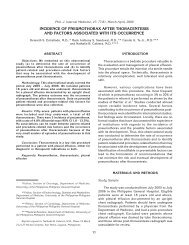
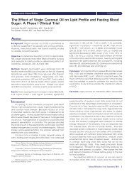
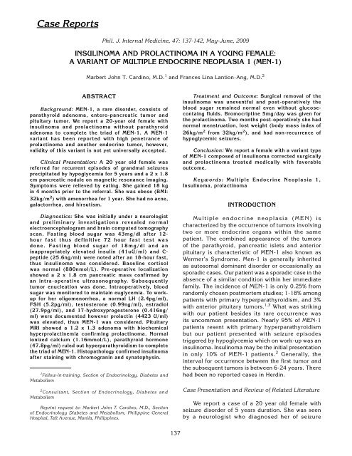
138 Card<strong>in</strong>o MJ T <strong>and</strong> Lantion-Ang FLdisorder <strong>and</strong> was started with Carbamazep<strong>in</strong>e withescalat<strong>in</strong>g doses. Bra<strong>in</strong> computed tomography scan<strong>and</strong> electroencephalogram were normal. Topiramatewas added to the anti-seizure regimen howeverpatient still had 4-6 seizure episodes per year. Therewere episodes of true hypoglycemia precipitat<strong>in</strong>gthe seizure attacks thus a computed tomographyscan of the abdomen was done <strong>and</strong> showed a 2 x1.8cm pancreatic mass thus she was referred toEndocr<strong>in</strong>ology for evaluation.Fig 1.MRI of the Abdomen Show<strong>in</strong>g A 2 x 1.8cmPancreatic Nodule Enhanced By ContrastOn review, our patient started to have symptomsof generalized weakness associated with palpitations,hunger pangs <strong>and</strong> dizz<strong>in</strong>ess resolved with food <strong>in</strong>takefor 5 years. These symptoms usually occurredon wak<strong>in</strong>g up. Because of this pattern, she beganga<strong>in</strong><strong>in</strong>g weight until 6 months later, she had onsetof generalized tonic seizures. Everytime she wasbrought to the emergency room, her blood sugarwas low. After giv<strong>in</strong>g <strong>in</strong>travenous glucose, patientrega<strong>in</strong>ed consciousness. She ga<strong>in</strong>ed 18 kg <strong>in</strong> 4months prior to referral to our service.Menarche was at 11 years old. Menstrual flowwas regular until a year prior to consult she hadamenorrhea. She had no dysmenorrheal, abnormaldischarges <strong>and</strong> history of contraceptive <strong>in</strong>take.Developmental history was at par with age. Shehad good school performance <strong>and</strong> even graduatedcollege. She had no history of blurr<strong>in</strong>g of vision, <strong>and</strong>chronic headaches. She had no family history oftumors, diabetes <strong>and</strong> hypertension.On exam<strong>in</strong>ation, she had normal vital signs.She was obese with a BMI of 32 kg/m, 2 waistcircumference of 95 cm, hip circumference of 99cm, <strong>and</strong> waist to hip ratio was 0.95. There was noacanthosis nigricans, thyromegaly, <strong>and</strong> xanthomas.Silver striae were noted <strong>in</strong> the <strong>in</strong>ner thighs. She hadno visual field cuts on confrontation test.Fig 2.Photograph of the Patient Dur<strong>in</strong>g Her Admission atPGHIn the approach to hypoglycemic patients, wehave to classify the etiology of the hypoglycemia.Our patient had fast<strong>in</strong>g hypoglycemia. Differentialdiagnoses for fast<strong>in</strong>g hypoglycemia <strong>in</strong>cludeddrugs e.g. sulfonylurea, critical illness, adrenal<strong>and</strong> pituitary <strong>in</strong>sufficiency <strong>and</strong> <strong>in</strong>sul<strong>in</strong>oma. Shewas not hypotensive, lethargic <strong>and</strong> weak mak<strong>in</strong>gthe consideration of adrenal <strong>in</strong>sufficiency unlikely.Patient had no <strong>in</strong>take of sulfonylurea.The signs <strong>and</strong> symptoms of patients weresuggestive of <strong>in</strong>sul<strong>in</strong>oma, thus serum <strong>in</strong>sul<strong>in</strong>, C-peptide <strong>and</strong> r<strong>and</strong>om blood sugar were taken aftera 72-hour fast test. Basel<strong>in</strong>e counter-regulatoryhormone levels were taken <strong>and</strong> then blood sugarwas monitored every 2 hours while patient fasted.Basel<strong>in</strong>e 8 oÊclock morn<strong>in</strong>g serum cortisol (880nmol/L) was normal. A 72 hour fast test was done. Onthe 18th hour, she manifested with neuroglycopenicsymptoms. Capillary blood sugar was 38 mg/dl.A low r<strong>and</strong>om blood sugar at 18mg/dl with an<strong>in</strong>appropriately high serum <strong>in</strong>sul<strong>in</strong> levels at 41.08uU/ml (normal: 2.6-24 uU/ml) were documented.C peptide was elevated at 25.6 ng/ml (normal: 1-5ng.ml) <strong>in</strong>dicative of endogenous hyper<strong>in</strong>sul<strong>in</strong>emia.Serum <strong>in</strong>sul<strong>in</strong> <strong>and</strong> C-peptide are secreted <strong>in</strong>equimolar amounts. In our patient, the serum <strong>in</strong>sul<strong>in</strong>to C-peptide was 1.6 reflective of degradation of theC-peptide. The catheps<strong>in</strong> B degrades the C term<strong>in</strong>alam<strong>in</strong>o acids of C-peptide which is be<strong>in</strong>g determ<strong>in</strong>ed<strong>in</strong> the assay, result<strong>in</strong>g to higher <strong>in</strong>sul<strong>in</strong> to C-peptideratio. 4
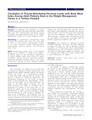
<strong>Insul<strong>in</strong>oma</strong> <strong>and</strong> <strong>Prolact<strong>in</strong>oma</strong> <strong>in</strong> A <strong>Young</strong> <strong>Female</strong> 139Table I. Review of Cases with Insul<strong>in</strong> to C-peptide Ratio <strong>in</strong><strong>Insul<strong>in</strong>oma</strong> Patients 4AuthorLevels <strong>in</strong> <strong>Insul<strong>in</strong>oma</strong>Katabami 7.2G<strong>in</strong> 6.3Sakai 6.1Scarlet 5.9Saudek 5Lebowitz 4.9Kondo 4.2Service 3.6Ciavarella 2.9Turner 2Fujikura 2Our Case 1.6Intraoperative ultrasound showed a 2 x 1.8 cmsolid nodule at the antero-<strong>in</strong>ferior portion of the bodyof the pancreas <strong>and</strong> subsequently an enucleation ofthe nodule was done. Post-operatively, blood sugarrose to 208 mg/dl with<strong>in</strong> 30 m<strong>in</strong>utes from enucleationof the tumor. No symptoms of acute pancreatitis weredocumented <strong>and</strong> serum amylase was slightly elevatedonly at 178 U/ml (normal 83-110 U/ml). There wereno calcifications noted. Post-operative biopsy revealedan <strong>in</strong>sul<strong>in</strong>oma. Cells were arranged <strong>in</strong> anastomos<strong>in</strong>gcords separated by a vascular stroma, <strong>in</strong>terspersedwith sparse collagenous stroma. Chromogran<strong>in</strong>-A<strong>and</strong> synaptophys<strong>in</strong> sta<strong>in</strong><strong>in</strong>g of the tumor were positive(see figure 3). These sta<strong>in</strong><strong>in</strong>g results confirmed thatthe tumor was an <strong>in</strong>sul<strong>in</strong>oma.Fig 3.A) 2x2 cm Pancreatic Nodule Post-Pancreatic Enucleation. B) Hematoxyl<strong>in</strong> <strong>and</strong> Eos<strong>in</strong> Sta<strong>in</strong><strong>in</strong>g Show<strong>in</strong>g Uniform Cells<strong>in</strong> Anastomos<strong>in</strong>g Cords. C) Panoramic View of the Tumor Cells <strong>in</strong> Clumps with Sparse Collagenous Stroma. D) PositiveSynaptophys<strong>in</strong> Sta<strong>in</strong><strong>in</strong>g of the Pancreatic Nodule. E) Positive Chromogran<strong>in</strong> Sta<strong>in</strong><strong>in</strong>g of the Pancreatic Nodule.
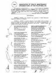
140 Card<strong>in</strong>o MJ T <strong>and</strong> Lantion-Ang FLDur<strong>in</strong>g the recuperation period, she had norecurrence of the hypoglycemic episodes, seizures<strong>and</strong> other neuroglycopenic symptoms. She wasdischarged improved on the 5th post-operative day.Two weeks from discharge, patient had a repeat<strong>in</strong>sul<strong>in</strong> determ<strong>in</strong>ation after a 12 hour fast <strong>and</strong> resultsshowed blood sugar of 100 mg/dl <strong>and</strong> suppressedserum <strong>in</strong>sul<strong>in</strong> of 0.5 uU/ml.On the 2nd month of follow-up, patient had norecurrence of any seizure episodes. No hypoglycemicattacks were claimed. Patient had significantly lostweight with a BMI of 26 kg/m 2 from a previous BMIof 32kg/m 2 .(LH), follicule stimulat<strong>in</strong>g hormone (FSH), thyroidstimulat<strong>in</strong>g hormone (TSH), adenocorticotrophichormone (ACTH), estradiol, progesterone, growthhormone, <strong>in</strong>sul<strong>in</strong> like growth factor-1 (IGF-1) whichwere all normal except for elevated serum prolact<strong>in</strong>at 4423 U/ml (refer to appendix 1 for the results <strong>and</strong>reference values). Patient had normal secondarysexual characteristics. She had well-developed breast<strong>and</strong> female genital tract (Tanner V). She had coarseaxillary <strong>and</strong> pubic hairs. She had no palpable neckmasses. She had a normal free thyrox<strong>in</strong>e (FT4)<strong>and</strong> thyroid stimulat<strong>in</strong>g hormone (TSH). To assessthe parathyroid axis, serum calcium, <strong>in</strong>organicphosphorus <strong>and</strong> parathyroid hormone were taken <strong>and</strong>were all normal (refer to appendix 1 for the results<strong>and</strong> reference values).In the stepwise approach for the diagnosis ofhyperprolact<strong>in</strong>emia, physiologic <strong>and</strong> drug-<strong>in</strong>ducedetiologies must be ruled out. Pregnancy test wasnegative <strong>and</strong> she had no previous <strong>in</strong>take of neuroleptics<strong>and</strong> dopam<strong>in</strong>e blockers. She had no galactorrhea.She had amenorrhea of a year duration. Ultrasoundof the ovaries showed polycystic ovaries. Polycysticovary syndrome (PCOS) may also be associated withhyperprolact<strong>in</strong>emia <strong>and</strong> may present with menstrualirregularities caus<strong>in</strong>g the dist<strong>in</strong>ction of betweenthe 2 conditions difficult. She had no evidence ofhyper<strong>and</strong>rogenism <strong>and</strong> the LH <strong>and</strong> FSH were normalmak<strong>in</strong>g the consideration of PCOS unlikely. A pituitaryMRI was done. It showed a macroadenoma measur<strong>in</strong>g1.2 x 1.5 x 1.3 cm enhanced by contrast thusprolact<strong>in</strong>oma was considered. Hyperprolact<strong>in</strong>emiafrom a pituitary stalk compression was less likelybecause the prolact<strong>in</strong> level was very high (4423 U/ml). Medical therapy rema<strong>in</strong>s to be the cornerstoneof therapy for both the microprolact<strong>in</strong>oma <strong>and</strong>macroprolact<strong>in</strong>oma. Surgical therapy is reserved fortumor growth despite therapy <strong>and</strong> drug resistance tomore than 2 dopam<strong>in</strong>e agonist drugs. Bromocript<strong>in</strong>e2.5mg three times a day was started.Fig 4.Patient at 2 Months Post-Pancreatic Encucleation.<strong>Insul<strong>in</strong>oma</strong> is common <strong>in</strong> patients 30-60 yearsold. It is a tumor of the beta cell of the pancreaticislets. The <strong>in</strong>cidence is very low at 4 cases per 1million-years. 5 Diagnosis of <strong>in</strong>sul<strong>in</strong>oma requires thedocumentation of hypoglycemia associated withhypersecretion of <strong>in</strong>sul<strong>in</strong> <strong>and</strong> presence of pancreaticnodules by imag<strong>in</strong>g techniques. Cases <strong>in</strong>volv<strong>in</strong>gthe younger age group are associated with multipleendocr<strong>in</strong>e disorders. Four percent of <strong>in</strong>sul<strong>in</strong>oma mayhave multiple endocr<strong>in</strong>e neopasia-1. 2 Generally,they occur <strong>in</strong> <strong>in</strong>dividuals younger than 20 years old.To assess for <strong>in</strong>volvement of the pituitary axis thefollow<strong>in</strong>g were taken: serum lute<strong>in</strong>iz<strong>in</strong>g hormoneOn the 2nd month of follow-up, she hadresumption of her regular menstruation. She hadno occurrence of blurr<strong>in</strong>g of vision. A repeat serumprolact<strong>in</strong> of 405 U/ml was documented 6 monthsafter. Because of the significant drop <strong>in</strong> the prolact<strong>in</strong>to physiologic level <strong>and</strong> the normalization of hermenstruation, bromocript<strong>in</strong>e was decreased to2.5 mg twice a day. <strong>Prolact<strong>in</strong>oma</strong> may be the firstmanifestation of MEN-1 <strong>in</strong> fewer than 10% of patients<strong>and</strong> less than 3% of patients with anterior pituitarytumors will have MEN-1. 6A multiple endocr<strong>in</strong>e neoplasia was considered<strong>in</strong> this case because of the <strong>in</strong>volvement of 2 or moreendocr<strong>in</strong>e gl<strong>and</strong>s. Multiple endocr<strong>in</strong>e neoplasia
<strong>Insul<strong>in</strong>oma</strong> <strong>and</strong> <strong>Prolact<strong>in</strong>oma</strong> <strong>in</strong> A <strong>Young</strong> <strong>Female</strong> 141(MEN) is characterized by occurrence of tumors<strong>in</strong>volv<strong>in</strong>g two or more endocr<strong>in</strong>e organs with<strong>in</strong> thesame patient. The comb<strong>in</strong>ed appearance of thetumors of the parathyroid, pancreatic islets <strong>and</strong>anterior pituitary is characteristic of MEN-1 alsoknown as WermerÊs Syndrome. However, 94.5% ofMEN-1 presented with primary hyperparathyroidismwhich was absent <strong>in</strong> our case. A variant of MEN-1with high penetrance of prolact<strong>in</strong>oma plus anotherendocr<strong>in</strong>e tumor has been described.Fig 5.Pituitary Microadenoma Measur<strong>in</strong>g 1.2 x 1.5 x 1.3 cm, Enhanced on Contrast with Pituitary MRIDISCUSSIONIn a review of cases of MEN-1 at the US-NationalInstitute of Health, 94.5% of cases had primaryhyperparathyroidism as the <strong>in</strong>itial presentation. 6 Thedistribution of cases is depicted below.second decade which is compatible with our casehowever it is also at this age bracket where we expectpeak <strong>in</strong>cidence of primary hyperparathyroidism as acomponent of MEN-1 which was not evident <strong>in</strong> ourcase. Please refer to the figure below.Parathyroid tumorGastr<strong>in</strong>oma<strong>Insul<strong>in</strong>oma</strong><strong>Prolact<strong>in</strong>oma</strong>Fig 6.Schemic Representation of 220 Patients Seen withMEN-1Fig 7.Adapted from Marx et al, Annals of InternalMedic<strong>in</strong>e, 1998 6As we shall see from the representation above,94.5% of cases willl have primary hyperparathyroidism.Twelve percent have comb<strong>in</strong>ed parathyroid,pancreatic <strong>and</strong> pituitary tumors. 2 A comb<strong>in</strong>ation ofboth a pancreatic <strong>and</strong> pituitary tumor occurs <strong>in</strong> 0.5%only. 2The peak of onset of MEN-1 occurs dur<strong>in</strong>g theThe gene for MEN-1 is localized <strong>in</strong> chromosome11q13 by mapp<strong>in</strong>g studies. Men<strong>in</strong> is nuclear prote<strong>in</strong>which is the by-product of the gene mutation <strong>in</strong>volved<strong>in</strong> transcription regulation, genome <strong>in</strong>stability <strong>and</strong>cell division. The utility of MEN-1 mutation is forearly detection amongst immediate family members<strong>and</strong> first degree relatives. However, our case was
142 Card<strong>in</strong>o MJ T <strong>and</strong> Lantion-Ang FLmost likely sporadic. Our patientÊs immediate familymembers were screened with a fast<strong>in</strong>g blood sugar<strong>and</strong> serum calcium <strong>and</strong> none had abnormal results.None <strong>in</strong> the family also compla<strong>in</strong>ed with blurr<strong>in</strong>gof vision, nausea <strong>and</strong> vomit<strong>in</strong>g suggestive of aspace occupy<strong>in</strong>g lesions. Chromosomal test<strong>in</strong>g formutations <strong>in</strong>volv<strong>in</strong>g MEN-1 gene is not available <strong>in</strong> thePhilipp<strong>in</strong>es. Pick-up rates by research centers <strong>in</strong> theUnited States are at best 24% for sporadic cases <strong>and</strong>68% for familial cases. 7 Cl<strong>in</strong>ical criteria for genetictest<strong>in</strong>g for sporadic MEN-1 <strong>and</strong> rarer comb<strong>in</strong>ations ofMEN-1 have not been well-def<strong>in</strong>ed. 7 Utility of MEN-1mutation identification does not necessarily lead tomajor therapeutic <strong>in</strong>tervention even <strong>in</strong> the youngerage groups, 7 unlike RET identification <strong>in</strong> MEN-2 weretotal thyroidectomy should be done long before theonset of its manifestation for prevention <strong>and</strong> cure ofmedullary thyroid cancer. In the absence of MEN-1mutational analysis, the follow<strong>in</strong>g laboratory testsare recommended among suspected carriers ofthe disease accord<strong>in</strong>g to the consensus guidel<strong>in</strong>esby the <strong>in</strong>ternational work<strong>in</strong>g group of cl<strong>in</strong>icalendocr<strong>in</strong>ologists however this does not replace themutation test<strong>in</strong>g. 8Table II. Program of Tests <strong>and</strong> Schedule for Suspected MEN-1carrier 8Tumor Age Biochemical test Imag<strong>in</strong>g(annual)Parathyroid adenoma 8 Calcium, PTH NoneGastr<strong>in</strong>oma 20 Gastr<strong>in</strong> None<strong>Insul<strong>in</strong>oma</strong> 5 FBS, Insul<strong>in</strong> MRIAnterior Pituitary Tumor 5 Prolact<strong>in</strong>, IGF-1 MRIForegut Carc<strong>in</strong>oid 20 None CT-ScanCONCLUSIONWe presented a 20 year old female with varianttype of MEN-1 present<strong>in</strong>g with hypoglycemic seizures<strong>and</strong> amenorrhea. On work-up, she had <strong>in</strong>sul<strong>in</strong>oma <strong>and</strong>prolact<strong>in</strong>oma. Her <strong>in</strong>sul<strong>in</strong>oma was removed by surgery<strong>and</strong> her macroprolact<strong>in</strong>oma treated medically. Shehad favorable outcome with surgical removal of the<strong>in</strong>sul<strong>in</strong>oma <strong>and</strong> medical therapy for the prolact<strong>in</strong>oma.<strong>Young</strong> patients diagnosed with <strong>in</strong>sul<strong>in</strong>oma should bescreened for other endocr<strong>in</strong>e tumors that def<strong>in</strong>e multipleendocr<strong>in</strong>e neoplasia. Because this is a rare syndrome,a high <strong>in</strong>dex of suspicion is a must to diagnose thisproblem <strong>and</strong> address the associated morbidity result<strong>in</strong>gfrom hormone excess.REFERENCES1. Kouvaraki M: Genotype-phenotype Analysis <strong>in</strong> MEN-1. Archof Surgery ; 137: 641, 2002.2. Farren D: Cl<strong>in</strong>ical Studies of MEN-1 <strong>in</strong> 220 Patients. QJMed;89:653, 1999.3. Carty, et al.: Variable Penetrance <strong>and</strong> Spectrum ofManifestations of MEN-1. Arch of Surgery; 124:1106,1998.4. Fujikura: A Case of Secret<strong>in</strong> Sensitive <strong>Insul<strong>in</strong>oma</strong> with LowC-Peptide. Endocr<strong>in</strong>e Journal; 54: 113, 2007.5. Hiramoto, et al.: Intraoperative Ultrasound <strong>and</strong> Localizationof Occult <strong>Insul<strong>in</strong>oma</strong>. Arch of Surgery 136: 1020, 2001.6. Marx, et al.: MEN-1: Cl<strong>in</strong>ical <strong>and</strong> Genetic Topics. Annals ofInternal Medic<strong>in</strong>e; 129:484, 1998.7. Ellard S, Hattersley AT, Brewer CM, Vaidya B: Detection ofAn MEN-1 Gene Mutation Depends on Cl<strong>in</strong>ical Features<strong>and</strong> Supports Current Referral Criteria for DiagnositcMolecular Genetic Test<strong>in</strong>g. Cl<strong>in</strong> Endocr<strong>in</strong>ol; 62:169,2005.8. Br<strong>and</strong>i, et al.: Consensus Guidel<strong>in</strong>es for MEN-1 <strong>and</strong> MEN-2.JCEM 86: 5658, 2001.APPENDIXTable I. Hormonal <strong>and</strong> Blood Chemistries of PatientLaboratory Tests Results Normal ValueR<strong>and</strong>om Blood Sugar 18 80-110mg/dlSerum Insul<strong>in</strong> 41 2.6-24 uU/mlC-peptide 25.6 1.1-5ng/mlIonized calcium 1.16 1.0-1.3 mmol/LPhosphorus 3.5 2.5-4.8 mg/dlIntact PTH 47.8 10-65 pg/mlFT4 0.8 0.8-2 ng/mlTSH 0.45 0.39-6.16 ulU/mlTestosterone 0.99 0.26-1.3 ng/mlEstradiol 27.9 12-166 pg/ml17-OH-progesterone 0.416 0.2-1.5 ng/mlLH 2.4 1.5-5 pg/mlFSH 5.26 3.5-12.5 pg/mlACTH (11 pm) 15.5 0-46 pg/mlIGF-1 34nmol/L 8-51 nmol/LGastr<strong>in</strong> 17 < 15 pg/mlProlact<strong>in</strong> 4423 127-637 U/ml