

Released August 2007 - The Indian Society for Parasitology
Released August 2007 - The Indian Society for Parasitology
Released August 2007 - The Indian Society for Parasitology

You also want an ePaper? Increase the reach of your titles
YUMPU automatically turns print PDFs into web optimized ePapers that Google loves.
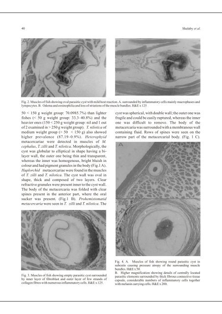
40 Shalaby et al.Fig. 2. Muscles of fish showing oval parasitic cyst with mild host reaction. A. surrounded by inflammatory cells mainly macrophases andlympocytes. B. Odema and eosinophilia and loss of striations of the muscle bundles. H&E x 12550 < 150 g weight group: 70.0985.7%) than lighter cyst was spherical, with double wall; the outer one wasfishes (< 50 g weight group: 33.3–40.8%) and the fragile and could be easily ruptured, whereas the innerheavier ones (150 < 250 g weight group: nil and 1 out one was difficult to remove. <strong>The</strong> body of theof 2 examined in > 250 g weight group). T. nilotica of metacercaria was surrounded with a membranous wallmedium weight group (> 50 < 150 g) also showed containing fluid. Rows of spines were seen on thehigher prevalence (87.19–0.9%). Heterophyid narrow part of the metacercarial body. (Fig. 1 C).metacercariae were detected in muscles of M.cephalus, T. zilli and T. nilotica. Morphologically, thecyst was globular to elliptical in shape having a bilayerwall, the outer one being thin and transparent,whereas the inner was homogenous, bright bluish incolour and had pigment granules in the body (Fig.1 A).Haplorchid metacercariae were found in the musclesof T. zilli and T. nilotica. <strong>The</strong> cyst wall was oval inshape, thick and composed of two layers. Clearrefractive granules were present inner to the cyst wall.<strong>The</strong> body of the metacercaria was folded with clearspines present in the anterior part, where the oralsucker was present. (Fig.1 B). Prohemistomatidmetacercaria were seen in T. zilli and T. nilotica. <strong>The</strong>Fig. 3. Muscles of fish showing empty parasitic cyst surroundedby inner layer of fibroblast and outer layer of few strands ofcollagen fibres with numerous inflammatory cells. H&E x 125.Fig. 4. A. Muscles of fish showing round parasitic cyst insubcutis causing pressure atropy of the surrounding musclebundles. H&E x 50.B. Higher magnification showing details of centrally locatedparasitic elements surrounded by thick fibrous connective tissuecapsule, considerable numbers of inflammatory cells togetherwith melanin carrying cells. H&E x 200.


